. 2019 Apr 3;102(1):105-119.e8.
doi: 10.1016/j.neuron.2019.01.035. Epub 2019 Feb 18.

Neuron-Specific Genome Modification in the Adult Rat Brain Using CRISPR-Cas9 Transgenic Rats

Affiliations

Neuron-Specific Genome Modification in the Adult Rat Brain Using CRISPR-Cas9 Transgenic Rats

Susanne Bäck et al. Neuron. .

Abstract

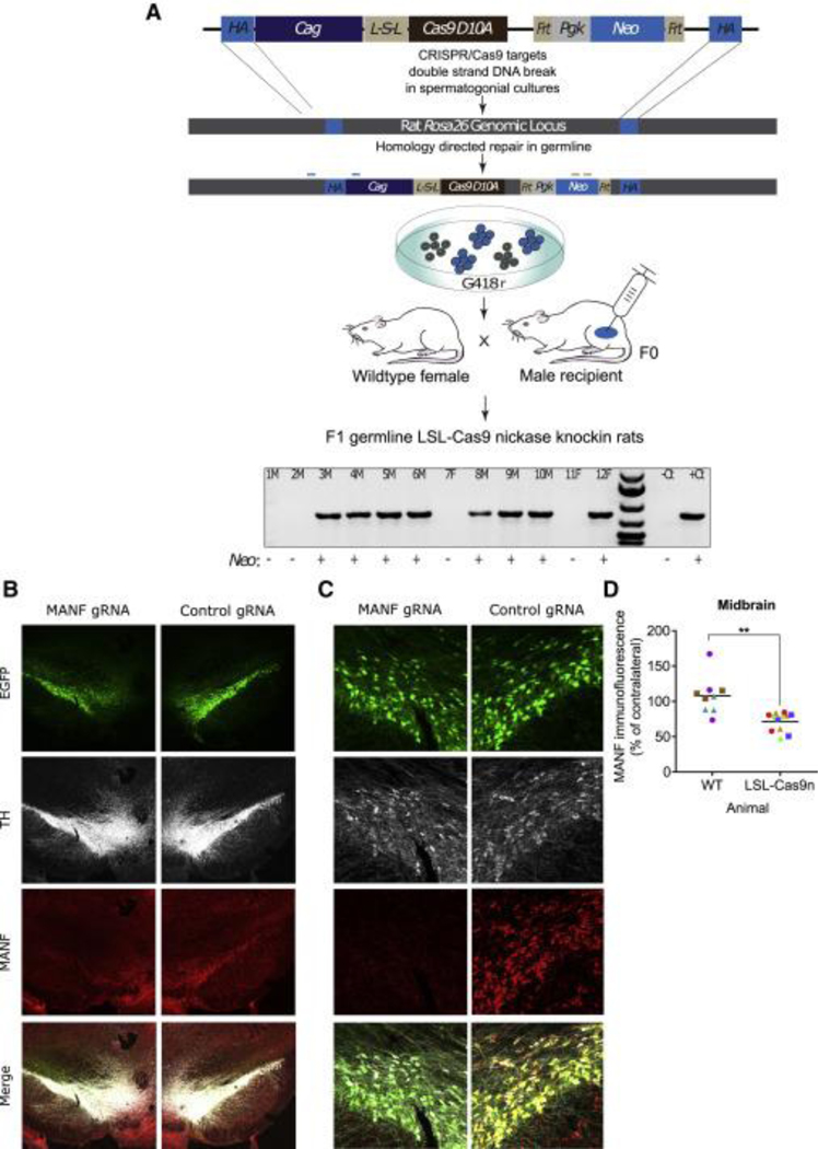
Historically, the rat has been the preferred animal model for behavioral studies. Limitations in genome modification have, however, caused a lag in their use compared to the bevy of available transgenic mice. Here, we have developed several transgenic tools, including viral vectors and transgenic rats, for targeted genome modification in specific adult rat neurons using CRISPR-Cas9 technology. Starting from wild-type rats, knockout of tyrosine hydroxylase was achieved with adeno-associated viral (AAV) vectors expressing Cas9 or guide RNAs (gRNAs). We subsequently created an AAV vector for Cre-dependent gRNA expression as well as three new transgenic rat lines to specifically target CRISPR-Cas9 components to dopaminergic neurons. One rat represents the first knockin rat model made by germline gene targeting in spermatogonial stem cells. The rats described herein serve as a versatile platform for making cell-specific and sequence-specific genome modifications in the adult brain and potentially other Cre-expressing tissues of the rat.

Keywords: AAV; CRISPR-Cas9; MANF; brain; genome editing; lsl-Cas9; spermatogonial stem cells; transgenic rat.


Figures

Figure 1.
Figure 1.
Experimental Approaches for Induction of CRISPR-Cas9-Mediated Knockout of Genes in the Rat Midbrain Targeted gene disruption in the adult rat midbrain was achieved using four different approaches. (A) WT rats received co-injections of AAV vectors expressing Cas9 or gRNAs specific to rat tyrosine hydroxylase (Th) gene. (B) The midbrain of transgenic rats (DAT-iCre) expressing Cre in their dopaminergic (DAT-ex- pressing) neurons was targeted with Cas9 and Cre-dependent Th gRNA to modify the Th gene only in dopaminergic neurons. (C) Co-injections of AAV iCre and AAV gRNAs for Th or Manf were performed to modify the Th or Manf genes in transgenic rats expressing Cre- dependent Cas9 or nickase. (D) A cross of the DAT-iCre and LSL-Cas9 rats generated rats with Cas9 expression limited to dopaminergic neurons, allowing for sequence-specific alterations of the genome in dopaminergic neurons with an injection of a single AAV carrying gRNAs.
Figure 2.
Figure 2.
Co-delivery of AAV Th gRNA and AAV Cas9 or AAV Nickase into WT Rats Leads to Decreased TH Immunoreactivity (A and D) Representative images of the midbrain (MB; top) and striatum (bottom) from WT rats 6 weeks following a co-injection of (A) AAV Cas9 or (D) AAV Cas9 nickase with either AAV control gRNAs (right side [R]) or AAV Th gRNAs (left side [L]) into the MB. GFP fluorescence, as a marker of gRNA delivery, and TH immunoreactivity, as a marker of TH knockout efficacy, were assessed. (B and E) Quantification of TH immunoreactivity in the SN shown as Th gRNA-injected compared to control gRNA-injected side with or without a co-injection of AAV Cas9. Both datasets were normalized to DAPI following 2-, 4-, or 6-week incubations. Each data point represents a coronal section (3–4 sections/animal), and each color represents a distinct animal (n = 3–5/group). (B) One-way ANOVA, F3,10 = 34.8, p < 0.0001; a versus b: p < 0.0001, Tukey’s multiple comparisons test. (E) One-way ANOVA, F3,33 = 10.59, p < 0.0001; a versus b: p < 0.01; a versus c: p < 0.05, a versus d: p = 0.226, b and c versus d: p < 0.001, Tukey’s multiple comparisons test. (C and F) Quantification of TH immunoreactivity in the striatum, as described for (B and E) (3–4 coronal sections/animal from 3–5 animals/group). (C) One-way ANOVA, F3,10 = 52.72, p < 0.0001; a versus b: p < 0.05 (4 wk); a versus b: p < 0.01 (6 wk); a versus c: p < 0.001; b versus c: p < 0.0001 (4 wk); b versus c: p < 0.0001 (6 wk), Tukey’s multiple comparison test. (F) One-way ANOVA, F3,37 = 31.20, p < 0.0001; a versus b and c, p < 0.0001; a versus d, p = 0.986; b versus d, p = 0.0001; c versus d, p < 0.0001, Tukey’s multiple comparisons test. Scale bars, 500 mm. The same group of animals injected with control gRNA only are used for reference in the midbrain (B and E) and the striatum (C and F).
Figure 3.
Figure 3.
Characterization of DAT-iCre Rats (A and B) Representative images of (A) in situ RNA hybridization and (B) immunohistochemical staining of the midbrain in WT and DAT-iCre transgenic rats.(A) Green, Cre; red, dopamine transporter (Dat); blue, DAPI staining. (B) Green, Cre; red, tyrosine hydroxylase (TH). Scale bars, 20 mm.
Figure 4.
Figure 4.
Unilateral Injection of LSL-gRNA in Rats Expressing Cre in Dopaminergic Neurons Enables Region- and Cell-Type-Specific Th Editing (A and C) Representative images demonstrate unilateral loss of TH immunoreactivity in the (A) midbrain and (C) striatum of DAT-iCre rats (bottom), but not WT animals (top), 6 weeks following co-delivery of AAV Cas9 and AAV LSL-Th gRNA to the left (L) SN. GFP fluorescence represents transduction by AAV. (B) Quantification of optical density of TH immunoreactivity relative to DAPI in the ipsilateral compared to the contralateral SN of animals described in (A). Each point represents one analyzed coronal section (n = 3–4 sections/animal), and each color represents a distinct animal (n = 5–6/group, unpaired t test, t(9) = 4.166, **p = 0.0024). (D) Quantification of optical density of TH immunoreactivity relative to DAPI in the ipsilateral compared to the contralateral striatum of animals described in (C). Each point represents one analyzed coronal section (n = 3–4 sections/animal), and each color represents a distinct animal (n = 5–6/group, unpaired t test, t(11) = 2.896, *p = 0.0177). Scale bars, 500 mm
Figure 5.
Figure 5.
Characterization and Use of an LSL-Cas9 Transgenic Rat for Cre-Dependent Knockout of TH (A) Representative confocal images of colocalization of iCre recombinase and the FLAG-tagged Cas9 transgene in LSL-Cas9 rats. FLAG immunoreactivity is not observed following delivery of Flpo, a non-Cre recombinase. iRFP fluorescence indicates comparable delivery of Flpo- and iCre-encoding viruses. (B) Representative confocal images of unilateral TH loss in the SN of LSL-Cas9 rats 4 weeks after a midbrain injection of AAV iCre and AAV control gRNAs (right side, top) or AAV Th gRNAs (left side, bottom). Comparable EGFP fluorescence in control and Th gRNAs-injected hemispheres indicates comparable viral delivery between conditions. (C) A TH-immunostained striatal section from a rat injected as in (B) (L, left side; R, right side). (D) Quantification of optical density of TH immunoreactivity in the SN and striatum of animals described in (B) and (C). Each data point represents one analyzed coronal section (n = 3–4 sections/animal), and each color represents a different animal (n = 4). Scale bars represent 50 mm (A), 100 mm (B), and 500 mm (C).
Figure 6.
Figure 6.
Developing an Assay for Knockout of MANF In Vivo (A) A schematic of the gRNA-binding sites and the PCR assay used to amplify the 893 nt flanking the second exon of rat Manf. (B–E) Rat primary cortical neurons were transduced with AAV Cas9 and AAV Manf gRNAs or AAV control gRNAs and harvested 1 week later for determination of mutagenesis and knockout. (B) Co-transduction with AAV Cas9 and AAV Manf gRNAs resulted in resolvase-induced cleavage of the PCR product (arrows). (C) An alignment of seven independently isolated clones of the PCR fragment shows precise +A insertions among the alleles. Knockout of Manf was verified with (D) real- time qRT-PCR and (E) Wes analyses of Manf mRNA and protein levels, respectively. (D) Manf mRNA levels were normalized to the geometric mean of reference genes and presented as 2-ddCq ± upper and lower limits (n = 3, unpaired t test using dCq values, t(4) = 20.27, ****p < 0.0001). (E) The MANF protein band density was normalized to actin and presented as density relative to control gRNA (mean ± SE, n = 3, unpaired t test, t(4) = 4.999, **p = 0.0075). The arrow in the cropped blot points at the ~25-kDa MANF band. (F) Representative images of unilateral loss of MANF immunoreactivity in the SN of LSL-Cas9 rats four weeks after co-injection of AAV iCre and AAV control gRNAs or AAV Manf gRNAs. GFP fluorescence represents delivery of gRNA. Scale bar 100 mm.(G) Quantification of the optical density of MANF immunoreactivity in the SN of LSL-Cas9 or WT animals injected as described in (F). Each data point represents one analyzed coronal section (n = 3–4/animal), and each color represents data from a distinct animal (n = 4/group, unpaired t test, t(6) = 5.437, **p = 0.0016).
Figure 7.
Figure 7.
Production of LSL-Cas9 Nickase Knockin Rats by HDR in Spermatogonial Stem Cells (A) Transgenic LSL-nickase rats were produced using donor rat spermatogonia harboring an ~11.9 kb, CAG promoter-driven floxed-stop FLAG-tagged SpCas9(D10A) transgene that was precisely targeted to the rat Rosa26 germline locus by CRISPR-Cas9-mediated HDR. Targeted spermatogonia were genetically selected for in G418-containing culture medium and then injected into recipient rat testes (F0, founder males) to produce spermatozoa that transmitted the targeted transgene to F1 germline mutant progeny (LSL-nickase hemizygotes). Genotyping of F1 progeny derived from a recipient male transplanted with spermatogonia genetically modified with the LSL-nickase targeting construct demonstrated the expected 797-bp PCR amplicon using primers that hybridize to rRosa26 locus and CAG transgene, respectively; - and + at the bottom of the gel represent genotyping results from primers that hybridize to the neomycin (Neo) selection cassette; F, female F1 pup DNA; M, male F1 pup DNA; WT (-Ct) and transgenic (+Ct) rat DNA from F1 progeny were generated from an earlier litter. (B and C) Representative midbrain images at (B) 23 and (C) 203 magnification indicating unilateral loss of MANF immunoreactivity in transgenic LSL- Cas9 nickase rats 4 weeks following co-injection of AAV iCre and AAV Manf gRNAs or AAV control gRNAs into the SN. GFP fluorescence indicates viral transduction. (D) Quantification of the optical density of MANF immunoreactivity in the SN of animals described in (B) and (C). Each data point represents one coronal section (n = 3/animal), and each color repre- sents data from a distinct animal (n = 3–4/group, unpaired t test, t(5) = 5.553, **p = 0.0026).
Figure 8.
Figure 8.
Characterization and Use of the LSL-Cas9 3 DAT-iCre Double-Transgenic Rat for Knockout of MANF in the Midbrain Dopaminergic Neurons (A) Representative confocal images of TH and FLAG-tagged Cas9 colocalization in the SN of LSL-Cas9 3 DAT-iCre double-transgenic rats. (B) Representative midbrain images depicting unilateral loss of MANF immunoreactivity (white arrowheads) in TH+ dopaminergic cells in LSL-Cas9 3 DAT-iCre rats 4 weeks following injection of AAV control or AAV Manf gRNAs into the SN. (C) High-magnification confocal images acquired 4 weeks after AAV injection demonstrating selective loss of MANF immunoreactivity in TH+ cells that received Manf gRNAs (right), but not in those that received control gRNAs (left). GFP fluorescence indicates viral transduction. Open arrowheads signal to GFP+TH- cells in which MANF immunoreactivity is maintained in both control and experimental conditions. Closed arrowheads signal to GFP+TH+ cells in which MANF immunoreactivity is lost in the hemisphere receiving Manf gRNAs, but not control.

References

    1. Bae S, Kweon J, Kim HS, and Kim JS (2014). Microhomology-based choice of Cas9 nuclease target sites. Nat. Methods 11, 705–706. - PubMed
    1. Barrangou R, Fremaux C, Deveau H, Richards M, Boyaval P, Moineau S, Romero DA, and Horvath P. (2007). CRISPR provides acquired resis- tance against viruses in prokaryotes. Science 315, 1709–1712. - PubMed
    1. Brinkman EK, Chen T, Amendola M, and van Steensel B. (2014). Easy quantitative assessment of genome editing by sequence trace decomposition. Nucleic Acids Res. 42, e168. - PMC - PubMed
    1. Chapman KM, Medrano GA, Jaichander P, Chaudhary J, Waits AE, Nobrega MA, Hotaling JM, Ober C, and Hamra FK (2015). Targeted germline modifications in rats using CRISPR/Cas9 and spermatogonial stem cells. Cell Rep. 10, 1828–1835. - PMC - PubMed
    1. Cho SW, Kim S, Kim JM, and Kim JS (2013). Targeted genome engineering in human cells with the Cas9 RNA-guided endonuclease. Nat. Biotechnol. 31, 230–232. - PubMed

Publication types

MeSH terms

LinkOut - more resources