. 2019 Mar 21;73(6):1282-1291.e8.
doi: 10.1016/j.molcel.2019.01.028. Epub 2019 Feb 18.

An NAD+ Phosphorylase Toxin Triggers Mycobacterium tuberculosis Cell Death

Affiliations

An NAD+ Phosphorylase Toxin Triggers Mycobacterium tuberculosis Cell Death

Diana Mendes Freire et al. Mol Cell. .

Abstract

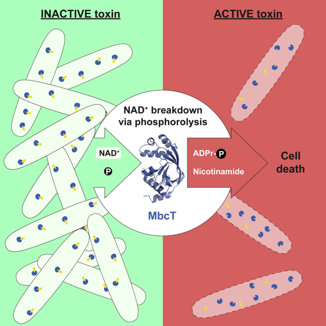
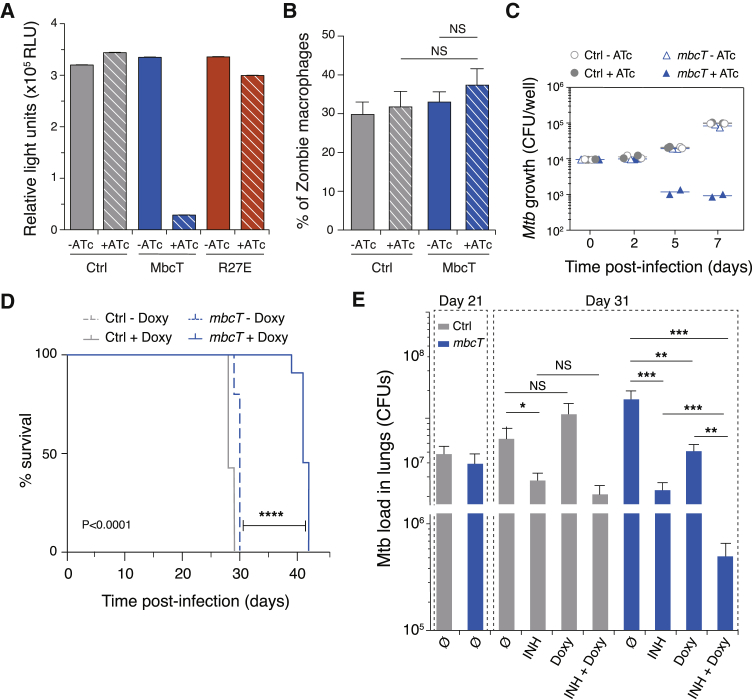
Toxin-antitoxin (TA) systems regulate fundamental cellular processes in bacteria and represent potential therapeutic targets. We report a new RES-Xre TA system in multiple human pathogens, including Mycobacterium tuberculosis. The toxin, MbcT, is bactericidal unless neutralized by its antitoxin MbcA. To investigate the mechanism, we solved the 1.8 Å-resolution crystal structure of the MbcTA complex. We found that MbcT resembles secreted NAD+-dependent bacterial exotoxins, such as diphtheria toxin. Indeed, MbcT catalyzes NAD+ degradation in vitro and in vivo. Unexpectedly, the reaction is stimulated by inorganic phosphate, and our data reveal that MbcT is a NAD+ phosphorylase. In the absence of MbcA, MbcT triggers rapid M. tuberculosis cell death, which reduces mycobacterial survival in macrophages and prolongs the survival of infected mice. Our study expands the molecular activities employed by bacterial TA modules and uncovers a new class of enzymes that could be exploited to treat tuberculosis and other infectious diseases.

Keywords: MbcTA; NAD; bacterial cell death; mycobacterium; toxin-antitoxin system; tuberculosis.


Figures

None
Graphical abstract
Figure 1
Figure 1
Rv1989c-Rv1990c Is a Bactericidal TA System in Mtb (A) WT Mtb or MtbΔTA transformed with pGMC derivatives carrying Rv1990c, Rv1989c, or Rv1989c-Rv1990c as indicated were grown in 7H9 ADC Tween medium. Growth on solid medium was tested by spotting serial 10-fold dilutions on 7H11 OADC agar (−ATc) or the same medium supplemented with anhydrotetracycline (+ATc) to induce transcription from the P1 promoter. Plates were observed after 20 days at 37°C. Shown data are representative of three independent experiments. (B) Growth of WT Mtb or MtbΔTA transformed with pGMC derivatives carrying Rv1990c, Rv1989c, or Rv1989c-Rv1990c in liquid medium as monitored by turbidity measurements (McFarland units). ATc was added at day 0 to induce ectopic gene expression when indicated. Data are represented as mean of three technical replicates ± SD. Shown data are representative of three independent experiments. (C) Survival of MtbΔTA strains carrying pGMC-TetR-P1-Rv1989c as measured by CFU scoring of liquid cultures. ATc was added at time 0 to induce expression of Rv1989c. Samples were collected at the seven different time points, cells were washed with growth medium to remove ATc and 10-fold serial dilutions were spotted on agar plates for CFU counting. Data are represented as mean of three independent replicates ± SD. (D) MtbΔTA strains carrying pGMC-TetR-P1-Rv1989c or an empty vector control were grown for 4 days in the presence of ATc (+ATc) or left untreated (−ATc). Cells were labeled with the LIVE/DEAD BacLight stains (Syto 9; propidium iodide (PI)) and analyzed by fluorescence-activated cell sorting (FACS). The empty vector control was either left unlabeled (negative control) or heat killed by incubation for 1 h at 100°C (positive control). Quadrants were established using the negative (no stain) and positive (heat-killed Mtb) controls as references. Shown data are representative of two independent experiments. (E) Bar diagram showing the fraction of MtbΔTA cells permeable to PI and Sypro 9 as determined by FACS analysis (see D). Data are represented as mean of two independent replicates ± SD. (F) Visualization of Syto9 (green) or PI (red) incorporation in MtbΔTA cells following ATc-induced expression of Rv1989c from pGMC-TetR-P1-Rv1989c by spinning disk confocal microscopy (see D). MtbΔTA cells transformed with empty vector were included as a negative control. PI incorporation is indicative of membrane damage. Representative maximum intensity Z projection images are shown. Scale bar, 5 μm. See also Figures S1 and S2.
Figure 2
Figure 2
Crystal Structure of the MbcTA Complex and Homology of MbcT to ARTs and NADases (A) Overall structure of the MbcTA heterododecamer consisting of three heterotetramers (3x[MbcT-MbcA]2) arranged around a 3-fold symmetry axis (as indicated by a black triangle). The dashed line box in the front view (left) represents one [MbcT-MbcA]2 heterotetramer formed by two MbcT (blue) and two MbcA (yellow) molecules as indicated in the side view (right). (B) Cartoon representation of the MbcTA complex and zoom-in of the putative NAD+-binding site. N and C termini and secondary structure elements are labeled (left). Part of the α2-β2 loop (G45GRW48) that lines the putative NAD+-binding site is displayed in red. Zoom (right): interactions based on a distance <3.8 Å as calculated by the PISA server (Krissinel and Henrick, 2004) are indicated by dotted lines. The orientation of the MbcTA complex was modified to optimize visualization of the MbcT/MbcA interaction. (C) Structure-based alignment of the conserved active-site motifs found in three sequence regions of mono- and poly-ADP-ribosyltransferases (ARTs) and NAD+ glycohydrolases (NADases). The C-alpha atoms of Tyr28 in MbcT do not strictly superimpose with those of Tyr97 and Tyr907 in Dtox and PARP1, respectively (see also D). Numbering of the residues refers to UniProt entries: MbcT (UniProt: Y1989), diphtheria toxin (Dtox) (UniProt: P00588), cholera toxin (Ctoxin) (UniProt: P01555), human poly [ADP-ribose] polymerase 1 (PARP1) (UniProt: P09874), the C-terminal toxin domain (tuberculosis necrotizing NAD+ glycohydrolase toxin TNT) of the Mtb outer membrane channel protein CpnT (UniProt: O05442), P. aeruginosa NAD+ glycohydrolase Tse6 (UniProt: Q9I739), and Streptococcus pyogenes NAD+ glycohydrolase SPN (UniProt: D7S065). (D) Structural comparison of the active site in MbcT, Dtox in complex with NAD+ (PDB: 1TOX), PARP1 (PDB: 4DQY), and CpnT (PDB: 4QLP). Conserved residues are colored according to their localization in the three distinct regions (cf. C). See also Figures S3 and S4.
Figure 3
Figure 3
Enzymatic Activity of MbcT (A) Representative autoradiograph of a TLC plate showing MbcT-mediated depletion of 32P-NAD+ over time and simultaneous accumulation of 32P-ADP-ribose (Appr) and a secondary reaction product (white arrow). The dotted line indicates the position where samples were applied to the plate. Similar results were obtained in two independent experiments. (B) Representative HPLC chromatograms of the reaction products of NAD+ (0.5 mM) with MbcT (0.7 μM) in the presence (orange) or absence (black) of sodium phosphate (30 mM). The white arrow indicates the reaction product formed in addition to nicotinamide (NAA) (cf. A). MbcT-R27E does not degrade NAD+ in sodium phosphate (30 mM) buffer (gray line). NAD+, Appr and NAA were identified by retention time comparisons with standards. The observed Appr is an impurity found in the commercial substrate. Similar results were obtained in three independent experiments. (C) 1H-31P HSQC31 spectra of the reaction products of NAD+ (5 mM) with MbcT (10 mM) and, for reference, of pure Appr (5 mM). Phosphate atoms from the ADP-ribose moiety are colored green, whereas the phosphate atom derived from orthophosphate is highlighted in orange. (D) Proposed reaction mechanism of MbcT-mediated NAD+ phosphorolysis yielding ADP-ribose-1″-phosphate (Appr1p) and NAA. (E) Kinetics of NAD+ phosphorolysis by MbcT (50 nM). Km and Vmax values were determined by nonlinear regression analysis with the Michaelis-Menten equation. (F) Comparison of initial velocity (V0) of NAD+ phosphorolysis of WT MbcT (50 nM) and MbcT-R27E (50 nM) in the presence or absence of sodium phosphate (50 mM). The initial velocities were determined at a substrate concentration of 100 μM. For data in (E) and (F), data are represented as mean of eight and four independent replicates ± SD, respectively. See also Figures S5 and S6.
Figure 4
Figure 4
MbcT Activity Can Be Bactericidal In Vivo (A) Relative intracellular NAD+ levels were measured in MtbΔTA cells transformed with empty vector (Ctrl) or with pGMC derivatives expressing WT MbcT or MbcT-R27E, as indicated. Cultures were grown in the absence (−ATc) or presence of ATc (+ATc) to induce mbcT gene expression from the P1 promoter. Cellular NAD+ was extracted 24 h after induction and measured by a coupled bioluminescence assay. Data are represented as mean of three independent replicates ± SD. (B) Viability of human macrophages infected with WT Mtb strains carrying a pGMC-TetR-P1-mbcT construct (MbcT), was measured by flow cytometry after labeling with Zombie Aqua Viability Kit. Infected macrophages were cultivated in the presence (+ATc) or absence of ATc (−ATc) to induce mbcT gene expression. MtbΔTA carrying an empty vector was included as control (Ctrl). Data are represented as mean of three independent replicates ± SD. (C) ATc-induction of mbcT in MtbΔTA results in mycobacterial killing inside human monocyte-derived macrophages. Cells were infected at a multiplicity of infection of 0.1 bacteria/cell with MtbΔTA strains carrying pGMC-TetR-P1-mbcT (mbcT) or empty vector (Ctrl). Toxin expression was induced 2 days of infection by addition of ATc, when indicated. (D) ATc-induction of mbcT expression in MtbΔTA reduces mycobacterial virulence and improves host survival in immune-deficient SCID mice. Mice were infected with MtbΔTA strains transformed with pGMC-TetR-P1-mbcT (mbcT) or empty vector (Ctrl). Toxin expression was induced by addition of doxycycline (Doxy) in the drinking water of the animals from 7 days onward prior to infection. Mouse survival was followed over time using ten mice per condition. Statistical analysis was performed using the log-rank (Mantel-Cox) test (∗∗∗∗<0.0001). (E) Number of CFU isolated from the lungs of immune-competent C57BL/6 mice infected with MtbΔTA carrying pGMC-TetR-P1-mbcT (mbcT) or empty vector (Ctrl). At day 21, mice were given isoniazid (INH) or Doxy by daily gavage for 10 days, as indicated. Data are represented as mean of at least four independent replicates ± SD (n = 4–8 mice/group). NS or stars indicate significance as determined by a Student’s t test (<0.05; ∗∗<0.01; ∗∗∗<0.001).

References

    1. Adams P.D., Afonine P.V., Bunkóczi G., Chen V.B., Davis I.W., Echols N., Headd J.J., Hung L.-W., Kapral G.J., Grosse-Kunstleve R.W. PHENIX: a comprehensive Python-based system for macromolecular structure solution. Acta Crystallogr. D Biol. Crystallogr. 2010;66:213–221. - PMC - PubMed
    1. Aravind L., Zhang D., de Souza R.F., Anand S., Iyer L.M. The natural history of ADP-ribosyltransferases and the ADP-ribosylation system. Curr. Top. Microbiol. Immunol. 2015;384:3–32. - PMC - PubMed
    1. Becq J., Gutierrez M.C., Rosas-Magallanes V., Rauzier J., Gicquel B., Neyrolles O., Deschavanne P. Contribution of horizontally acquired genomic islands to the evolution of the Tubercle bacilli. Mol. Biol. Evol. 2007;24:1861–1871. - PubMed
    1. Blanchet C.E., Spilotros A., Schwemmer F., Graewert M.A., Kikhney A., Jeffries C.M., Franke D., Mark D., Zengerle R., Cipriani F. Versatile sample environments and automation for biological solution X-ray scattering experiments at the P12 beamline (PETRA III, DESY) J. Appl. Cryst. 2015;48:431–443. - PMC - PubMed
    1. Botella L., Vaubourgeix J., Livny J., Schnappinger D. Depleting Mycobacterium tuberculosis of the transcription termination factor Rho causes pervasive transcription and rapid death. Nat. Commun. 2017;8:14731. - PMC - PubMed

Publication types

MeSH terms

LinkOut - more resources