Review
. 2019 Mar 4;216(3):501-516.
doi: 10.1084/jem.20181886. Epub 2019 Feb 21.

Reactive species and pathogen antioxidant networks during phagocytosis

Affiliations
Review

Reactive species and pathogen antioxidant networks during phagocytosis

Lucía Piacenza et al. J Exp Med. .

Abstract

The generation of phagosomal cytotoxic reactive species (i.e., free radicals and oxidants) by activated macrophages and neutrophils is a crucial process for the control of intracellular pathogens. The chemical nature of these species, the reactions they are involved in, and the subsequent effects are multifaceted and depend on several host- and pathogen-derived factors that influence their production rates and catabolism inside the phagosome. Pathogens rely on an intricate and synergistic antioxidant armamentarium that ensures their own survival by detoxifying reactive species. In this review, we discuss the generation, kinetics, and toxicity of reactive species generated in phagocytes, with a focus on the response of macrophages to internalized pathogens and concentrating on Mycobacterium tuberculosis and Trypanosoma cruzi as examples of bacterial and parasitic infection, respectively. The ability of pathogens to deal with host-derived reactive species largely depends on the competence of their antioxidant networks at the onset of invasion, which in turn can tilt the balance toward pathogen survival, proliferation, and virulence over redox-dependent control of infection.


Figures

Figure 1.
Figure 1.
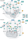
Reactive species at the phagosome. (A) In macrophages (phagosome pH ≤ 5), NADPH oxidase activation (blue sphere) leads to O2•− generation toward the phagocytic vacuole (1). O2•− dismutates to H2O2 (2) and/or protonates to generate HO2 radical (3). O2•− may reach the pathogen by means of anion channels, whereas both H2O2 and HO2 can diffuse across membranes. Immune-stimulated macrophages expressing iNOS (red sphere) produce NO (4), which diffuses to the phagosome while reacting fast with O2•− (5) to yield peroxynitrite anion (ONOO). ONOO can protonate to peroxynitrous acid (ONOOH); it also permeates the parasite and reacts with CO2 (6) to yield NO2 and CO3•− radicals (inset). Both ONOO and ONOOH (7) promote the oxidation and nitration of membrane lipids and proteins (8). (B) In neutrophils (phagosome pH ≥ 7.5), MPO-derived HOCl is the dominant oxidant generated in this phagocyte (3) and promotes oxidation and chlorination reactions (9). Although the de novo production of NO (4) by human neutrophils has rarely been documented, NO may arise from exogenous sources existing in inflammatory foci and permeate the neutrophil plasma membrane. The other numbers in B denote the same processes described above for A.
Figure 2.
Figure 2.
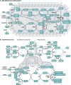
Pathogen antioxidant networks: The examples of Mtb and T. cruzi. (A) Mtb. NADPH oxidase-derived (O2•−) and iNOS-derived (NO) radicals can react with specific enzymes (Fe- or Cu-containing SODs to form H2O2; trHbN to form nitrate) or recombine to form peroxynitrite. H2O2, peroxynitritous acid (ONOOH), and organic hydroperoxides (ROOH) are reduced particularly by KatG and peroxiredoxins (Prx). Most Mtb Prx (AhpC, TPx, PrxQB) can use Trx as reducing substrate. AhpC can also be reduced by AhpD, an adaptor protein that links antioxidant with metabolic enzymes and NADH. NADPH—derived from the pentose phosphate pathway (PPP), isocitrate dehydrogenase (IDH), malic enzyme (ME), and H+-transhydrogenases (TH)—is the final electron donor for TrxR and MshR. TrxR reduces Trx B and C (collectively indicated herein as Trx’s). The one-Cys Prx AhpE is reduced by Mrx-1 either directly (data not shown) or through the formation of a mixed disulfide with MSH, which is reduced by Mrx-1 in a monothiolic mechanism that leads to mycothiolated Mrx-1 (Mrx-1-SM) and is resolved by MSH/MshR/NADPH. MsrA and B reduce S– and R–Met-SO, respectively, and accept electrons from the Trx/TrxR/NADPH system. (B) T. cruzi. Enzymatic and nonenzymatic redox-active molecules use reducing equivalents from NADPH—derived from the PPP, IDH, ME, and TH—are funneled through the T(SH)2, GSH, ascorbate (ASC), and/or TXN-I/II redox systems. H2O2 is metabolized by APxCcP at the endoplasmic reticulum, mitochondria, and plasma membrane using ASC/Cyt cII as the electron donors. Dehidroascorbate (DHA) is reduced by a direct reaction with T(SH)2. Organic hydroperoxides (ROOH) are substrates for GPX-II that uses GSH. T(SH)2 reduces oxidized GSH (GSSG), while TR reduces TS2. Mitochondrial O2•− formation by the electron transport chain (mainly at complex III) is detoxified by Fe-SODA with H2O2 generation. NO can reach the mitochondria and inhibit respiration at complex IV (CIV; with an enhanced O2•− generation). Fe-SODA outcompetes for O2•− inhibiting ONOO formation. Mitochondrial peroxiredoxin (MPX) decomposes H2O2 and/or ONOO, probably using reduced TXN-II and T(SH)2 as the reducing substrate (dashed arrows). Met-SO is repaired by the action of MsrB at the expense of TXN. In the cytosol, cytosolic peroxiredoxin (CPX) detoxifies ROOH. T(SH)2 is synthesized from two molecules of GSH and one spermidine in a reaction catalyzed by the enzyme trypanothione synthetase (TS). Met-SO is repaired by the presence of MsrA and B in the different compartments.

References

    1. Albesa-Jové D., Chiarelli L.R., Makarov V., Pasca M.R., Urresti S., Mori G., Salina E., Vocat A., Comino N., Mohorko E., et al. 2014. Rv2466c mediates the activation of TP053 to kill replicating and non-replicating Mycobacterium tuberculosis. ACS Chem. Biol. 9:1567–1575. 10.1021/cb500149m - DOI - PubMed
    1. Albrett A.M., Ashby L.V., Dickerhof N., Kettle A.J., and Winterbourn C.C.. 2018. Heterogeneity of hypochlorous acid production in individual neutrophil phagosomes revealed by a rhodamine-based probe. J. Biol. Chem. 293:15715–15724. 10.1074/jbc.RA118.004789 - DOI - PMC - PubMed
    1. Alegria T.G., Meireles D.A., Cussiol J.R., Hugo M., Trujillo M., de Oliveira M.A., Miyamoto S., Queiroz R.F., Valadares N.F., Garratt R.C., et al. 2017. Ohr plays a central role in bacterial responses against fatty acid hydroperoxides and peroxynitrite. Proc. Natl. Acad. Sci. USA. 114:E132–E141. 10.1073/pnas.1619659114 - DOI - PMC - PubMed
    1. Allmann S., Morand P., Ebikeme C., Gales L., Biran M., Hubert J., Brennand A., Mazet M., Franconi J.M., Michels P.A., et al. 2013. Cytosolic NADPH homeostasis in glucose-starved procyclic Trypanosoma brucei relies on malic enzyme and the pentose phosphate pathway fed by gluconeogenic flux. J. Biol. Chem. 288:18494–18505. 10.1074/jbc.M113.462978 - DOI - PMC - PubMed
    1. Alvarez M.N., Trujillo M., and Radi R.. 2002. Peroxynitrite formation from biochemical and cellular fluxes of nitric oxide and superoxide. Methods Enzymol. 359:353–366. 10.1016/S0076-6879(02)59198-9 - DOI - PubMed

Publication types