. 2019 Apr 24;39(17):3175-3187.
doi: 10.1523/JNEUROSCI.1731-18.2019. Epub 2019 Feb 21.

Calcium Channel Subunit α2δ4 Is Regulated by Early Growth Response 1 and Facilitates Epileptogenesis

Affiliations

Calcium Channel Subunit α2δ4 Is Regulated by Early Growth Response 1 and Facilitates Epileptogenesis

Karen M J van Loo et al. J Neurosci. .

Abstract

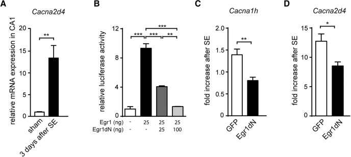
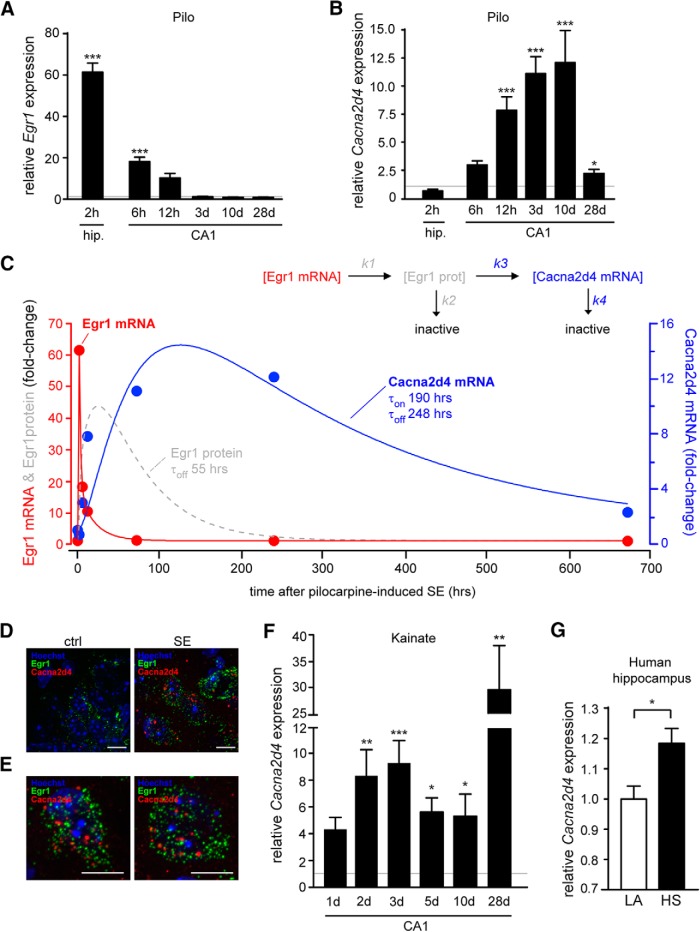
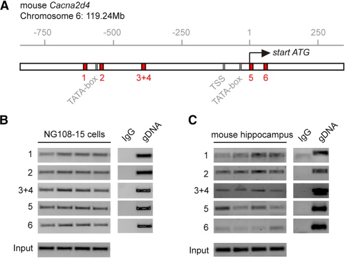
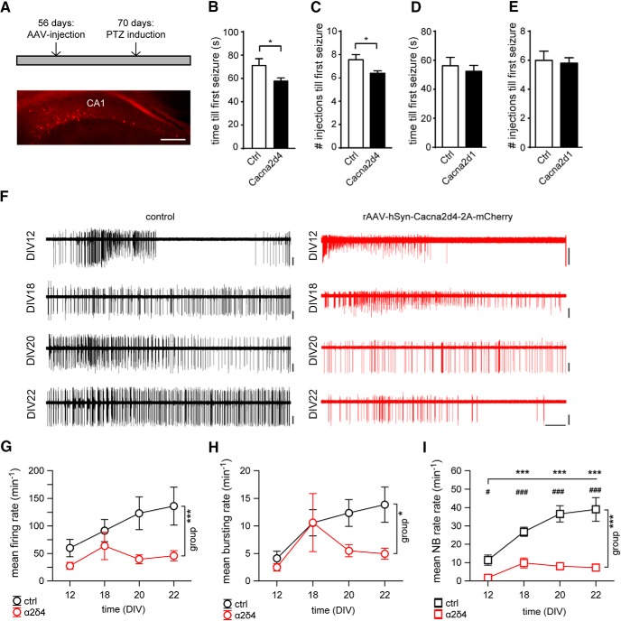
Transient brain insults, including status epilepticus (SE), can trigger a period of epileptogenesis during which functional and structural reorganization of neuronal networks occurs resulting in the onset of focal epileptic seizures. In recent years, mechanisms that regulate the dynamic transcription of individual genes during epileptogenesis and thereby contribute to the development of a hyperexcitable neuronal network have been elucidated. Our own results have shown early growth response 1 (Egr1) to transiently increase expression of the T-type voltage-dependent Ca2+ channel (VDCC) subunit CaV3.2, a key proepileptogenic protein. However, epileptogenesis involves complex and dynamic transcriptomic alterations; and so far, our understanding of the transcriptional control mechanism of gene regulatory networks that act in the same processes is limited. Here, we have analyzed whether Egr1 acts as a key transcriptional regulator for genes contributing to the development of hyperexcitability during epileptogenesis. We found Egr1 to drive the expression of the VDCC subunit α2δ4, which was augmented early and persistently after pilocarpine-induced SE. Furthermore, we show that increasing levels of α2δ4 in the CA1 region of the hippocampus elevate seizure susceptibility of mice by slightly decreasing local network activity. Interestingly, we also detected increased expression levels of Egr1 and α2δ4 in human hippocampal biopsies obtained from epilepsy surgery. In conclusion, Egr1 controls the abundance of the VDCC subunits CaV3.2 and α2δ4, which act synergistically in epileptogenesis, and thereby contributes to a seizure-induced "transcriptional Ca2+ channelopathy."SIGNIFICANCE STATEMENT The onset of focal recurrent seizures often occurs after an epileptogenic process induced by transient insults to the brain. Recently, transcriptional control mechanisms for individual genes involved in converting neurons hyperexcitable have been identified, including early growth response 1 (Egr1), which activates transcription of the T-type Ca2+ channel subunit CaV3.2. Here, we find Egr1 to regulate also the expression of the voltage-dependent Ca2+ channel subunit α2δ4, which was augmented after pilocarpine- and kainic acid-induced status epilepticus. In addition, we observed that α2δ4 affected spontaneous network activity and the susceptibility for seizure induction. Furthermore, we detected corresponding dynamics in human biopsies from epilepsy patients. In conclusion, Egr1 orchestrates a seizure-induced "transcriptional Ca2+ channelopathy" consisting of CaV3.2 and α2δ4, which act synergistically in epileptogenesis.

Keywords: CaV3.2; Cacna2d4; early growth response 1; epileptogenesis; pilocarpine and kainic acid-induced status epilepticus; transcriptional Ca2+ channelopathy.


Figures

Figure 1.
Figure 1.
Interference with Egr1 antagonizes pilocarpine-induced augmentation of CaV3.2 and Cacna2d4. A, Relative mRNA expression of Cacna2d4 3 d after SE (n = 5). **p = 0.0018 (t test). B, Luciferase activity of the CaV3.2 promoter-luciferase reporter gene (Kulbida et al., 2015) after transfection with Egr1 (25 ng) and Egr1dN (25 or 100 ng) in NG108-15 cells (n = 3). One-way ANOVA: p = 0.48, F(3,8) = 0.91. Tukey's multiple-comparisons test: **p < 0.01; ***p < 0.001. C, D, mRNA expression of Cacna1h (C) and Cacna2d4 (D) of mice injected with rAAV-hSyn-GFP (GFP: sham: n = 8; SE: n = 11) and rAAV-hSyn-Egr1dN-2A-mCherry (Egr1dN: sham: n = 9; SE: n = 10) 3 d after pilocarpine-induced SE. Cacna1h: **p = 0.0012 (t test). Cacna2d4: *p = 0.012 (t test).
Figure 2.
Figure 2.
Increase in Cacna2d4 expression in two models for epilepsy. Egr1 (A) and Cacna2d4 (B) mRNA expression of total hippocampi 2 h after pilocarpine-induced SE (n = 8) and hippocampal CA1 6 h (n = 6), 12 h (n = 5), 3 d (n = 5), 10 d (n = 5), and 28 d (n = 6) after pilocarpine-induced SE (Egr1: 2 h: 61-fold increase; p < 0.0001, 6 h: 18-fold; p = 0.0002. Cacna2d4: 12 h: 7.8-fold; p = 0.0005, 3 d: 11.1-fold; p = 0.0002, 10 d: 12.1-fold; p < 0.0001, 28 d; 2.3-fold). p = 0.03. *p < 0.05. ***p < 0.001. C, Coupled differential equations of Egr1 protein and Cacana2d4 mRNA levels (inset) reveal slow and long-lasting transcriptional regulation following a spike-like increase in Egr1 mRNA. A, B, Red and blue circles represent the mean values of mRNA levels, respectively, following SE. Red line indicates the approximation of Egr1 mRNA by a biexponential function. Dashed gray line indicates relative levels of the assumed intermediary, Egr1 protein. The output of the model well predicts the time course of Cacna2d4 levels between SE and the chronic phase (blue line). For more details, see Materials and Methods. D, Representative pictures of RNAscope hybridization in hippocampal sections of control (left) and pilocarpine-induced SE (right) mice 6 d after SE using probes targeting Egr1 (green) and Cacna2d4 (red). Hoechst was used to stain the nuclei. Scale bars, 10 μm. E, Two representative cells expressing both Egr1 (green) and Cacna2d4 (red) mRNA in a hippocampal slice from a pilocarpine-induced SE mice 6 d after SE. Nuclei stained with Hoechst (blue). Scale bars, 10 μm. F, Cacna2d4 mRNA expression of hippocampal CA1 1, 2, 3, 5, 10, and 28 d after KA-induced SE: 1 d: 4.3-fold (sham: n = 8; SE: n = 7), 2 d: 8.3-fold increase; p = 0.0027 (sham: n = 6; SE: n = 5), 3 d: 9.3-fold; p < 0.0001 (sham: n = 11; SE: n = 10), 5 d: 5.6-fold; p = 0.02 (sham: n = 8; SE: n = 7); 10 d: 5.3-fold; p = 0.03 (sham: n = 6; SE: n = 8), 28 d: 29-fold; p = 0.0036 (sham: n = 16; SE: n = 17). *p < 0.05. **p < 0.01. ***p < 0.001). G, Cacna2d4 expression in hippocampal tissue of patients with lesion-associated TLE (LA; n = 35) versus hippocampi from patients with HS (n = 79). *p = 0.025 (t test).
Figure 3.
Figure 3.
Egr1 binds the Cacna2d4 promoter in vitro and in vivo. A, Schematic representation of the predicted promoter region of the mouse Cacna2d4 gene (750 bp upstream and 250 bp downstream of the start ATG). Predicted TATA boxes (gray boxes), TSSs (black box), and Egr1-binding sites (red boxes) are indicated. B, PCR analysis of the six Egr1 binding sites in the Cacna2d4 promoter region in NG108-15 cells. Five ChIP PCR assays were designed, spanning the Egr1 binding sites. Because binding sites 3 and 4 are located close to each other, only one assay was developed for these two binding sites. PCR amplicons were generated on ChIP-input DNA and anti-Egr1 ChIP immunoprecipitates. C, ChIP analysis of Egr1 binding to the Cacna2d4 promoter in vivo. PCR amplicons were generated of anti-Egr1 ChIP immunoprecipitates from mouse hippocampi transduced with rAAV-hSyn-Egr1-2A-Cherry.
Figure 4.
Figure 4.
Cacna2d4 has a proconvulsive effect and alters spontaneous network activity. A, PTZ induction in adult mice. Adult mice were injected with rAAV-hSyn-Cacna2d4-mCherry or rAAV-hSyn-tdTomato and tested for PTZ sensitivity 14 d later. Bottom, Representative picture of an rAAV-hSyn-Cacna2d4-mCherry-injected hippocampus. Scale bar, 200 μm. B, C, Time until first seizure (B) and number of injections until first seizure (C) in rAAV-hSyn-tdTomato (ctrl; n = 9) and rAAV-hSyn-Cacna2d4–2A-mCherry (Cacna2d4; n = 10) injected animals after injection with PTZ every 10 min. Time until first seizure: *p = 0.04 (t test). Number of injections until first seizure: *p = 0.019 (t test). D, E, PTZ susceptibility in pLenti-hSyn-Cacna2d1-HA (Cacna2d1; n = 5) and pLenti-GFP (ctrl; n = 5) injected mice. Time until first seizure: p = 0.61 (t test). Number of injections until first seizure: p = 0.79 (t test). F, Representative traces of spontaneous neuronal firing derived by an individual electrode at different DIV as indicated in control (left) or α2δ-4 overexpressing hippocampal mouse culture (right) grown on 60 channel MEAs. Vertical scale bars: 50 μV; horizontal scale bars: 20 s. G, H, The mean firing (G) and bursting (H) rates in either naive mouse cultures (n = 2 MEAs), or in cultures overexpressing α2δ4 (n = 3 MEAs; infection at DIV12 after baseline recording; 2 μl per 1 ml of medium). The developmental increase in the neuronal activity is less prominent in α2δ4-overexpressing cultures. Group effect: firing rate, ***p < 0.001, F(1,399) = 11.14 (two-way ANOVA). Bursting rate: *p < 0.05, F(1,184) = 4.57 (two-way ANOVA). I, The developmental increase of the mean rate of spontaneous network bursts (NB; time effect: p < 0.001 F(2,192) = 14.48, two-way ANOVA) was evident under control conditions (***p < 0.001 within-group comparison with respective values at DIV12, Duncan's post hoc test), but not in α2δ4 overexpressing cultures (group effect: p < 0.001, F(1,192) = 108.96; group × time interaction: p = 0.001, F(1,192) = 6.08; two-way ANOVA). The effect of α2δ4 overexpression is particularly prominent 8–10 d after AAV infection. Between-group comparison at respective DIV: #p < 0.05 (Duncan's post hoc test); ###p < 0.001 (Duncan's post hoc test).

Similar articles

Cited by

References

    1. Barclay J, Balaguero N, Mione M, Ackerman SL, Letts VA, Brodbeck J, Canti C, Meir A, Page KM, Kusumi K, Perez-Reyes E, Lander ES, Frankel WN, Gardiner RM, Dolphin AC, Rees M (2001) Ducky mouse phenotype of epilepsy and ataxia is associated with mutations in the Cacna2d2 gene and decreased calcium channel current in cerebellar Purkinje cells. J Neurosci 21:6095–6104. 10.1523/JNEUROSCI.21-16-06095.2001 - DOI - PMC - PubMed
    1. Becker AJ. (2018) Review: animal models of acquired epilepsy: insights into mechanisms of human epileptogenesis. Neuropathol Appl Neurobiol 44:112–129. 10.1111/nan.12451 - DOI - PubMed
    1. Becker AJ, Pitsch J, Sochivko D, Opitz T, Staniek M, Chen CC, Campbell KP, Schoch S, Yaari Y, Beck H (2008) Transcriptional upregulation of Cav3.2 mediates epileptogenesis in the pilocarpine model of epilepsy. J Neurosci 28:13341–13353. 10.1523/JNEUROSCI.1421-08.2008 - DOI - PMC - PubMed
    1. Beckmann AM, Davidson MS, Goodenough S, Wilce PA (1997) Differential expression of egr-1-like DNA-binding activities in the naive rat brain and after excitatory stimulation. J Neurochem 69:2227–2237. 10.1046/j.1471-4159.1997.69062227.x - DOI - PubMed
    1. Bernard C, Anderson A, Becker A, Poolos NP, Beck H, Johnston D (2004) Acquired dendritic channelopathy in temporal lobe epilepsy. Science 305:532–535. 10.1126/science.1097065 - DOI - PubMed

Publication types

MeSH terms