. 2019 Feb 22;363(6429):eaav7969.
doi: 10.1126/science.aav7969.

Latrophilin GPCRs direct synapse specificity by coincident binding of FLRTs and teneurins

Affiliations

Latrophilin GPCRs direct synapse specificity by coincident binding of FLRTs and teneurins

Richard Sando et al. Science. .

Erratum in

Abstract

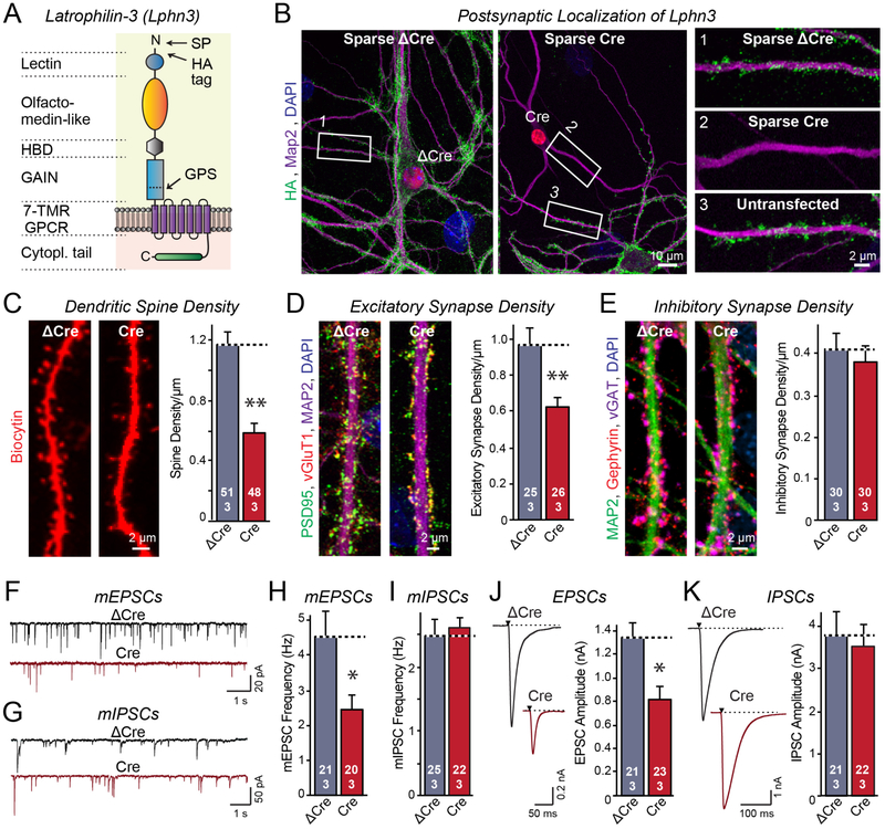
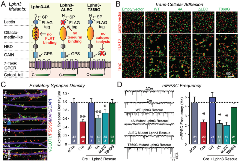
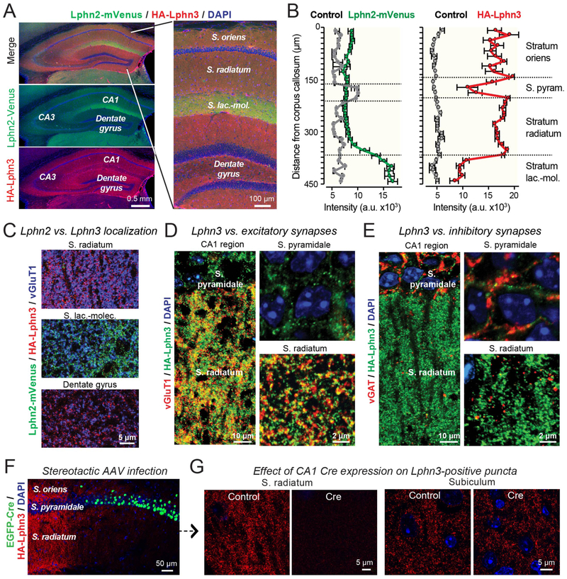
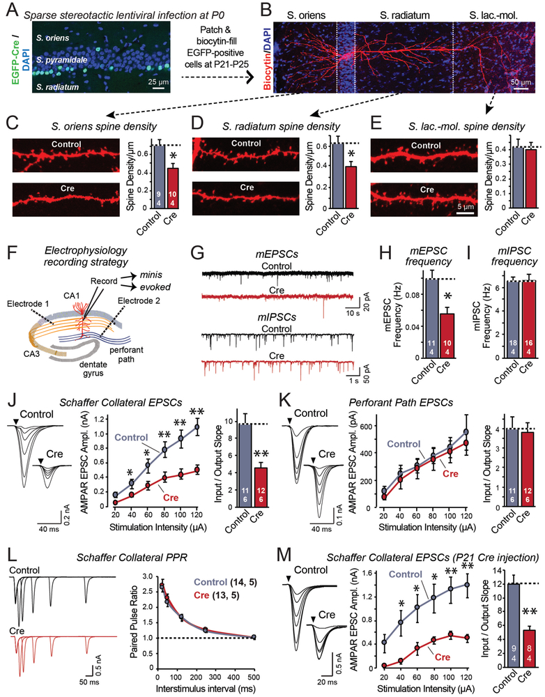
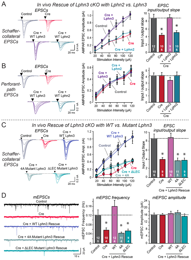
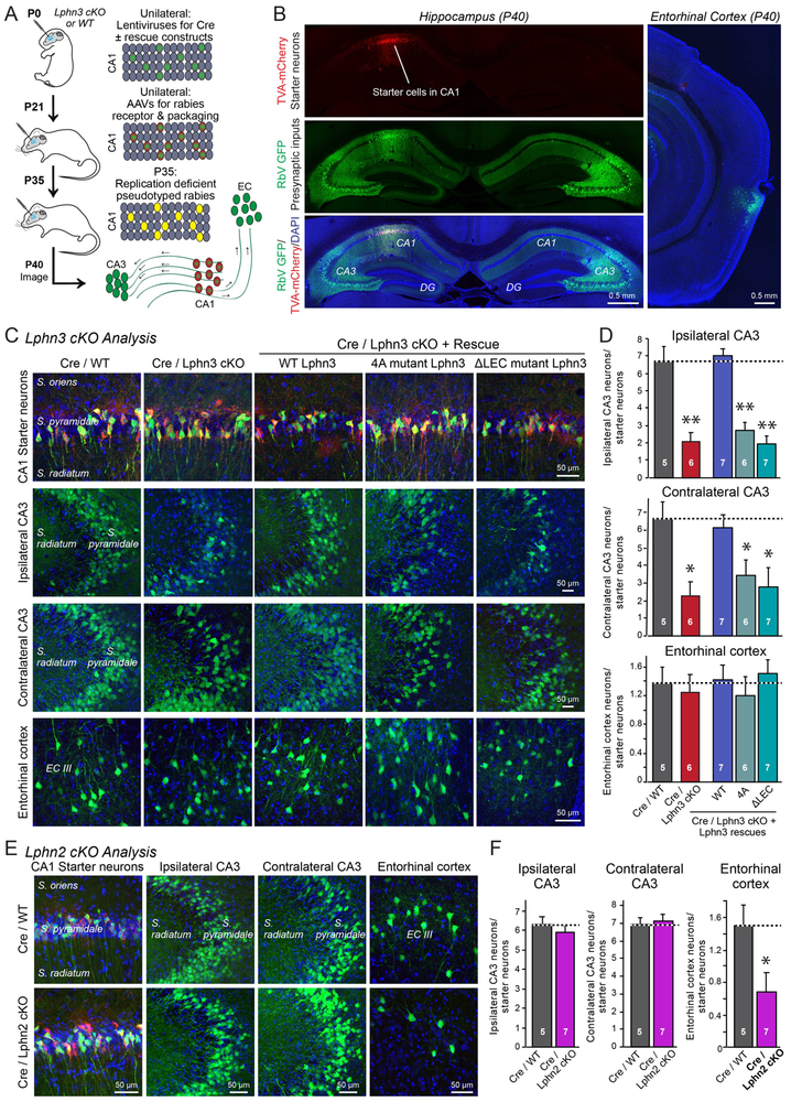
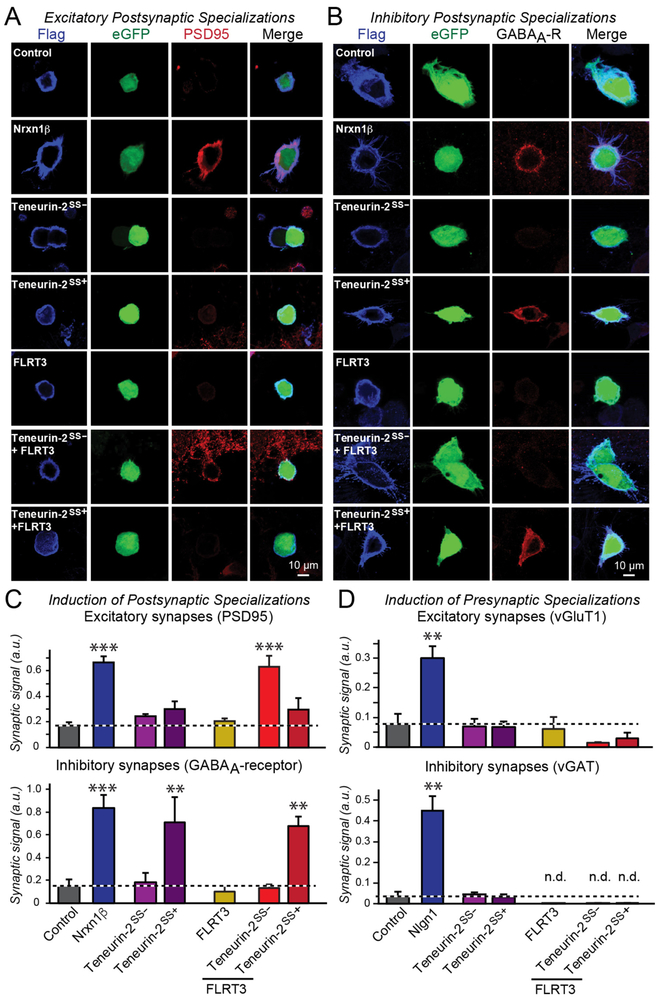
Bidirectional signaling by cell adhesion molecules is thought to mediate synapse formation, but the mechanisms involved remain elusive. We found that the adhesion G protein-coupled receptors latrophilin-2 and latrophilin-3 selectively direct formation of perforant-path and Schaffer-collateral synapses, respectively, to hippocampal CA1-region neurons. Latrophilin-3 binds to two transcellular ligands: fibronectin leucine-rich repeat transmembrane proteins (FLRTs) and teneurins. In transgenic mice in vivo, both binding activities were required for input-specific synapse formation, which suggests that coincident binding of both ligands is necessary for synapse formation. In cultured neurons in vitro, teneurin or FLRT alone did not induce excitatory synapse formation, whereas together they potently did so. Thus, postsynaptic latrophilins promote excitatory synapse formation by simultaneous binding of two unrelated presynaptic ligands, which is required for formation of synaptic inputs at specific dendritic localizations.


Conflict of interest statement

Competing interests: The authors declare no conflict of interest.

Figures

Figure 1:
Figure 1:. Latrophilin-3 (Lphn3) is essential for formation of a subset of excitatory synapses in cultured hippocampal neurons
A. Schematic of the Lphn3 domain structure with location of the HA-tag in Lphn3 cKO mice. B. Lphn3 is postsynaptic as revealed by sparse expression of Cre-recombinase in transfected hippocampal neurons that were cultured from Lphn3 cKO mice. C-E. Postsynaptic Lphn3 deletion in cultured hippocampal neurons decreases the density of dendritic spines and the number of excitatory but not of inhibitory synapses. F-I. Postsynaptic Lphn3 deletion suppresses the frequency of spontaneous mEPSCs but not mIPSCs monitored in the presence of tetrodotoxin. J & K. Postsynaptic deletion of Lphn3 decreases the amplitudes of evoked EPSC but not IPSCs. Data are means ± SEM (numbers of cells/experiments are indicated in bars). Statistical significance was assessed by two-tailed t-tests (** denotes p<0.01; * denotes p<0.05).
Figure 2:
Figure 2:. Lphn3-dependent synapse formation in cultured hippocampal neurons requires both teneurin- and FLRT-binding by Lphn3 but not Lphn3 autoproteolysis
A. Domain structures of Lphn3 mutants that selectively block binding of Lphn3 to FLRTs (Lphn3-4A) or teneurins (Lphn3-ΔLEC), or that abolish Lphn3 autoproteolysis (Lphn3-T869G). B. Lphn3 mutants are efficiently transported to the cell surface and mediate cell adhesion via the Lphn3 binding partners whose binding site was not mutated (for quantifications, see Figure S2). C & D. Wild-type Lphn3 and Lphn3-T869G lacking autoproteolysis activity efficiently rescue the loss of synapse density (C) or the decrease in mEPSC frequency (D) in Lphn3-deficient neurons, whereas the Lphn3 mutants that do not bind to FLRTs or to teneurins do not rescue. Data are means ± SEM (numbers of cells/experiments are indicated in bars). Statistical significance was assessed by one-way ANOVA (** denotes p<0.01; * denotes p<0.05).
Figure 3:
Figure 3:. Postsynaptic Lphn2 and Lphn3 are targeted to distinct non-overlapping dendritic domains of pyramidal CA1 neurons
A & B. Lphn3 is broadly distributed in the hippocampal formation except for the S. lacunosum-moleculare of the CA1 region, whereas Lphn2 is present only in the S. lacunosum-moleculare. Overview images (A) depict the entire hippocampal formation (left) or the CA1 region and dentate gyrus (right) viewed in a cryosection from knockin mice expressing mVenus-tagged Lphn2 (16) and HA-tagged Lphn3. The section was stained for Lphn2-mVenus, HA-tagged Lphn3, and nuclei (DAPI). Quantifications of relative Lphn2 and Lphn3 levels (B) show that Lphn2 is specifically targeted in the CA1 region to the S. lacunosum-moleculare, while Lphn3 is present in the S. oriens and S. radiatum but not the S. lacunosum-moleculare (means ± SEM; n=5 mice/experimental group; controls are wild-type mice). C. High-magnification images showing that the S. radiatum of the CA1 region and the dentate gyrus contain only Lphn2-positive puncta but not Lphn3-positive puncta, whereas the S. moleculare-lacunosum contains only Lphn2-positive puncta but not Lphn3-positive puncta. Images were taken from a cryosection obtained and stained as described for panel A. D. Low- and high-magnification images showing that Lphn3-positive puncta in the S. radiatum overlap with excitatory vGluT1-positive synapses (left, overview of the S. pyramidale and S. radiatum; right, higher magnification images). E. Low- and high-magnification images showing that Lphn3-positive puncta in the S. radiatum do not overlap with inhibitory vGAT-positive synapses (left, overview of the S. pyramidale and S. radiatum; right, higher magnification images). Note that most inhibitory synapses are perisomatic in the S. pyramidale. F. AAV-mediated Cre-recombination deletes Lphn3 in a large area of the CA1 region of HA-Lphn3 cKO mice. Image shows hippocampal section of a mouse that had been stereotactically injected with AAVs encoding eGFP-tagged Cre-recombinase. Section was labeled for Lphn3 and eGFP. G. High-resolution images of the S. radiatum and the subiculum from control mice and mice after AAV-mediated deletion of Lphn3 in CA1 region (see F). Note that the deletion of Lphn3 abolishes all Lphn3-positive puncta in the CA1 region, excluding the presence or Lphn3-positive presynaptic inputs, but the CA1 region deletion has no effect on Lphn3-positive puncta in the subiculum, ruling out a presence of Lphn3 on CA1-derived presynaptic outputs.
Figure 4:
Figure 4:. Lphn3 is selectively essential for Schaffer-collateral synapse formation in the CA1 region
A. Unilateral infection of the neonatal hippocampal CA1 region with lentiviruses expressing EGFP-tagged Cre-recombinase causes sparse deletion of Lphn3 in Lphn3 cKO mice. Image shows a hippocampal section stained for DAPI; infected neurons are identified via their EGFP fluorescence. B. Filling of individual neurons with biocytin via a patch pipette enables analysis of dendritic morphology. Image shows representative pyramidal neuron filled with biocytin; Cre-recombined and uninfected control neurons were examined in opposite hemispheres of the same mice. C-E. Quantifications of biocytin-filled, patched CA1 neurons show that postsynaptic Lphn3 deletion decreases dendritic spine densities in the S. oriens and S. radiatum but not the S. lacunosum-moleculare. F. Diagram of the electrophysiological recording configuration in acute slices with sparse lentiviral expression of Cre-recombinase. G-I. Postsynaptic Lphn3 deletion in CA1 region neurons decreases the mEPSC but not mIPSC frequency (G, representative mEPSC and mIPSC traces; H & I, summary graphs of the mean mEPSC (H) and mIPSC frequency (I)). J. Postsynaptic Lphn3 deletion in CA1 neurons decreases the Schaffer-collateral synaptic strength (left, representative traces; right, summary graph of EPSC amplitude). Input-output curves were used to control for differences in stimulus strength. K. Postsynaptic Lphn3 deletion in CA1 neurons had no effect on the synaptic strength of entorhinal cortex inputs (left, representative traces; right, summary graph of EPSC amplitude). L. Measurements of paired-pulse ratios of Schaffer-collateral EPSCs as a function of the interstimulus interval show that the Lphn3 deletion has no effect on release probability (left, representative traces; right, summary plot). M. Same as J, but measured in acute slices from mice that were sparsely infected with Cre-expressing lentiviruses at P21, and analyzed at P40. Numerical data are means ± SEM (numbers of cells/mice are indicated in bars). Statistical significance was assessed by two-tailed t-tests (** denotes p<0.01; * denotes p<0.05).
Figure 5:
Figure 5:. Neither wild-type Lphn2 nor mutant Lphn3 lacking FLRT- or teneurin-binding activity rescue the decrease in Schaffer-collateral synaptic strength induced by Lphn3 deletion.
Experiments were performed by whole-cell patch-clamp recordings from CA1 neurons in acute slices from Lphn3 cKO mice that were lentivirally-infected at P0 as described in Figure 4A and 4F. A & B. Wild-type Lphn3 but not wild-type Lphn2 fully rescues the decrease in Schaffer-collateral synaptic strength induced by loss of Lphn3 (A), while none of the manipulations affects entorhinal cortex-derived synapses (B). C. Although wild-type Lphn3 fully rescues the decrease in Schaffer-collateral synaptic strength induced by Lphn3 deletion, mutant Lphn3 lacking FLRT-binding (4A) or teneurin-binding (ΔLEC) was unable to rescue. D. Wild-type Lphn3 also fully rescues the decrease in mEPSC frequency induced by Lphn3 deletion, but again mutant Lphn3 lacking FLRT-binding (4A) or teneurin-binding (ΔLEC) was unable to rescue, with none of the manipulations having any effect on mEPSC amplitude. Numerical data are means ± SEM (numbers of cells/experiments are indicated in bars). Statistical significance was assessed by one-way ANOVA (* denotes p<0.05).
Figure 6:
Figure 6:. Rabies virus-mediated retrograde tracing demonstrates selective functions and ligand-dependence of Lphn2 and Lphn3 in hippocampal synaptic connectivity
A. Experimental approach. The CA1 region of newborn Lphn3 cKO or control mice was sparsely infected unilaterally with lentiviruses encoding Cre-recombinase without or with co-expression of Lphn3 rescue constructs. At P21, the same CA1 region was infected with AAVs encoding the mCherry-tagged receptor and packaging proteins for pseudotyped rabies viruses. At P35, the same CA1 region was infected with the pseudotyped rabies virus encoding eGFP, and at P40, the expression of eGFP transcribed from trans-synaptically transferred rabies virus was analyzed in the ipsi- and contralateral hippocampal CA3 region and the ipsilateral entorhinal cortex. B. Exemplary images of synaptic inputs mapped by pseudotyped rabies virus administered into the hippocampal CA1 region. Red mCherry-TVA expression marks neurons (starter cells) in which the rabies virus was originally introduced, while green eGFP expression marks neurons with synaptic inputs onto the starter cells. C. Exemplary higher-magnification images of starter neurons (top row) and neurons providing synaptic inputs onto starter neurons (middle and bottom rows) after Lphn3 manipulations. D. Quantifications of presynaptic inputs onto postsynaptic CA1 neurons as a function of Lphn3 manipulations, determined by rabies virus tracing and normalized for starter cell numbers. E & F. Representative images (E) and summary graphs of the presynaptic inputs onto postsynaptic CA1 neurons (F) as a function of Lphn2 deletions. Experiments were performed analogous to those of C and D. Note that the Lphn2 deletion selectively impairs entorhinal cortex inputs into the CA1 region. Data in D and F are means ± SEM (number of mice analyzed are indicated in bars). Statistical significance was assessed by one-way ANOVA (** denotes p<0.01; * denotes p<0.05).
Figure 7:
Figure 7:. Teneurin-2 and FLRT3 expressed in HEK293T cells induce postsynaptic specializations in co-cultured neurons only when FLRT3 is co-expressed with the latrophilin-binding splice variant of teneurin-2
A & B. Representative images of in vitro synapse-formation assays in which the indicated cell-adhesion molecules are co-expressed with eGFP in HEK293T cells that are then co-cultured with cortical neurons, and subsequently immunostained for the postsynaptic excitatory synapse marker PSD95 (A) or inhibitory synapse marker GABAAα2-receptor (B). For other in vitro synapse formation immunostaining experiments, see Figure S6. C. Summary graphs showing that only combined expression of Teneurin-2SS− that binds to latrophilins and of FLRT3 that also binds to latrophilins induced excitatory postsynaptic specializations. In contrast, expression of Teneurin-2SS+ alone, but not of Teneurin-2SS−, induced inhibitory postsynaptic specializations. In these experiments, Nrxn1β is used as a positive control that equally induced excitatory and inhibitory postsynaptic specializations (Graf et al., 2004). D. Summary graphs showing that FLRT3 and teneurin-2 splice variants, alone or in combination, do not induce presynaptic excitatory or inhibitory specializations, whereas Nlgn1, used as a positive control, potently does (n.d. = non-detectable). Data in C & D are means ± SEM (numbers of cells/experiments are indicated in bars). Statistical significance was assessed by one-way ANOVA (*** denotes p<0.001; ** denotes p<0.01; * denotes p<0.05).

Similar articles

Cited by

References

    1. Sanes JR, Yamagata M, Many paths to synaptic specificity. Annu. Rev. Cell Dev. Biol. 25, 161–195 (2009). - PubMed
    1. Nusser Z, Creating diverse synapses from the same molecules. Curr. Opin. Neurobiol. 51, 8–15 (2018). - PubMed
    1. Südhof TC, Towards an understanding of synapse formation. Neuron, in press (2018). - PMC - PubMed
    1. O'Sullivan ML, de Wit J, Savas JN, Comoletti D, Otto-Hitt S, Yates JR 3rd, Ghosh A, FLRT proteins are endogenous latrophilin ligands and regulate excitatory synapse development. Neuron. 73, 903–910 (2012). - PMC - PubMed
    1. Silva JP, Lelianova VG, Ermolyuk YS, Vysokov N, Hitchen PG, Berninghausen O, Rahman MA, Zangrandi A, Fidalgo S, Tonevitsky AG, Dell A, Volynski KE, Ushkaryov YA, Latrophilin 1 and its endogenous ligand Lasso/teneurin-2 form a high-affinity transsynaptic receptor pair with signaling capabilities. Proc. Natl. Acad. Sci. USA. 108, 12113–12118 (2011). - PMC - PubMed

Publication types

MeSH terms