. 2019 Apr 19;294(16):6621-6634.
doi: 10.1074/jbc.RA118.006727. Epub 2019 Feb 21.

Mitochondrial localization, import, and mitochondrial function of the androgen receptor

Affiliations

Mitochondrial localization, import, and mitochondrial function of the androgen receptor

Prachi Bajpai et al. J Biol Chem. .

Abstract

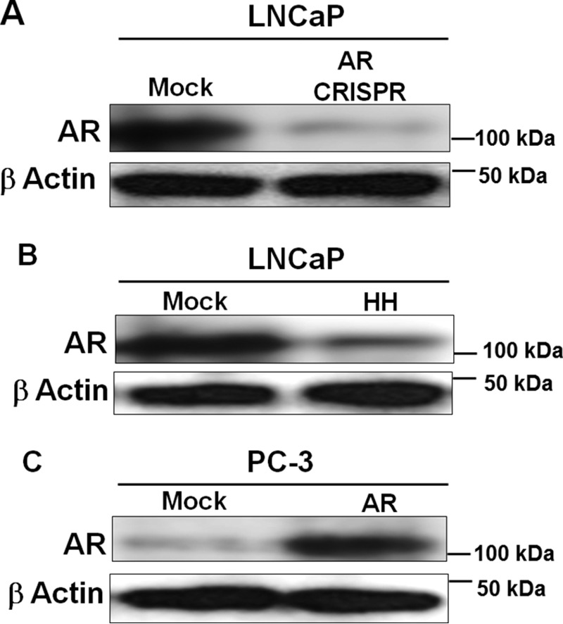
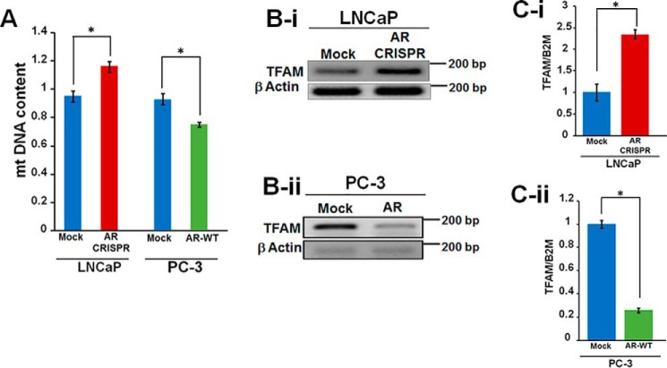
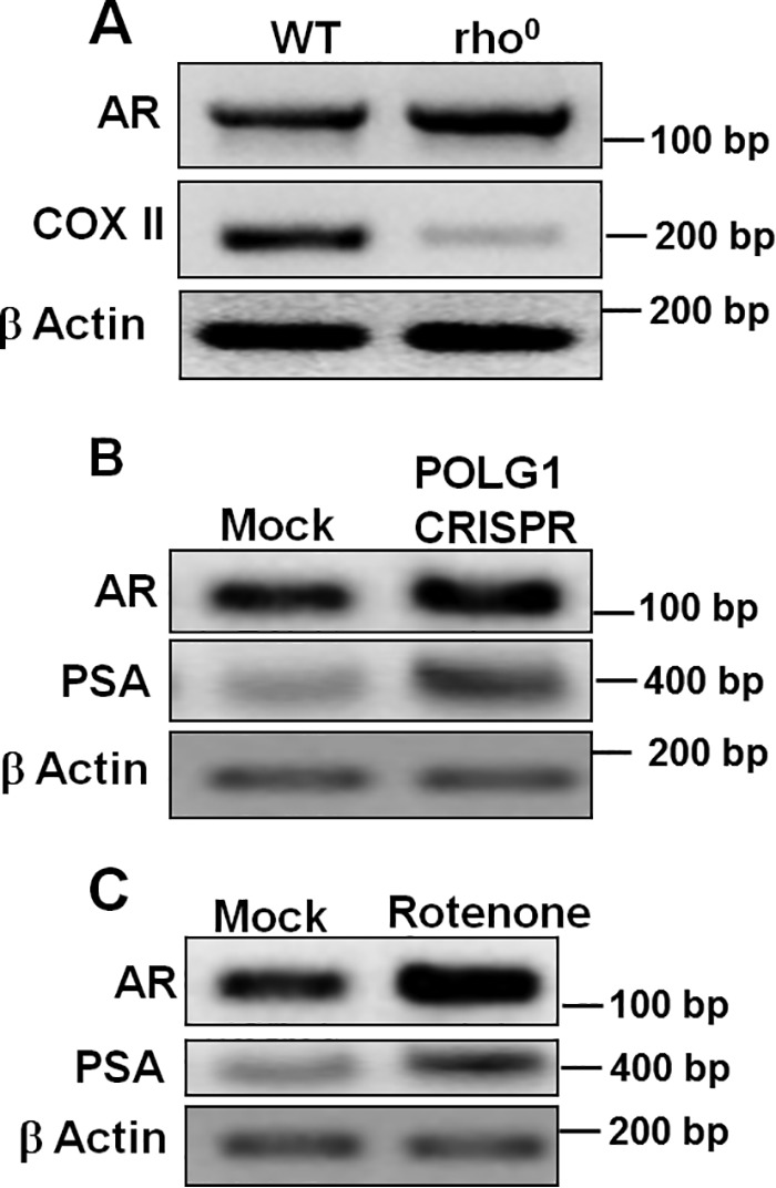
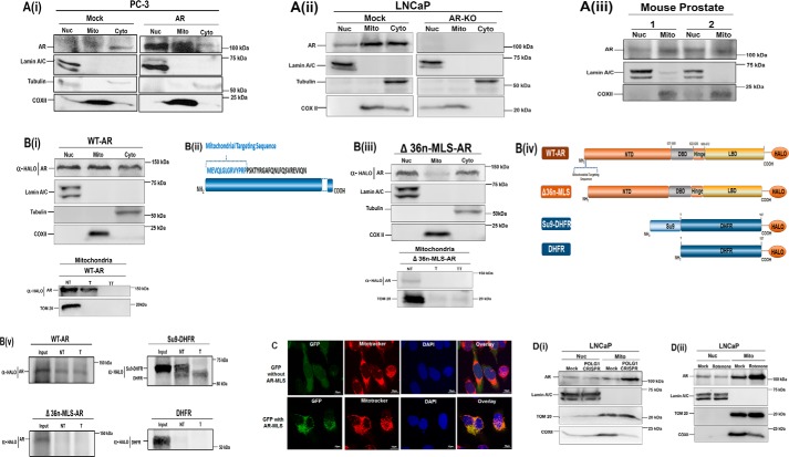
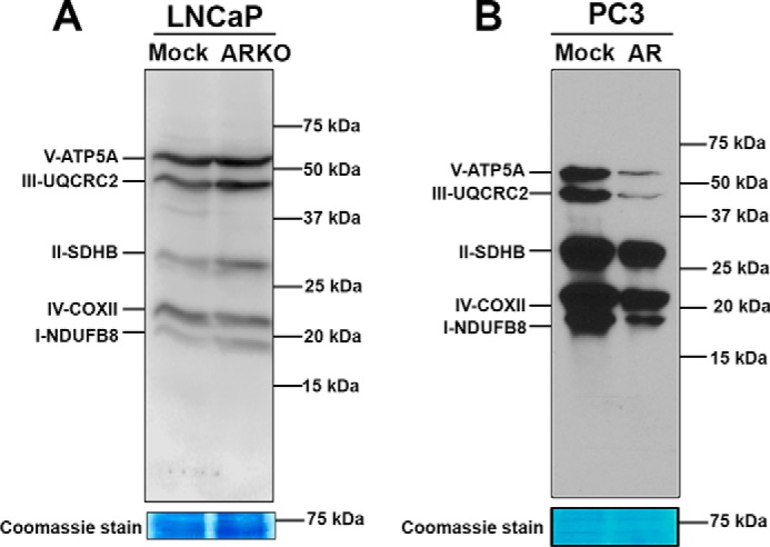
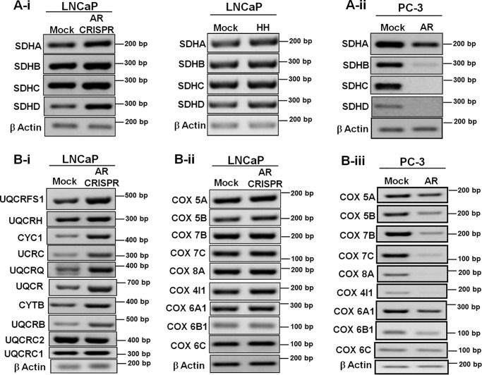
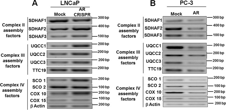
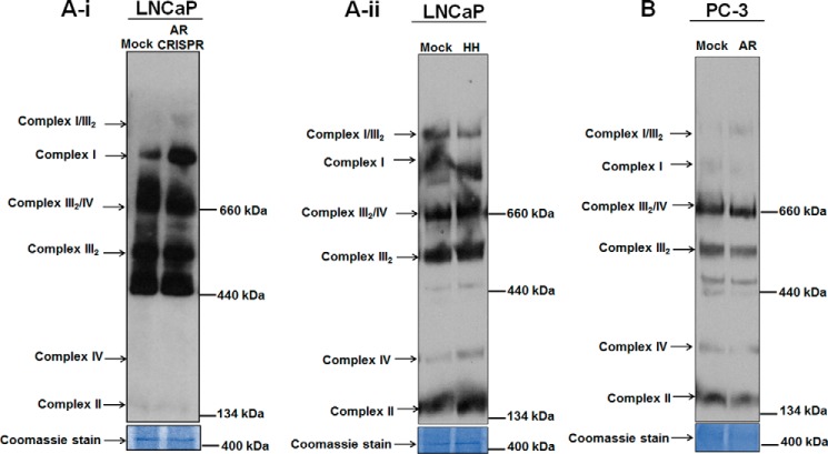
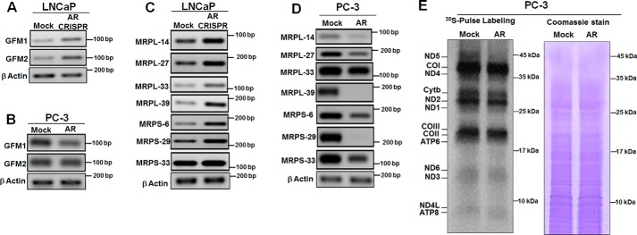
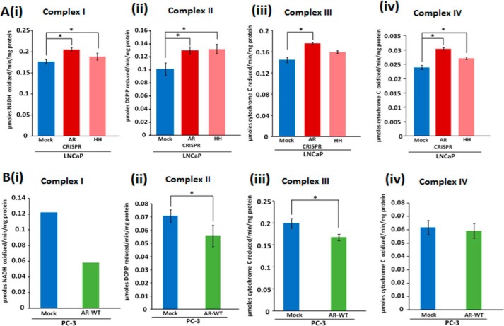
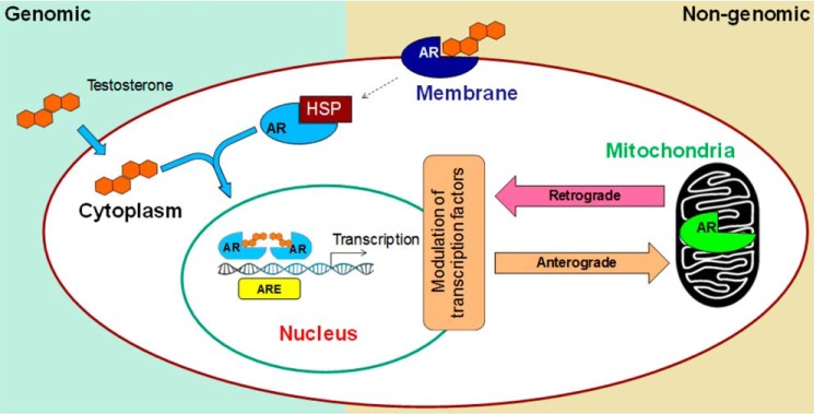
Nuclear localization of androgen receptor (AR) directs transcriptional regulation of a host of genes, referred to as genomic signaling. Additionally, nonnuclear or nongenomic activities of the AR have long been described, but understanding of these activities remains elusive. Here, we report that AR is imported into and localizes to mitochondria and has a novel role in regulating multiple mitochondrial processes. Employing complementary experimental approaches of AR knockdown in AR-expressing cells and ectopic AR expression in AR-deficient cells, we demonstrate an inverse relationship between AR expression and mitochondrial DNA (mtDNA) content and transcription factor A, mitochondrial (TFAM), a regulator of mtDNA content. We show that AR localizes to mitochondria in prostate tissues and cell lines and is imported into mitochondria in vitro We also found that AR contains a 36-amino-acid-long mitochondrial localization sequence (MLS) capable of targeting a passenger protein (GFP) to the mitochondria and that deletion of the MLS abolishes the import of AR into the mitochondria. Ectopic AR expression reduced the expression of oxidative phosphorylation (OXPHOS) subunits. Interestingly, AR also controlled translation of mtDNA-encoded genes by regulating expression of multiple nuclear DNA-encoded mitochondrial ribosomal proteins. Consistent with these observations, OXPHOS supercomplexes were destabilized, and OXPHOS enzymatic activities were reduced in AR-expressing cells and restored upon AR knockdown. Moreover, mitochondrial impairment induced AR expression and increased its translocation into mitochondria. We conclude that AR localizes to mitochondria, where it controls multiple mitochondrial functions and mitonuclear communication. Our studies also suggest that mitochondria are novel players in nongenomic activities of AR.

Keywords: androgen receptor; castration-resistant; cell signaling; gene transcription; genomic signaling; mitochondria; mitochondrial localization sequence; nongenomic signaling; nuclear receptor; oxidative phosphorylation; prostate cancer; retrograde signaling.


Conflict of interest statement

The authors declare that they have no conflicts of interest with the contents of this article

Figures

Figure 1.
Figure 1.
Cellular models employed in this study. A and B, genetic knockout of AR in LNCaP cells was achieved using CRISPR-Cas9 (A), and pharmacological inhibition was achieved by HH treatment for 48 h (B). C, AR overexpression in PC-3 cells was achieved by transfecting these cells with AR cDNA. Western blots show AR down-regulation in LNCaP cells (A and B) and up-regulation in PC3 cells (C).
Figure 2.
Figure 2.
Androgen receptor expression regulates mtDNA content and TFAM level. A, AR inactivation in LNCaP cells increases mtDNA content, whereas ectopic AR expression in PC3 cells reduces mtDNA content. The ratio of mtDNA to nuclear DNA was used as an index for measuring the mtDNA content. B-i, AR knockout in LNCaP cells increased TFAM expression; B-ii, AR expression in PC-3 cells decreased TFAM expression. TFAM expression was analyzed by semiquantitative PCR. C-i and -ii, real-time quantitative PCR analysis of TFAM expression: validation of semiquantitative PCR results showing increased TFAM expression in AR knockout LNCaP cells (C-i) and decreased TFAM expression in PC-3 cells ectopically expressing AR (C-ii). Statistical significance was calculated by Student's t test, and significant differences (p < 0.05) are marked with asterisks. All experiments were done in triplicates. Error bars, S.D.
Figure 3.
Figure 3.
Mitochondrial dysfunction increases androgen receptor expression. A, rho0 (mtDNA-depleted) cell line showed increased AR expression when compared with parental cells. CRISPR targeting POLG1 (B) and rotenone treatment (25 nm for 24 h) (C) were used to induce mitochondrial stress in LNCaP cells, both of which showed increased expression of AR and prostate specific antigen.
Figure 4.
Figure 4.
Mitochondrial Localization of the androgen receptor. A-i, exogenous expression of WT-AR in PC-3 cells. PC-3 cells transiently transfected with AR construct showed a prominent presence of AR in the nuclear and mitochondrial fractions in addition to its presence in the cytosolic fraction. A-ii, endogenous expression of AR in LNCaP cells and LNCaP CRISPR-Cas9 AR knockout (KO) cells. Shown is Western blot analysis of nuclear, mitochondrial, and cytosolic fractions to analyze endogenous AR expression in LNCaP cells (mock) and AR knockout by CRISPR-Cas9 in LNCaP cells (AR-KO). 80 μg of protein was resolved on 10% SDS gel. A-iii, endogenous expression of AR in mitochondrial fractions of mouse prostate tissue was analyzed by fractionation followed by Western blotting. Blots were probed with antibodies against AR, lamin A/C (nuclear control), tubulin (cytosolic control), and COXII (mitochondrial control) to analyze cross-contamination with nuclear, cytosolic, and mitochondrial fractions, respectively. B-i, WT-AR expression in AR knockout cells. LNCaP CRISPR-Cas9 AR knockout cells (KO) cells were transfected with WT-AR cloned in pHTC-Halo-Tag vector. Western blotting with subcellular nuclear, cytosolic, and mitochondrial fractions was done to analyze WT-AR expression (top). To confirm intramitochondrial localization of WT-AR, mitochondrial fractions of WT-AR were treated with trypsin (T). Additionally, one set was treated with 1% Triton X-100 (v/v) (bottom) before trypsin treatment. WT-AR containing MLS translocates to mitochondria and is resistant to trypsin (T). The WT-AR mitochondrial protein became sensitive to trypsin following disruption of the mitochondrial membrane by treatment with Triton X-100 (TT), further confirming that it is localized in the mitochondrial matrix compartment. B-ii, schematic of predicted MLS in the AR protein. B-iii, mutant Δ36n-MLS-AR expression in AR knockout cells. LNCaP CRISPR-Cas9 AR knockout cells (KO) cells were transfected with Δ36n-MLS-AR cloned in pHTC-Halo-Tag vector. Western blotting demonstrates that AR expression in mitochondrial fractions was drastically reduced upon truncating 36 amino acids from the N terminus (Δ36n-MLS-AR). This Δ36n-MLS-AR mutant lacked mitochondrial localization and was detected in nuclear and cytosolic fractions (top). The blot was probed with lamin A/C (nuclear control), COXII (mitochondrial control), and tubulin (cytosolic control). Further, trypsin treatment in mitochondrial fractions of Δ36n-MLS-AR, lacking MLS, was sensitive to trypsin, indicating that it was membrane-bound and did not translocate to mitochondria. NT, no trypsin; T, trypsin treatment; TT, trypsin treatment with 1% Triton X-100 (v/v). B-iv, schematic showing cDNA constructs cloned in pHTC-Halo-Tag vector used for in vitro transcription and translation and Western blot analysis as presented in B-i and B-ii. B-v, in vitro mitochondrial import of WT and Δ36n-MLS-AR mutant AR. We carried out an in vitro import experiment in a mouse brain mitochondrial system after translating AR cDNA in rabbit reticulocyte lysate (RRL). Limited trypsin treatment showed protection to trypsin in WT-AR, accounting for the intramitochondrial localization of AR. 36n-MLS-AR mutant was sensitive to trypsin treatment, indicating that AR protein lacking 36 amino acids of MLS did not translocate to mitochondria. Su9-DHFR and DHFR were used as positive and negative controls, respectively. Su9-DHFR contains a classic MTS, a presequence of subunit 9 of N. crassa F0F1-ATPase that has been fused to DHFR. Upon successful import, this MTS is cleaved after entry into mitochondria; thus, only the cleaved protein is present inside mitochondria after import and protected from trypsin treatment. Because DHFR is a cytosolic protein, it was used as a negative control in the experiment. Western blots were probed with anti-Halo-Tag antibody. C-i, mitochondrial localization of GFP by AR-MLS. pEGFP-N2 containing MLS derived from AR shows strong GFP localization in mitochondria (bottom). pEGFP-N2 lacking MLS was used as a negative control (top) and lacked co-localization with Mitotracker signal. D, increased translocation of AR into mitochondria under stress conditions generated by POLG1 CRISPR knockout and rotenone treatment (a complex I inhibitor) was observed. Western blotting showed nuclear and mitochondrial fractions isolated from LNCaP exposed to mitochondrial stress. The mitochondrial stress was induced by (i) POLG1 CRISPR and (ii) rotenone (25 nm for 24 h) inhibition of OXPHOS complex I. The blot was probed with lamin A/C (nuclear control) and TOM20 (mitochondrial control) to analyze cross-contamination. COXII was used as a marker of mitochondrial dysfunction.
Figure 5.
Figure 5.
Androgen receptor regulation of OXPHOS subunits. Western blotting showing the effect of AR on expression of OXPHOS subunits. AR knockdown LNCaP cells showed an increase in OXPHOS subunit expression (A), and AR expression in PC3 cells led to a decrease in expression (B). Blots were probed with OXPHOS antibody mixture. Coomassie staining shows equal loading of proteins.
Figure 6.
Figure 6.
Expression of nucleus-encoded OXPHOS subunits modulated by AR. Expression analyses of OXPHOS subunits by PCR are shown. Complex II subunits were up-regulated in AR knockdown LNCaP cells and in HH-treated LNCaP cells (A-i) and down-regulated in AR-expressing PC-3 cells (A-ii). B-i and -ii, expression of complex III and IV subunits was up-regulated in AR knockdown LNCaP cells; B-iii, complex IV subunit expression was down-regulated in AR-expressing PC-3 cells.
Figure 7.
Figure 7.
Regulation of OXPHOS assembly factors by AR. PCR was done for expression analysis. Various OXPHOS assembly factors were up-regulated in AR knockout LNCaP cells (A) and down-regulated in AR-expressing PC-3 cells (B).
Figure 8.
Figure 8.
Androgen receptor affects stability of OXPHOS supercomplexes. BN-PAGE was performed with mitochondrial fractions from AR knockdown, HH-treated LNCaP cells, and PC3 cells expressing AR ectopically were probed with OXPHOS antibody mixture. A-i and -ii, more stabilized supercomplexes were observed upon AR knockdown in LNCaP and after pharmacological inhibition of AR by HH in treated LNCaP cells. B, destabilized supercomplexes were observed in AR-expressing PC3 cells.
Figure 9.
Figure 9.
Androgen receptor regulates mitochondrial translation. Gene expression was analyzed by RT-PCR. Expression of GFM1 and GFM2 genes was up-regulated after AR knockout in LNCaP cells (A), whereas in PC3 cells expressing AR it was down-regulated (B). Expression of mitoribosomal genes was up-regulated in AR knockout in LNCaP cells (C), while AR ectopic expression in PC-3 cells negatively regulates the expression of mitoribosomal genes (D). E, a pulse-chase labeling experiment showed the electrophoretic pattern of the de novo synthesized translational products of complex I (ND1, ND2, ND3, ND4, ND4L, ND5, and ND6 subunits); complex III (cytochrome b subunit), complex IV (COI, COII, and COIII subunits), and complex V (ATP6 and ATP8 subunits). Coomassie Blue staining of the same gel (total protein) shows equal loading of protein.
Figure 10.
Figure 10.
Androgen receptor regulates mitochondrial respiratory complex activities. A-i to -iv, increased complex I to IV activities upon AR knockdown; B-i to -iv, decreased activities upon AR expression. The statistical significance (p < 0.05) is marked with asterisks. Error bars, S.D.
Figure 11.
Figure 11.
Schematic showing a novel role of mitochondria in nongenomic AR signaling. In the traditional genomic pathway, AR undergoes conformational change and dimerization upon ligand binding, followed by its migration to the nucleus. Subsequent binding to the androgen response elements induces target gene transcription. In the nongenomic pathway, membrane-bound AR signals in the cytoplasm, which via second messengers activates other transcription factors. Our studies suggest that mitochondria are novel players in the nongenomic action of AR. Signaling cascades triggered upon translocation of AR into mitochondria may, in a retrograde manner, affect nuclear gene transcription that may contribute to aggressive prostate cancer.

Similar articles

Cited by

References

    1. Siegel R. L., Miller K. D., and Jemal A. (2015) Cancer statistics, 2015. CA Cancer J. Clin. 65, 5–29 10.3322/caac.21254 - DOI - PubMed
    1. Siegel R. L., Miller K. D., and Jemal A. (2016) Cancer statistics, 2016. CA Cancer J. Clin. 66, 7–30 10.3322/caac.21332 - DOI - PubMed
    1. Torre L. A., Siegel R. L., Ward E. M., and Jemal A. (2016) Global cancer incidence and mortality rates and trends–an update. Cancer Epidemiol. Biomarkers Prev. 25, 16–27 10.1158/1055-9965.EPI-15-0578 - DOI - PubMed
    1. Ritch C. R., and Cookson M. S. (2016) Advances in the management of castration resistant prostate cancer. BMJ 355, i4405 10.1136/bmj.i4405 - DOI - PubMed
    1. Jernberg E., Bergh A., and Wikström P. (2017) Clinical relevance of androgen receptor alterations in prostate cancer. Endocr. Connect. 6, R146–R161 10.1530/EC-17-0118 - DOI - PMC - PubMed

Publication types

MeSH terms