. 2019 Apr 19;294(16):6439-6449.
doi: 10.1074/jbc.RA119.007577. Epub 2019 Feb 21.

Functional reconstitution of vacuolar H+-ATPase from Vo proton channel and mutant V1-ATPase provides insight into the mechanism of reversible disassembly

Affiliations

Functional reconstitution of vacuolar H+-ATPase from Vo proton channel and mutant V1-ATPase provides insight into the mechanism of reversible disassembly

Stuti Sharma et al. J Biol Chem. .

Abstract

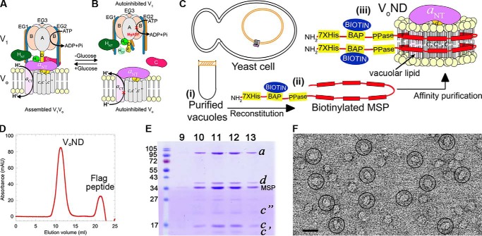
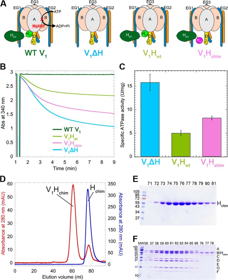
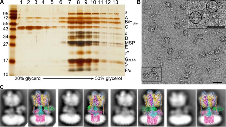
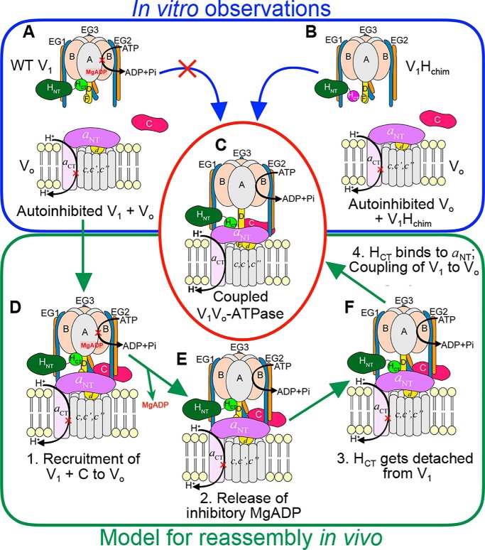
The vacuolar H+-ATPase (V-ATPase; V1Vo-ATPase) is an ATP-dependent proton pump that acidifies subcellular compartments in all eukaryotic organisms. V-ATPase activity is regulated by reversible disassembly into autoinhibited V1-ATPase and Vo proton channel subcomplexes, a process that is poorly understood on the molecular level. V-ATPase is a rotary motor, and recent structural analyses have revealed different rotary states for disassembled V1 and Vo, a mismatch that is likely responsible for their inability to reconstitute into holo V-ATPase in vitro Here, using the model organism Saccharomyces cerevisiae, we show that a key impediment for binding of V1 to Vo is the conformation of the inhibitory C-terminal domain of subunit H (HCT). Using biolayer interferometry and biochemical analyses of purified mutant V1-ATPase and Vo proton channel reconstituted into vacuolar lipid-containing nanodiscs, we further demonstrate that disruption of HCT's V1-binding site facilitates assembly of a functionally coupled and stable V1Vo-ATPase. Unlike WT, this mutant enzyme was resistant to MgATP hydrolysis-induced dissociation, further highlighting HCT's role in the mechanism of V-ATPase regulation. Our findings provide key insight into the molecular events underlying regulation of V-ATPase activity by reversible disassembly.

Keywords: biolayer interferometry; biophysics; lipid nanodisc; membrane protein; molecular motor; protein-protein interaction; reversible disassembly; vacuolar ATPase.


Conflict of interest statement

The authors declare that they have no conflicts of interest with the contents of this article

Figures

Figure 1.
Figure 1.
Purification and characterization of VoND. A and B, schematic of V-ATPase regulation by reversible disassembly. C, purification strategy. Yeast vacuoles are isolated by flotation on a Ficoll gradient (panel i). Detergent-solubilized vacuolar proteins are mixed with biotinylated MSP (panel ii) and reconstituted into lipid nanodiscs followed by α-FLAG affinity capture of VoND (panel iii). D, size-exclusion chromatography of VoND. E, peak fractions were resolved using SDS-PAGE. F, negative stain EM of purified VoND. Bar, 20 nm.
Figure 2.
Figure 2.
Purification and characterization of V1 mutants. A, schematic representation of the V1 mutants. B, time-dependent MgATPase activities of the V1 mutants measured in an ATP regenerating assay. C, specific activities of the V1 mutants ± S.E. from at least two independent purifications per mutant. D, size-exclusion chromatography of Hchim (blue trace) and V1ΔH reconstituted with Hchim (V1Hchim, red trace). E and F, SDS-PAGE of column fractions of Hchim (E) and V1Hchim (F).
Figure 3.
Figure 3.
V1Hchim and C associate with VoND to form coupled V1Vo-ATPase. A, VoND was immobilized on streptavidin-coated BLI sensors via biotinylated MSP (step 1). Sensors were then dipped into 0.4 μm of V1 mutants in presence of 1 μm C (association; step 2) followed by buffer (dissociation; step 3). Association with VoND was most efficient with V1Hchim (red trace). Sensors were then dipped in PreScission protease to verify that the BLI signal was not due to nonspecific binding (step 4). Inset shows an enlarged view of the association and dissociation steps. B, equimolar amounts of V1Hchim and VoND, and a 2-fold molar excess of C subunit were incubated at 22 °C, and the ConA-sensitive MgATPase activity was measured as a function of time. Each point represents the mean ± S.E. of two separate reconstitutions from two individual purifications. Inset, specific MgATPase activities of reconstituted V1HchimVoND and V1HwtVoND (± S.E. from two independent purifications) compared with purified V1VoND (29). C, following association of the V1HchimVoND complex, sensors were dipped in wells containing buffer (green) or buffer + 1 mm MgATP (blue) for dissociation rate measurement. The dissociation phase of WT V1VoND in buffer (red) and buffer +1 mm MgATP (orange) is included for comparison (data from Ref. 29).
Figure 4.
Figure 4.
Structural and functional characterization of the V1HchimVoND complex. A, reconstituted V1HchimVoND was subjected to glycerol gradient centrifugation, and the gradient fractions were analyzed by silver-stained SDS-PAGE. B, negative stain EM of V1HchimVoND showing homogeneous and monodisperse dumbbell-shaped molecules. Inset in the top right shows 2× zoomed area highlighted in the bottom left. C, a data set of ∼5800 particle projections was subjected to reference-free alignment and classification, and selected class averages were overlaid with projections of the cryoEM model of yeast V1Vo (Protein Data Bank code 3J9U). Bars in B, 50 nm.
Figure 5.
Figure 5.
Model for reassembly of autoinhibited V1 and Vo. A–C, our in vitro experiments have shown that although WT H containing V1 does not readily bind VoND (A), V1Hchim spontaneously associates with VoND (B) to form a structurally and functionally coupled V-ATPase, albeit at a slow rate. C, in vivo, however, V1 exists in the autoinhibited conformation (A), and the rate of assembly with Vo is significantly faster (within 5 min). D–F, for in vivo (re)assembly, we propose that the following steps occur: step 1, recruitment of V1 and subunit C to the vacuolar membrane (D); step 2, release of inhibitory MgADP (E); step 3, detachment of HCT from its inhibitory position on V1; and step 4, HCT binding to aNT (F). For further details, see text.

Similar articles

Cited by

References

    1. Forgac M. (2007) Vacuolar ATPases: rotary proton pumps in physiology and pathophysiology. Nat. Rev. Mol. Cell Biol. 8, 917–929 10.1038/nrm2272 - DOI - PubMed
    1. Karet F. E., Finberg K. E., Nelson R. D., Nayir A., Mocan H., Sanjad S. A., Rodriguez-Soriano J., Santos F., Cremers C. W., Di Pietro A., Hoffbrand B. I., Winiarski J., Bakkaloglu A., Ozen S., Dusunsel R., et al. (1999) Mutations in the gene encoding B1 subunit of H+-ATPase cause renal tubular acidosis with sensorineural deafness. Nat. Genet. 21, 84–90 10.1038/5022 - DOI - PubMed
    1. Thudium C. S., Jensen V. K., Karsdal M. A., and Henriksen K. (2012) Disruption of the V-ATPase functionality as a way to uncouple bone formation and resorption: a novel target for treatment of osteoporosis. Curr. Protein Pept. Sci. 13, 141–151 10.2174/138920312800493133 - DOI - PubMed
    1. Williamson W. R., and Hiesinger P. R. (2010) On the role of v-ATPase V0a1-dependent degradation in Alzheimer disease. Commun. Integr. Biol. 3, 604–607 10.4161/cib.3.6.13364 - DOI - PMC - PubMed
    1. Sun-Wada G. H., Toyomura T., Murata Y., Yamamoto A., Futai M., and Wada Y. (2006) The a3 isoform of V-ATPase regulates insulin secretion from pancreatic β-cells. J. Cell Sci. 119, 4531–4540 10.1242/jcs.03234 - DOI - PubMed

Publication types

MeSH terms

Substances

Associated data