. 2019 Feb 21;10(3):179.
doi: 10.1038/s41419-019-1427-2.

Angiomotin-p130 inhibits β-catenin stability by competing with Axin for binding to tankyrase in breast cancer

Affiliations

Angiomotin-p130 inhibits β-catenin stability by competing with Axin for binding to tankyrase in breast cancer

Jiao Yang et al. Cell Death Dis. .

Abstract

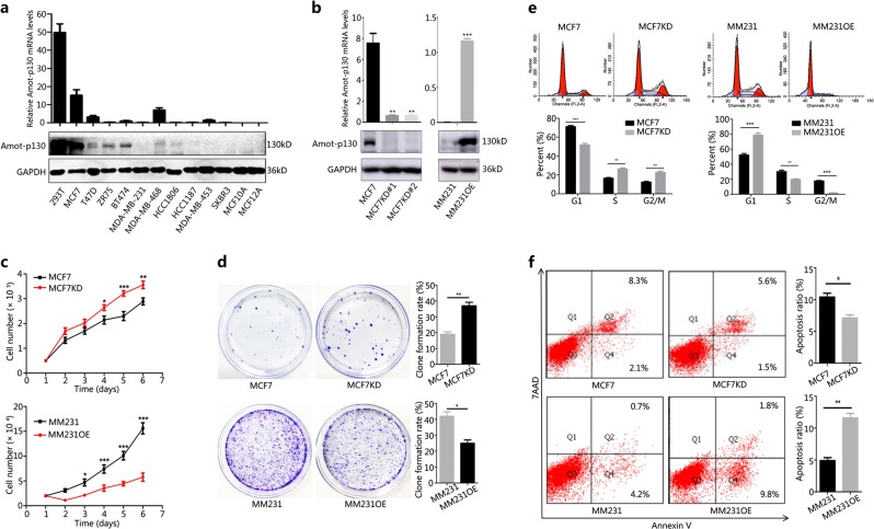
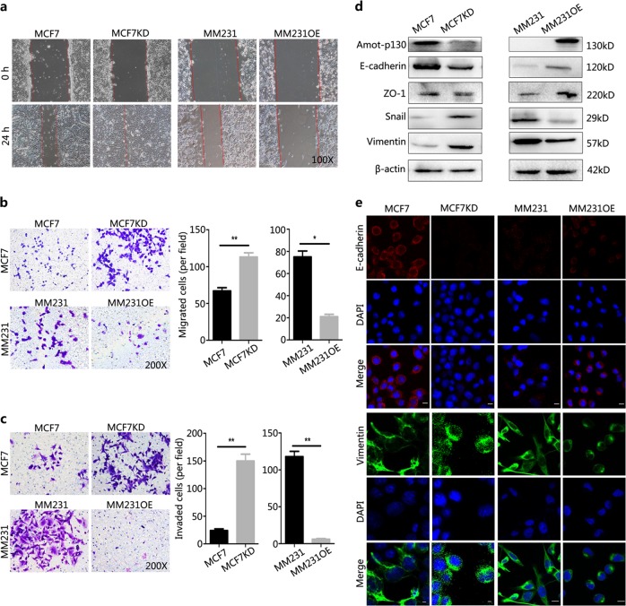
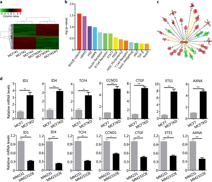
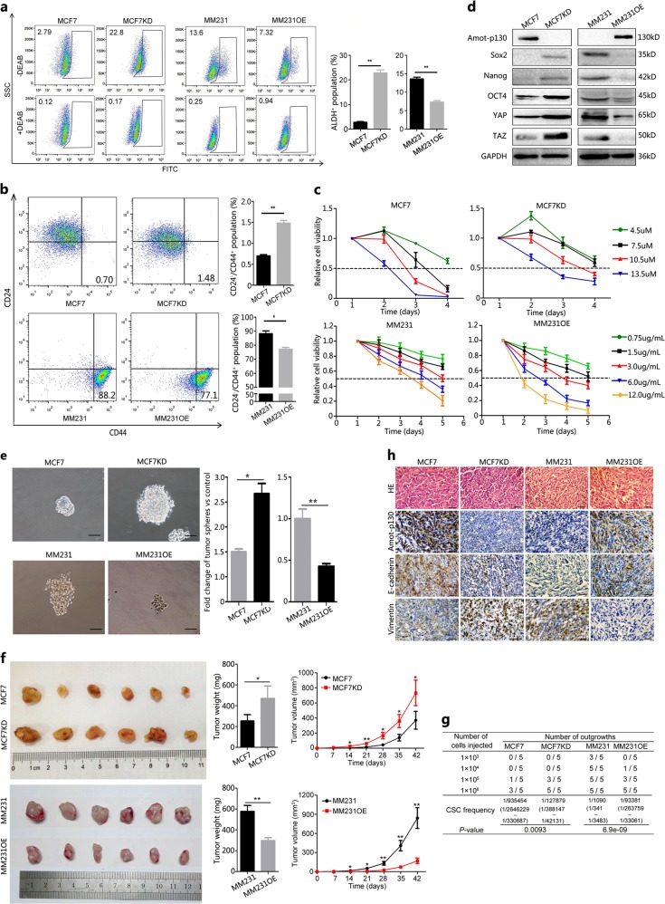
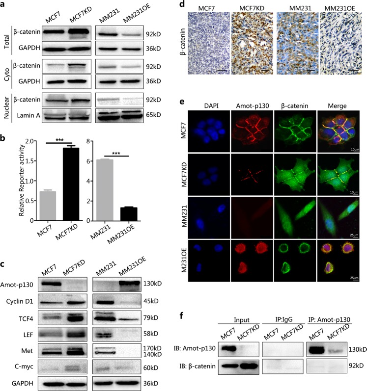
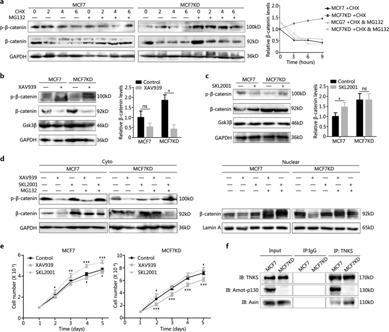
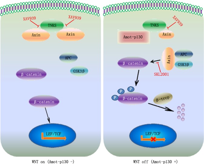
Growing evidence indicates that Angiomotin (Amot)-p130 and Amot-p80 have different physiological functions. We hypothesized that Amot-p130 is a tumor suppressor gene in breast cancer, in contrast with the canonical oncogenicity of Amot-p80 or total Amot. To clarify the role of Amot-p130 in breast cancer, we performed real-time quantitative PCR, western blotting, flow cytometry, microarray, immunofluorescence, immunoprecipitation, and tumor sphere-formation assays in vitro, as well as tumorigenesis and limited-dilution analysis in vivo. In this study, we showed that Amot-p130 inhibited the proliferation, migration, and invasion of breast cancer cells. Interestingly, transcriptional profiles indicated that genes differentially expressed in response to Amot-p130 knockdown were mostly related to β-catenin signaling in MCF7 cells. More importantly, most of the downstream partners of β-catenin were associated with stemness. In a further validation, Amot-p130 inhibited the cancer stem cell potential of breast cancer cells both in vitro and in vivo. Mechanistically, Amot-p130 decreased β-catenin stability by competing with Axin for binding to tankyrase, leading to a further inhibition of the WNT pathway. In conclusions, Amot-p130 functions as a tumor suppressor gene in breast cancer, disrupting β-catenin stability by competing with Axin for binding to tankyrase. Amot-p130 was identified as a potential target for WNT pathway-targeted therapies in breast cancer.


Conflict of interest statement

The authors declare that they have no conflict of interest.

Figures

Fig. 1
Fig. 1. Amot-p130 inhibits the proliferation of breast cancer cells.
a Expression levels of Amot-p130 in nine breast cancer cell lines and two immortalized breast epithelial cells (MCF10A and MCF12A) (using 293T cells as the positive control) as determined by (upper) RT-qPCR and (lower) western blotting. GAPDH was used as the loading control. b Interference efficiency of Amot-p130 was evaluated by (upper) RT-qPCR and (lower) western blotting. GAPDH was used as the loading control. c Cell proliferation was measured using cell count assay. d Cell proliferation was measured using plate clone formation. Clones were stained with 0.4% crystal violet (left). Numbers of clones are shown as the mean ± SD of three independent experiments (right). e Cell cycles were determined by flow cytometry (upper). The proportion of cells distributed in each cell cycle was calculated from three independent experiments (lower). f Cell apoptosis was determined by flow cytometry (left). Numbers of apoptotic cells were calculated from three independent experiments (right). MM231 is the abbreviation of MDA-MB-231. All P values were calculated by paired Student’s t test. *P < 0.05, **P < 0.01, ***P < 0.001. RT-qPCR, real-time quantitative PCR
Fig. 2
Fig. 2. Amot-p130 inhibits the EMT of breast cancer cells.
a A photomicrograph of a scratch created with a 200 μl pipette tip in the wound-healing assay at 0 and 24 h. b Cell migration and c cell invasion were assessed using transwell assay (left). The number of migrated or invaded cells was expressed as the mean ± SD of three independent experiments (right). d Expression of epithelial to mesenchymal transition (EMT) markers was determined by western blotting. β-actin was the loading control. e The subcellular localization of EMT markers was determined by immunofluorescence staining. Both E-cadherin and vimentin antibodies were diluted at a ratio of 1:200. DAPI was used for nuclear staining (blue). Scale bars = 25 µm. All P values were calculated by Student’s t test. *P < 0.05, **P < 0.01. DAPI, 4′,6-diamidino-2-phenylin
Fig. 3
Fig. 3. Identification of β-catenin as an effector of Amot-p130 in breast cancer cells.
a A visualized heatmap of differentially expressed genes in MCF7KD cells compared to MCF7 control cells. Red color indicates upregulation and green indicates downregulation (detailed data are uploaded as Supplementary Table S1). b Functionally associated pathways were identified by Ingenuity Pathway Analysis. Enriched pathways related to breast carcinogenesis were sorted by Fisher's exact P values. c An interactive network analysis of the key molecular β-catenin from the first ranked WNT pathway. d Expression validation of the genes from the interactive network by quantitative PCR. The means ± SD of relative fold changes from triplicate experiments were plotted. GAPDH was used as the control. P values were calculated by paired Student’s t test
Fig. 4
Fig. 4. Amot-p130 inhibits the CSC potential of breast cancer cells.
a ALDH-positive population was detected by flow cytometry (left). The proportions were calculated from three independent experiments (right). b CD24-low/CD44-high populations were detected by flow cytometry (left). The proportions were calculated from three independent experiments (right). c The tolerance of MCF7 cells to tamoxifen and MM231 cells to cisplatin was determined by MTT assay. d Stemness markers were assessed by western blotting. GAPDH was used as the loading control. e The formation of tumor spheres was recorded under microscopy (left). Scale bar = 50 µm. The proportions were calculated from three independent experiments (right). f Xenograft formation in NOD-SCID mice evaluated at 6 weeks after injection. Tumor volumes were determined using the formula volume = 1/2 × length × width2. g The number of outgrowths was recorded at the end of the 6th week using limited-dilution assays. h IHC analysis of protein expression of Amot-p130, E-cadherin, and vimentin in xenograft tissues. Scale bar = 200 µm. All P values were calculated by paired Student’s t test. *P < 0.05, **P < 0.01. IHC, immunohistochemistry; MTT, 3-(4,5-dimethylthiazol-2-yl)-2,5-diphenyltetrazolium bromide
Fig. 5
Fig. 5. Amot-p130 inhibits WNT pathway activation in breast cancer cells.
a Protein levels of total β-catenin, cytoplasmic β-catenin, and nuclear β-catenin as determined by western blotting. GAPDH was used as the loading control for the total and cytoplasmic protein. Lamin A was used as the loading control for nuclear protein. b β-Catenin-driven transcription activity was determined by TOP/FOP luciferase reporter assays. Normalization was based on internal Renilla luciferase actvity. The final reporter activity was measured as TOP/FOP ratio and was expressed as the mean ± SD of three independent experiments. c Protein levels of WNT downstream targets were determined by western blotting. d β-Catenin expression was determined by immunohistochemistry staining in xenograft tissues. Scale bar = 200 µm. e Co-localization of Amot-p130 (red) and β-catenin (green) in cell–cell contacts was indicated by immunofluorescence confocal microscopy. DAPI was used for nuclear staining (blue). f The interaction between Amot-p130 and β-catenin was evaluated in MCF7 cells by co-immunoprecipitation assay. IgG was used as the negative control. ***P < 0.001. DAPI, 4′,6-diamidino-2-phenylin
Fig. 6
Fig. 6. Amot-p130 regulates β-catenin stability by competing with Axin for binding to TNKS.
a β-Catenin levels in cells treated with CHX (200 µg/ml) for the indicated time, with or without MG132 (20 µM) for 2 h, were determined using western blotting (left). GAPDH was used as the loading control. The curve showed the relative trend of β-catenin changes (right). b β-Catenin levels in cells treated with XAV939 10 µg/ml for 24 h were determined using western blotting (left). The quantitation of β-catenin was expressed as the mean ± SD of three independent experiments (right). c β-catenin levels in cells treated with SKL2001 30 µM for 24 h were determined using western blotting (left). The quantitation of β-catenin was expressed as the mean ± SD of three independent experiments (right). d Protein levels of the total, cytoplasmic, and nuclear β-catenin in cells treated with XAV939 or SKL2001, alone or in combination with MG132, were determeined using western blotting. GAPDH was used as the loading control for the total and cytoplasmic protein. Lamin A was used as the loading control for nuclear protein. e MCF7 cell proliferation under XAV939 or SKL2001 treatment was determined by MTT assay. f Relative quantitation of Amot-p130 and Axin pulled down by TNKS in co-immunoprecipitation assay. IgG pull-down was used as the negative control. *P < 0.05, **P < 0.01, ***P < 0.001; ns, no significance. MTT, 3-(4,5-dimethylthiazol-2-yl)-2,5-diphenyltetrazolium bromide; TNKS, tankyrase
Fig. 7
Fig. 7. Model showing the regulation of the WNT pathway by Amot-p130.
Axin/TNKS binding contributes to the nuclear translocation of β-catenin and the further activation of the WNT pathway in Amot-p130 negative cells (left). Amot-p130/TNKS binding allows Axin to form β-catenin-destruction complexes, leading to the phosphorylation and ubiquitination-mediated degradation of β-catenin, thereby inactivating WNT signaling in Amot-p130 positive cells (right). TNKS, tankyrase

Similar articles

Cited by

References

    1. Fan L, et al. Breast cancer in China. Lancet Oncol. 2014;15:e279–e289. doi: 10.1016/S1470-2045(13)70567-9. - DOI - PubMed
    1. Boris Troyanovsky TL, Månsson Goumlran, Matvijenko O, Holmgren L. Angiomotin: an angiostatin binding protein that regulates endothelial cell migration and tube formation. J. Cell. Biol. 2001;152:1247–1254. doi: 10.1083/jcb.152.6.1247. - DOI - PMC - PubMed
    1. Zhang H, Fan Q. MicroRNA-205 inhibits the proliferation and invasion of breast cancer by regulating AMOT expression. Oncol. Rep. 2015;34:2163–2170. doi: 10.3892/or.2015.4148. - DOI - PubMed
    1. Zeng H, et al. Angiomotin regulates prostate cancer cell proliferation by signaling through the Hippo-YAP pathway. Oncotarget. 2017;8:10145–10160. - PMC - PubMed
    1. Hsu YL, et al. Angiomotin decreases lung cancer progression by sequestering oncogenic YAP/TAZ and decreasing Cyr61 expression. Oncogene. 2015;34:4056–4068. doi: 10.1038/onc.2014.333. - DOI - PubMed

Publication types

MeSH terms