. 2019 Feb 21;10(3):176.
doi: 10.1038/s41419-019-1437-0.

miR-29a contributes to breast cancer cells epithelial-mesenchymal transition, migration, and invasion via down-regulating histone H4K20 trimethylation through directly targeting SUV420H2

Affiliations

miR-29a contributes to breast cancer cells epithelial-mesenchymal transition, migration, and invasion via down-regulating histone H4K20 trimethylation through directly targeting SUV420H2

You Wu et al. Cell Death Dis. .

Erratum in

Abstract

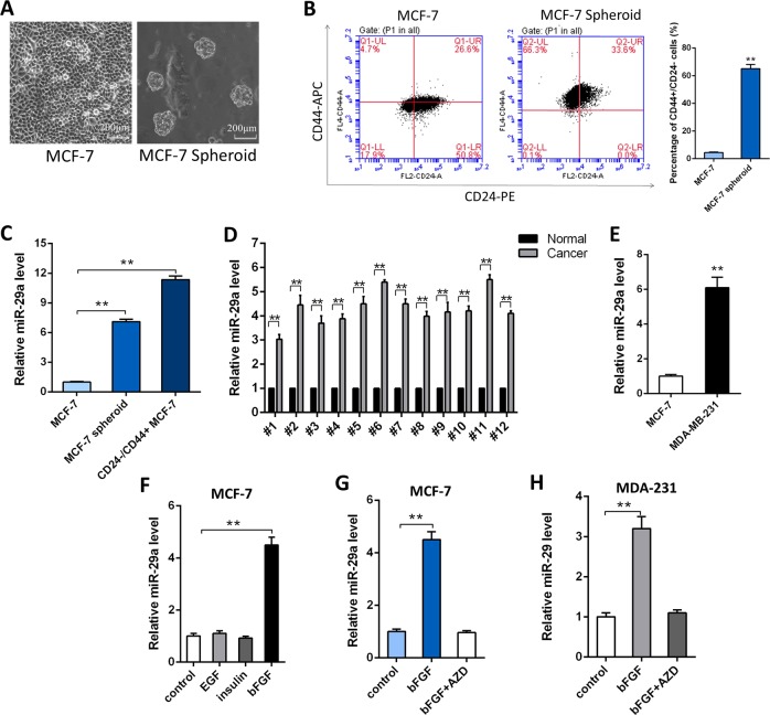
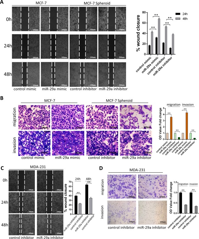
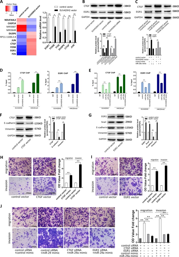
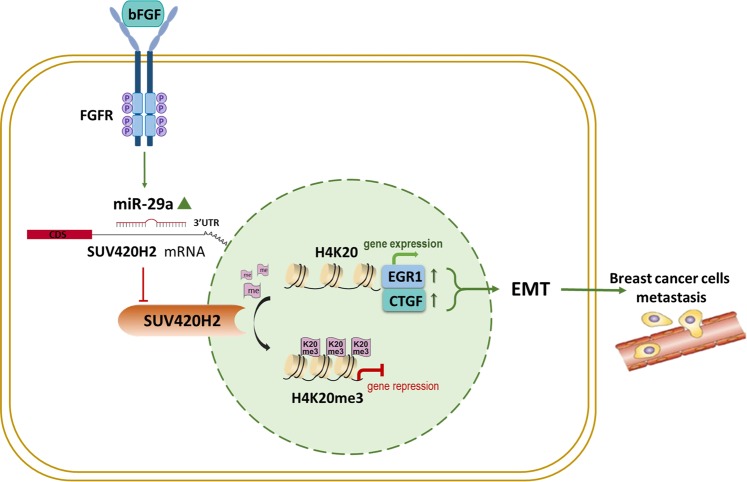
Breast cancer is the most prevalent cancer in women worldwide, which remains incurable once metastatic. Breast cancer stem cells (BCSCs) are a small subset of breast cancer cells which are essential in tumor formation, metastasis, and drug resistance. microRNAs (miRNAs) play important roles in the breast cancer cells and BCSCs by regulating specific genes. In this study, we found that miR-29a was up-regulated in BCSCs, in aggressive breast cancer cell line and in breast cancer tissues. We also confirmed suppressor of variegation 4-20 homolog 2 (SUV420H2), which is a histone methyltransferase that specifically trimethylates Lys-20 of histone H4 (H4K20), as the target of miR-29a. Both miR-29a overexpression and SUV420H2 knockdown in breast cancer cells promoted their migration and invasion in vitro and in vivo. Furthermore, we discovered that SUV420H2-targeting miR-29a attenuated the repression of connective tissue growth factor (CTGF) and growth response protein-1 (EGR1) by H4K20 trimethylation and promoted the EMT progress of breast cancer cells. Taken together, our findings reveal that miR-29a plays critical roles in the EMT and metastasis of breast cancer cells through targeting SUV420H2. These findings may provide new insights into novel molecular therapeutic targets for breast cancer.


Conflict of interest statement

The authors declare that they have no conflict of interest.

Figures

Fig. 1
Fig. 1. miR-29a is up-regulated in breast cancer cells.
a Bright field images of MCF-7 cells formation under monolayer culture condition and 3D culture condition (Magnification: ×20, scale bar: 200 μm), respectively. b The percentage of CD44+/CD24− population in MCF-7 cells and MCF-7 spheroid cells respectively. c miR-29a levels in MCF-7 cells, MCF-7 spheroid cells and CD44+/CD24− MCF-7 cells. d miR-29a levels in 12 pairs of human breast cancer tissues (Cancer) and corresponding distal non-cancerous tissues (Normal). e miR-29a levels in MCF-7 cells and MDA-MB-231 cells. f miR-29a levels in MCF-7 cells under stimulation of EGF, bFGF, and insulin. g, h miR-29a levels in MCF-7 cells (g) and MDA-MB-231 cells (h) under stimulation of bFGF, and bFGF plus AZD. **P < 0.01
Fig. 2
Fig. 2. miR-29a promotes breast cancer cells migration and invasion in vitro.
a, b Migration and invasion of MCF-7 cells transfected with control mimic or miR-29a mimic and MCF-7 spheroid cells transfected with control inhibitor or miR-29a inhibitor detected by wound healing assay (a), transwell migration assay and transwell invasion assay (b). c, d Migration and invasion of MDA-MB-231 cells transfected with control inhibitor or miR-29a inhibitor detected by wound healing assay (c), transwell migration assay and transwell invasion assay (d). **P < 0.01
Fig. 3
Fig. 3. Identification of SUV420H2 as a direct target gene of miR-29a in breast cancer cells.
a Left: Venn diagram analysis of three independent databases reveals four possible targets of miR-29a. Right: schematic description of the hypothetical duplexes formed by the interactions between the binding sites in the SUV420H2 3′-UTR and miR-29a. The predicted free energy value of each hybrid is indicated. The seed recognition sites are denoted, and all nucleotides in these regions are highly conserved across species, including human, mouse, and rat. b, c SUV420H2 protein (b) and mRNA (c) levels in MCF-7 cells, MCF-7 spheroid cells and CD44+/CD24− MCF-7 cells. d, e SUV420H2 protein (d) and mRNA (e) levels in 12 pairs of human breast cancer tissues (Cancer) and corresponding distal non-cancerous tissues (Normal). f Pearson’s correlation scatter plot of the fold change of miR-29a and SUV420H2 protein in human breast cancer tissues. g Pearson’s correlation scatter plot of the fold change of miR-29a and SUV420H2 mRNA in human breast cancer tissues. h, i SUV420H2 protein (h) and mRNA (i) levels in MCF-7 cells and MDA-MB-231 cells. j Direct recognition of the SUV420H2 3′-UTR by miR-29a. Firefly luciferase reporters containing either wild-type (WT) or mutant (MUT) miR-29a-binding sites in the SUV420H2 3′-UTR were co-transfected into MCF-7 cells with either the control mimic, control inhibitor, miR-29a mimic or miR-29a inhibitor. Twenty-four hours post-transfection, the cells were assayed using a luciferase assay kit. The results were calculated as the ratio of firefly luciferase activity normalized to the control cells. k, l SUV420H2 protein (i) and mRNA (j) levels in MCF-7 cells transfected with scrambled negative control RNA, miR-29a mimic, or miR-29a inhibitor. *P < 0.05; **P < 0.01
Fig. 4
Fig. 4. miR-29a promotes breast cancer cells migration and invasion via targeting SUV420H2.
a, b Migration and invasion of MCF-7 cells transfected with either the control vector, SUV420H2 vector, control siRNA, or SUV420H2 siRNA as indicated, detected by wound healing assay (a), transwell migration assay, and transwell invasion assay (b). c, d Migration and invasion of MCF-7 cells transfected with either the control mimic plus control vector, miR-29a mimic plus control vector or miR-29a mimic plus SUV420H2 vector detected by wound healing assay (c), transwell migration assay and transwell invasion assay (d). *P < 0.05; **P < 0.01
Fig. 5
Fig. 5. miR-29a-mediated SUV420H2 decrease trimethylation of H4K20 and promotes EMT progress in MCF-7 cells.
a SUV420H2 and H4K20me3 protein levels in MCF-7 cells transfected with either the scrambled negative control RNA, miR-29a mimic, or miR-29a inhibitor. b SUV420H2 and H4K20me3 protein levels in MCF-7 cells transfected with either the control vector, SUV420H2 vector, control siRNA, or SUV420H2 siRNA. c SUV420H2 and H4K20me3 protein levels in MCF-7 cells transfected with either the control mimic plus control vector, miR-29a mimic plus control vector, or miR-29a mimic plus SUV420H2 vector. d Immunofluorescent staining of E-cadherin and Vimentin in MCF-7 cells transfected with either the control mimic plus control vector, miR-29a mimic plus control vector, control mimic plus SUV420H2 vector, or miR-29a mimic plus SUV420H2 vector. **P < 0.01
Fig. 6
Fig. 6. miR-29a promotes breast cancer cells EMT, migration and invasion by overexpressing EGR1 and CTGF genes through down-regulation of H4K20me3.
a The heat map of significantly up- or down-regulated miRNAs between MCF-7 transfected with control vector and MCF-7 transfected with SUV420H2 vector. Each group contained three batches of individual samples, which were pooled and mixed. b Verification of EGR1, FOS, FOSB, JUN, DUSP6, and CTGF mRNA levels by qRT-PCR in the two groups. c CTGF and EGR1 protein levels in MCF-7 cells transfected with either the control vector, SUV420H2 vector, control siRNA, or SUV420H2 siRNA. d CTGF and EGR1 protein levels in MCF-7 cells transfected with either the control mimic plus control vector, miR-29a mimic plus control vector, or miR-29a mimic plus SUV420H2 vector. e, f ChIP-qPCR of SUV420H2/H4K20me3 of the promoter region of CTGF or EGR1 locus in MCF-7 cells transfected with either the control vector or SUV420H2 vector (e), and in MCF-7 cells transfected with either the control mimic plus control vector, miR-29a mimic plus control vector or miR-29a mimic plus SUV420H2 vector (f). Antibody enrichment was quantified relative to the amount of input DNA. Antibody directed against IgG was used as a negative control. g CTGF, E-cadherin, and Vimentin protein levels in MCF-7 cells transfected with control vector or CTGF vector. h EGR1, Snail, E-cadherin, and Vimentin protein levels in MCF-7 cells transfected with control vector or EGR1 vector. i–k Migration and invasion of MCF-7 cells transfected with control vector or CTGF vector (i); control vector or EGR1 vector (j); with either the control mimic plus control vector, miR-29a mimic plus control vector, miR-29a mimic plus CTGF vector, or miR-29a mimic plus EGR1 vector (k). *P < 0.05; **P < 0.01
Fig. 7
Fig. 7. Effects of SUV420H2-targeted miR-29a on the lung colonization of MCF-7 cells xenografts in mice.
a, b miR-29a levels (a), SUV420H2 protein and H4K20me3 levels (b) in MCF-7 cells transfected with either the control lentivirus, miR-29a lentivirus, SUV420H2 lentivirus or miR-29a lentivirus plus SUV420H2 lentivirus. c Experimental design. Immunocompromised mice were injected through tail vein with MCF-7 cells transfected with either the control lentivirus, miR-29a lentivirus, SUV420H2 lentivirus or miR-29a lentivirus plus SUV420H2 lentivirus. d Representative BLI images of four groups. The BLI was performed on days 7, 28, and 56 after injection. The intensity of BLI is represented by the color. e The numbers of tumor nodules in the lungs. Results were derived from five mice in each group. f, g Mouse lungs were subjected to H&E staining (f) and immunohistochemical staining for SUV420H2 and H4K20me3 (g), respectively. **P < 0.01
Fig. 8
Fig. 8. A working model for the role of SUV420H2-targeted miR-29a in breast cancer.
During breast tumorigenesis, miR-29a activated by bFGF will directly inhibit SUV420H2 expression, consequently triggering loss of H4K20me3 and EGR1 and CTGF genes expression, which eventually promotes EMT and results in the distal metastasis of human breast cancer

Similar articles

Cited by

References

    1. Bray F, et al. Global cancer statistics 2018: GLOBOCAN estimates of incidence and mortality worldwide for 36 cancers in 185 countries. CA Cancer J. Clin. 2018;68:394–424. - PubMed
    1. Mosmann T. Rapid colorimetric assay for cellular growth and survival: application to proliferation and cytotoxicity assays. J. Immunol. Methods. 1983;65:55–63. - PubMed
    1. Li B, et al. miR-221/222 promote cancer stem-like cell properties and tumor growth of breast cancer via targeting PTEN and sustained Akt/NF-ΰB/COX-2 activation. Chem. -Biol. Interact. 2017;277:33–42. - PubMed
    1. Takahashi, R. U. et al. Loss of microRNA-27b contributes to breast cancer stem cell generation by activating ENPP1. Nat. Commun.6, 7318 (2015). - PMC - PubMed
    1. Uhlmann S, et al. miR-200bc/429 cluster targets PLCgamma1 and differentially regulates proliferation and EGF-driven invasion than miR-200a/141 in breast cancer. Oncogene. 2010;29:4297–4306. - PubMed

Publication types

MeSH terms