. 2019 Feb 21;10(1):890.
doi: 10.1038/s41467-019-08719-8.

Extreme slow growth as alternative strategy to survive deep starvation in bacteria

Affiliations

Extreme slow growth as alternative strategy to survive deep starvation in bacteria

Declan A Gray et al. Nat Commun. .

Abstract

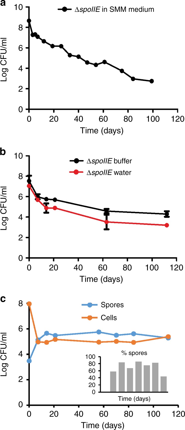
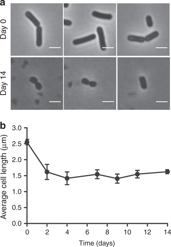
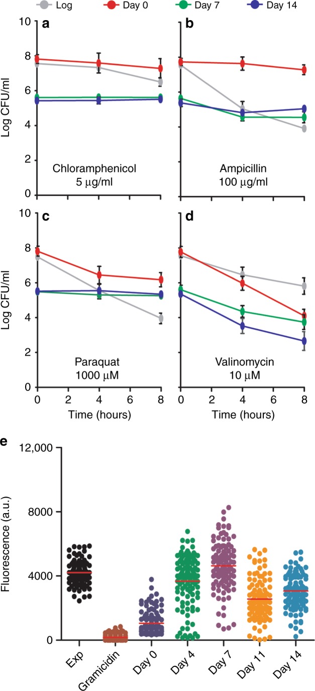
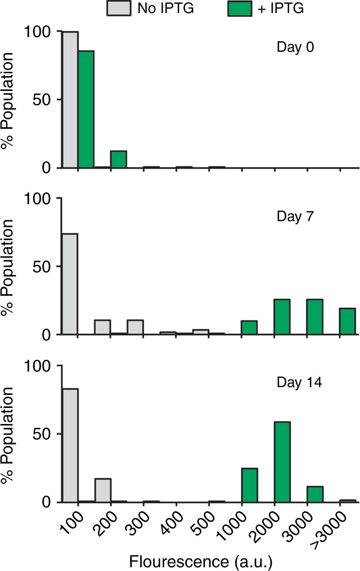
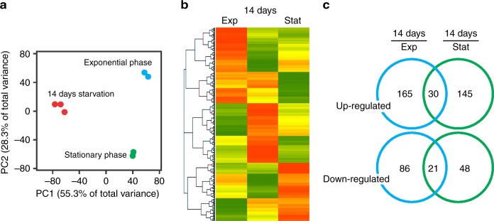
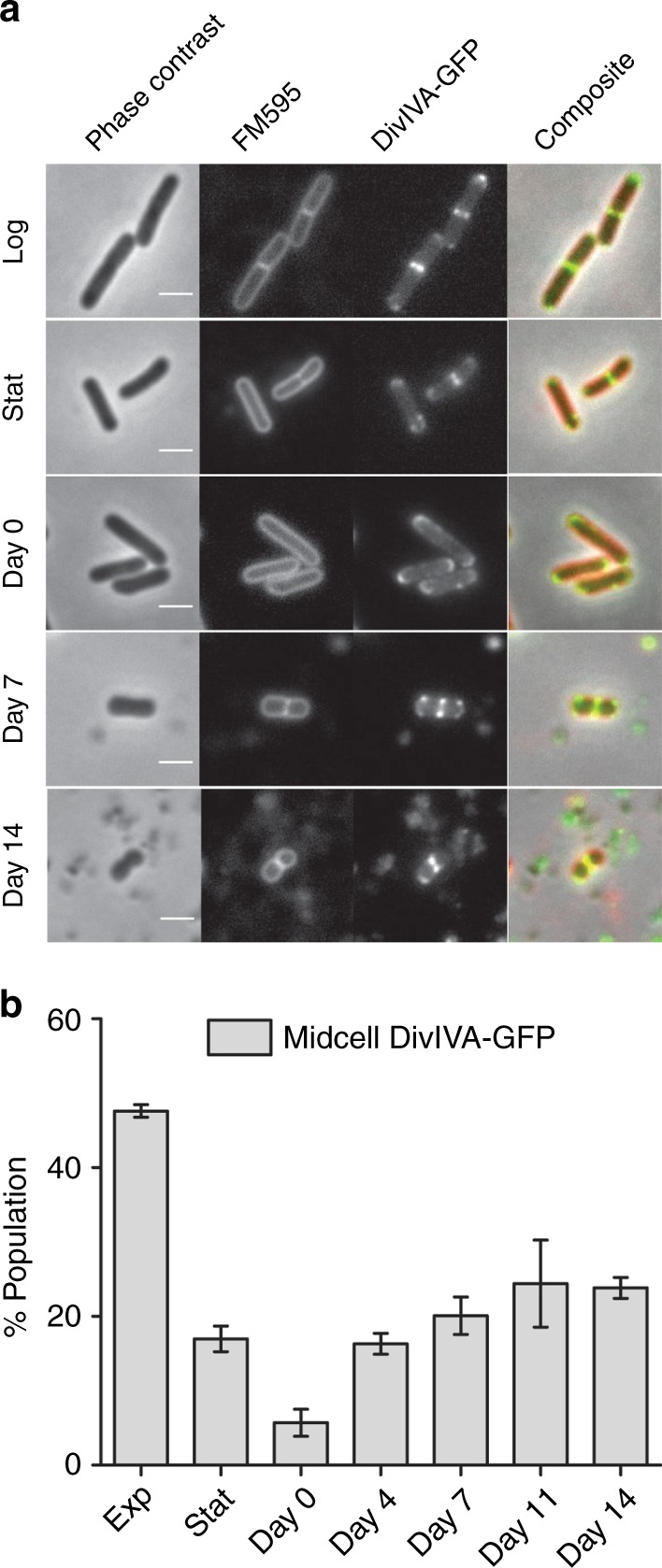
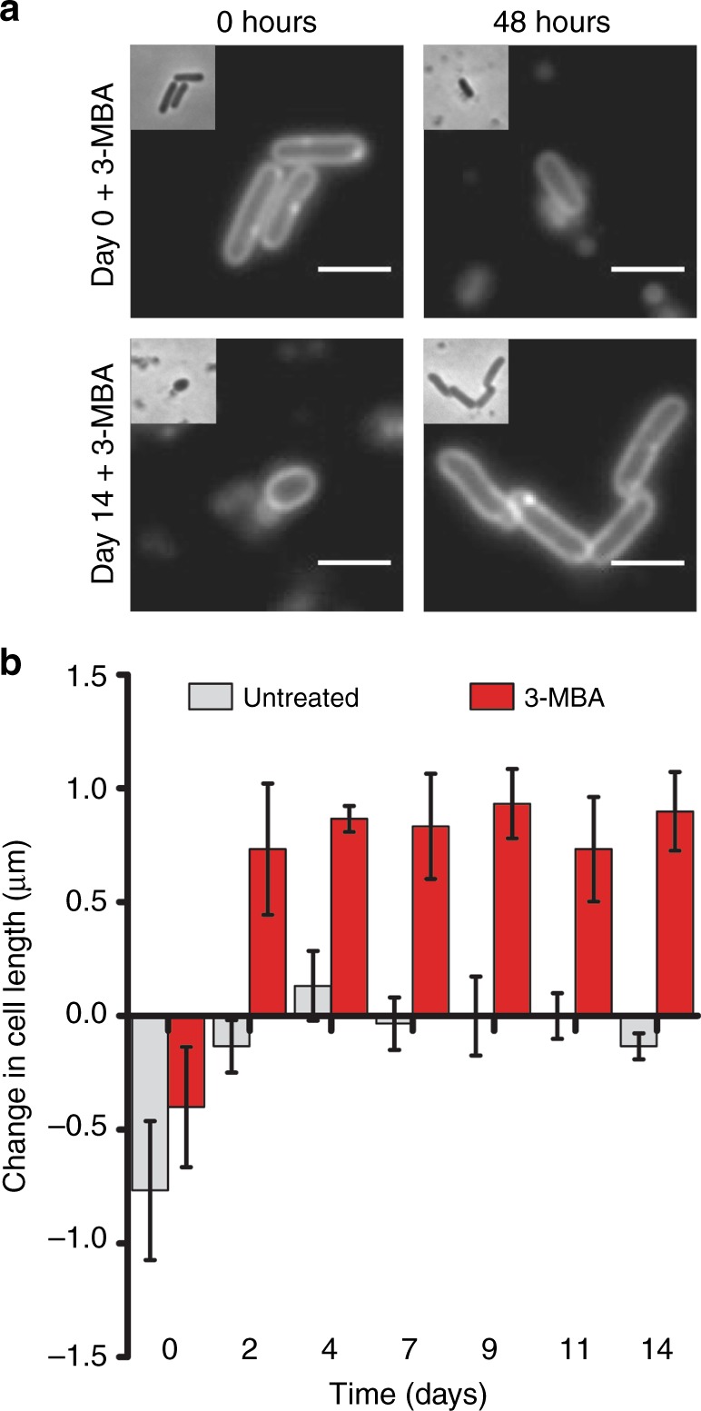
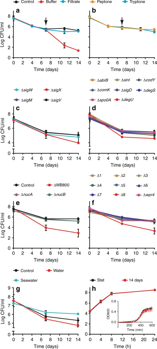
Bacteria can become dormant or form spores when they are starved for nutrients. Here, we find that non-sporulating Bacillus subtilis cells can survive deep starvation conditions for many months. During this period, cells adopt an almost coccoid shape and become tolerant to antibiotics. Unexpectedly, these cells appear to be metabolically active and show a transcriptome profile very different from that of stationary phase cells. We show that these starved cells are not dormant but are growing and dividing, albeit with a doubling time close to 4 days. Very low nutrient levels, comparable to 10,000-fold diluted lysogeny broth (LB), are sufficient to sustain this growth. This extreme slow growth, which we propose to call 'oligotrophic growth state', provides an alternative strategy for B. subtilis to endure nutrient depletion and environmental stresses. Further work is warranted to test whether this state can be found in other bacterial species to survive deep starvation conditions.


Conflict of interest statement

The authors declare no competing interests.

Figures

Fig. 1
Fig. 1
Long-term survival of non-sporulating B. subtilis. a Colony-forming units (CFU) of B. subtilis ΔspoIIE (strain DG001) incubated in Spizizen minimal medium (SMM). b CFU of ΔspoIIE cells that were first grown for 2 days in SMM, and subsequently filtered and incubated in either starvation buffer or water (=0 days time point). The CFU numbers of the first time point are therefore comparable to those of time point 2 days in graph (a). Averages and standard deviation from three independent experiments are depicted. The difference between the two graphs becomes significant after day 7 (p < 0.05, unpaired two-tailed t-test). c CFU of spores and cells in a wild-type B. subtilis culture (strain BSB1) incubated in starvation buffer. The percentage of spores is indicated in the bar diagram. Results of two replicate experiments are shown in Supplementary Figure 2A. See Methods for details on growth and starvation conditions
Fig. 2
Fig. 2
Cell length reduction during deep starvation. a Phase contrast images of B. subtilis ΔspoIIE cells in the beginning (day 0), and after 14 days of incubation in starvation buffer. Scale bar is 2 µm. b Average cell length. Average and standard deviation from three independent experiments are depicted. Approximately 100 cells were measured for every condition
Fig. 3
Fig. 3
Antibiotic sensitivity under starvation conditions. Cultures of ΔspoIIE were set up in accordance with the starvation assay. Samples were taken from cultures at days 0, 7, and 14, and treated with either: a 5 µg ml−1 chloramphenicol, b 100 µg ml−1 ampicillin, c 1 mM paraquat, or d 10 µM valinomycin, for 8 h. As a control, exponential phase cells were treated in the same manner as the starvation samples (gray lines). Graphs show the average and standard deviation of three independent experiments. e Membrane potential levels of cells after 0, 4, 7, 11, and 14 days’ deep starvation. Relative membrane potential levels were determined based on the uptake of the membrane potential sensitive fluorescent dye 3,3′-dipropylthiadicarbocyanine iodide. As controls, membrane potential levels of exponentially growing cells (OD600 0.2), and cells treated with the ionophore gramicidin ABC (10 µg ml−1) were determined. Fluorescence intensities of approximately 100 cells were quantified (median in red). Two biological replicates are presented in Supplementary Figure 4
Fig. 4
Fig. 4
gfp expression capacity during deep starvation. B. subtilis strain ΔspoIIE containing an inducible green fluorescent protein (GFP) reporter (strain DG017, amyE::Phyperspank-sfGFP) was incubated in starvation buffer for 0, 7, and 14 days, followed by incubation in the presence or absence of 1 mM isopropyl β-d-1-thiogalactopyranoside (IPTG) for 4 h, after which cells were imaged by fluorescence light microscopy. The fluorescence of approximately 100 cells was quantified for each condition. Cells were binned based on their fluorescence intensities (arbitrary units), and the number of cells in each bin were plotted as histograms. Two biological replicates can be found in Supplementary Figure 5
Fig. 5
Fig. 5
Transcriptome comparison. Transcriptomes (RNA-seq) of B. subtilisspoIIE were analyzed for cells incubated 14 days in starvation buffer (in triplicate), and for cells grown in Spizizen minimal medium (SMM) and harvested in exponential or stationary growth phase, respectively (each in duplicate). a Principal component analysis plot of the different samples. Samples were plotted against the first two principal components calculated from the gene expression values. The axes labels indicate percentage of total variance that is explained by each component. b Heat map of the averaged transcriptome profiles. Expression levels were transformed to Z-scores. Green indicates low expression (Z-score = −3.5), yellow indicates average expression (Z-score = 0), and red indicates high expression (Z-score = +3). c Venn diagram indicating the number of upregulated and downregulated genes in 14-day-starved cells compared to exponential growth phase cells (left) and stationary growth phase cells (right). Strain used: B. subtilis PG344 (spoIIE::erm)
Fig. 6
Fig. 6
Presence of cell division sites. B. subtilis ΔspoIIE cells encoding divIVA-gfp reporter were incubated for 14 days in starvation buffer. a At different time intervals samples were taken for microscopic analysis. Cells were stained with the fluorescent membrane dye FM5–95. Scale bar is 2 µm. b To avoid bias, the captured phase contrast images were used to select approximately 100 cells per sample, and the number of cells containing a midcell DivIVA-GFP signal, indicative of cell division, were counted. As a control, the number of division sites present in both exponentially growing cells (OD600 ~0.2), and stationary growth phase cells (overnight) were determined. Bar diagram represents average and standard deviation of three independent experiments. Strain used: B. subtilis DG001 (spoIIE::erm, divIVA:divIVA-GFP(cm))
Fig. 7
Fig. 7
Cell growth under deep starvation conditions. B. subtilis ΔspoIIE cells (strain DG001) were incubated for 14 days in starvation buffer. At regular time intervals samples were withdrawn and incubated with the cell division inhibitor 3-methoxybenzamide (3-MBA) for 48 h. a Cells were stained with the fluorescent membrane dye FM5-95 in order to determine cell length before and after 3-BMA treatment. Scale bar is 2 µm. b Average change in cell length was calculated for approximately 100 individual cells for each time point. Bar diagram depicts the average and standard deviation of three independent experiments
Fig. 8
Fig. 8
Nutrient and genetic factors, and escape. a Importance of nutrients released by lysed cells was determined by filtering B. subtilis ΔspoIIE cultures (strain DG001) after 7-day starvation followed by resuspension (arrow) in either fresh starvation buffer (buffer) or the cell filtrate (filtrate). b Effect of resuspension, after 7-day starvation, in starvation buffer supplemented with 0.5 µg ml−1 peptone or tryptone. c Survival of different extra-cytoplasmic function (ECF) sigma factor mutants. Only log 4–8 colony-forming units (CFU) ml−1 is shown to emphasize differences. d Survival of mutants lacking different stationary phase regulators. Only log 4–8 CFU ml−1 is shown to emphasize differences. e To determine whether proteases are important for survival, a strain lacking all eight secreted proteases, ∆WB800, was tested. The absence of the two extracellular nucleases produced by B. subtilis on survivability was also tested (∆nucA and ∆nucB). f Survival of strains carrying cumulative deletions of the eight secreted proteases (nprB, aprE, epr, bpr, nprE, mpr, vpr, and wprA, respectively). wprA was the last deleted gene (∆8) but a single wprA deletion (∆wprA) shows normal CFU levels. g Survival of oligotrophically growing cells in seawater. Cells were incubated for 14 days in starvation buffer, water, or artificial seawater (450 mM NaCl, 10 mM KCl, 9 mM CaCl2, 30 mM MgCl2, and 30 mM MgSO4). Only log 5–9 CFU ml−1 is shown to emphasize differences. h Escape from the oligotrophic growth state. SMM was inoculated with either stationary phase cells or 14-day-starved cells, and outgrowth was measure by either CFU or optical density (OD) measurements (inset). Graphs represent averages and standard deviations of three independent experiments. The ΔspoIIE background strain was used in all experiments

Comment in

  • Life in the slow lane.
    Hofer U. Hofer U. Nat Rev Microbiol. 2019 May;17(5):266-267. doi: 10.1038/s41579-019-0176-7. Nat Rev Microbiol. 2019. PMID: 30833717 No abstract available.

Similar articles

Cited by

References

    1. Nicholson WL, Munakata N, Horneck G, Melosh HJ, Setlow P. Resistance of Bacillus endospores to extreme terrestrial and extraterrestrial environments. Microbiol. Mol. Biol. Rev. 2000;64:548–572. doi: 10.1128/MMBR.64.3.548-572.2000. - DOI - PMC - PubMed
    1. Arias CR, Lafrentz S, Cai W, Olivares-Fuster O. Adaptive response to starvation in the fish pathogen Flavobacterium columnare: cell viability and ultrastructural changes. BMC Microbiol. 2012;12:266. doi: 10.1186/1471-2180-12-266. - DOI - PMC - PubMed
    1. Al Dahouk S, Jubier-Maurin V, Neubauer H, Kohler S. Quantitative analysis of the Brucella suis proteome reveals metabolic adaptation to long-term nutrient starvation. BMC Microbiol. 2013;13:199. doi: 10.1186/1471-2180-13-199. - DOI - PMC - PubMed
    1. Flint KP. The long-term survival of Escherichia coli in river water. J. Appl. Bacteriol. 1987;63:261–270. doi: 10.1111/j.1365-2672.1987.tb04945.x. - DOI - PubMed
    1. Kaprelyants AS, Kell DB. Dormancy in stationary-phase cultures of Micrococcus luteus: flow cytometric analysis of starvation and resuscitation. Appl. Environ. Microbiol. 1993;59:3187–3196. - PMC - PubMed

Publication types

Substances