. 2019 Feb 21;10(3):178.
doi: 10.1038/s41419-019-1431-6.

CXCL5 induces tumor angiogenesis via enhancing the expression of FOXD1 mediated by the AKT/NF-κB pathway in colorectal cancer

Affiliations

CXCL5 induces tumor angiogenesis via enhancing the expression of FOXD1 mediated by the AKT/NF-κB pathway in colorectal cancer

Chun Chen et al. Cell Death Dis. .

Abstract

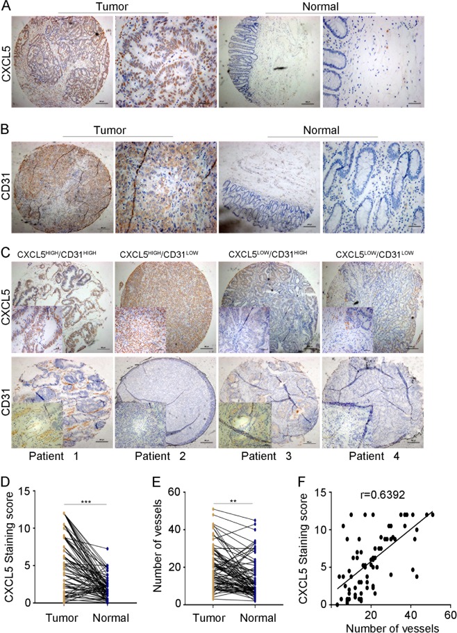
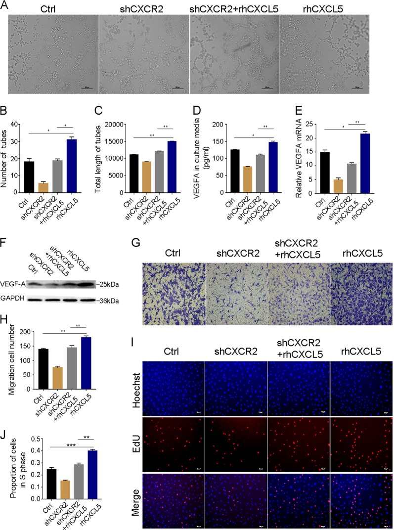
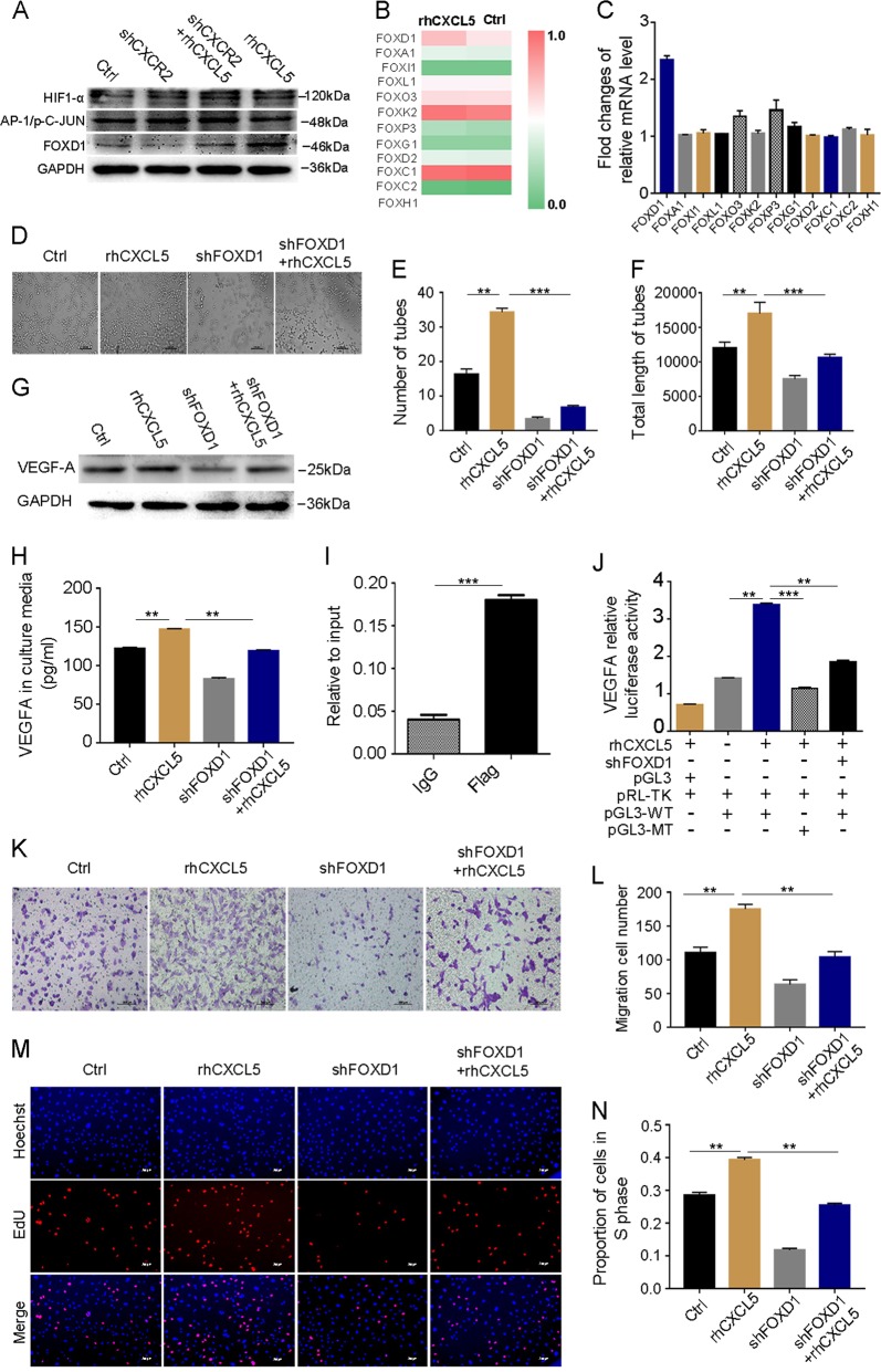
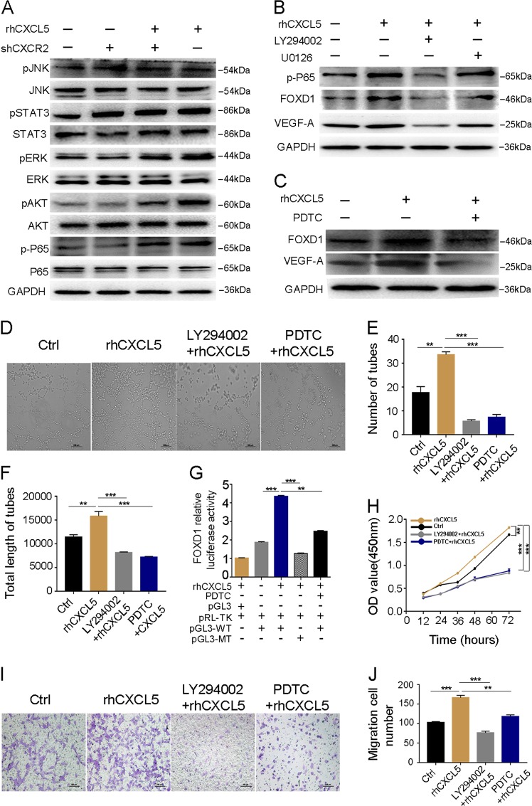
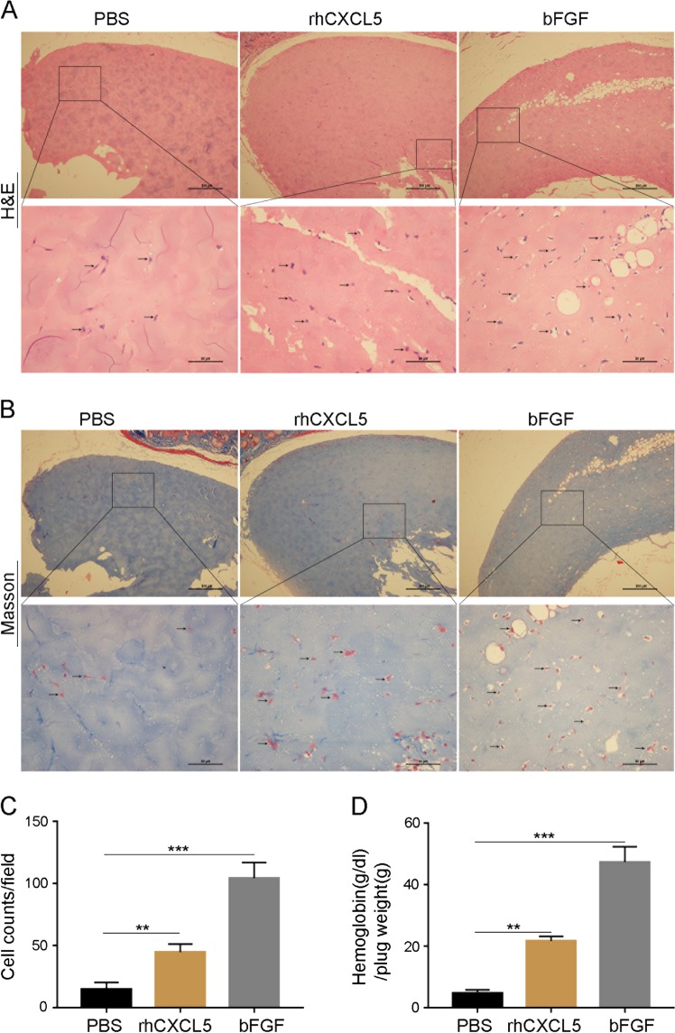
The mechanisms underlying the role of CXCL5 in tumor angiogenesis have not been fully defined. Here, we examined the effect of CXCL5 on tumor angiogenesis in colorectal cancer (CRC). Immunohistochemistry was used to monitor the expression of CXCL5 and CD31 in CRC patients' tissues. HUVEC cell lines stably transfected with shCXCR2 and shFOXD1 lentivirus plasmids were used in an in vitro study. Based on some molecular biological experiments in vitro and in vivo, we found that CXCL5 was upregulated in tumor tissues and that its level positively correlated with the expression of CD31. Next, we used recombinant human CXCL5 (rhCXCL5) to stimulate HUVECs and found that their tube formation ability, proliferation, and migration were enhanced by the activation of the AKT/NF-κB/FOXD1/VEGF-A pathway in a CXCR2-dependent manner. However, silencing of CXCR2 and FOXD1 or inhibition of the AKT and NF-κB pathways could attenuate the tube formation ability, proliferation, and migration of rhCXCL5-stimulated HUVECs in vitro. rhCXCL5 can promote angiogenesis in vivo in Matrigel plugs, and the overexpression of CXCL5 can also increase microvessel density in vivo in a subcutaneous xenotransplanted tumor model in nude mice. Taken together, our findings support CXCL5 as an angiogenic factor that can promote cell metastasis through tumor angiogenesis in CRC. Furthermore, we propose that FOXD1 is a novel regulator of VEGF-A. These observations open new avenues for therapeutic application of CXCL5 in tumor anti-angiogenesis.


Conflict of interest statement

Conflict of interest

The authors declare that they have no conflict of interest.

All the experiments involving in human specimens and animals were in accordance with the ethical code and recommendation issued by Ethics Committee of Human Experimentation and Chinese Animal Community and with the Helsinki Declaration of 1975, as revised in 2008.

Figures

Fig. 1
Fig. 1. High expression of CXCL5 and CD31 in CRC tissues.
a, d Immunohistochemistry images showing that CXCL5 is highly expressed in tissue microarray. b, e Immunohistochemistry images showing that CD31 is highly expressed in tissue microarray. c, f Correlation between CXCL5 and CD31 expression. CD31 expression is positively related with CXCL5 expression (r = 0.6392). Scale bars, 200 μm (magnification ×40) and 50 μm (magnification ×200). Data represent the mean ± SD, *P < 0.05, **P < 0.01, ***P < 0.001
Fig. 2
Fig. 2. rhCXCL5 promotes HUVEC tube formation, migration, and proliferation through the CXCR2 pathway.
a Images of HUVEC tube formation assay in different groups. Formation of tube-like networks was stimulated by the addition of rhCXCL5. Number of tubes and total length of tubes demonstrate the ability of angiogenesis in each group. Scale bars, 200 μm (magnification ×40). b, c Number of tubes and total length of tubes significantly increase in the rhCXCL5 group and decrease in the CXCR2-shRNA group. df VEGF-A protein and mRNA expression were detected by ELISA, western blot, and qPCR in different groups. rhCXCL5 increases VEGF-A expression which is inhibited by CXCR2-shRNA. g Images of transwell assay in different groups. Scale bar, 100 μm (magnification ×100). h Migration cell numbers are increased in the rhCXCL5 group compared with rhCXCL5-stimulated HUVEC-shCXCR2 group and control group. i EdU assay results in different groups. Scale bar, 200 μm (magnification ×40). j Proportion of cells in the S phase markedly increase in the rhCXCL5 group compared with rhCXCL5-stimulated HUVEC-shCXCR2 group and control group. Data represent the mean ± SD, *P < 0.05, **P < 0.01, ***P < 0.001
Fig. 3
Fig. 3. The CXCL5/CXCR2 induces the expression of VEGF-A dependent on FOXD1.
a Western blot analyses of HIF-1α, C-JUN, and FOXD1 expression in the different groups. b A heat map displays some FOX protein expression level after being treated with or without rhCXCL5 in HUVECs. c Fold changes of the relative mRNA level of VEGF-A-related FOX gene after being treated with or without rhCXCL5 in HUVECs. d Images of HUVEC tube formation assay in each group. Scale bar, 200 μm (magnification ×40). e, f Number of tubes and total length of tubes in different groups, FOXD1 silencing in HUVECs significantly decreases HUVEC tube formation. g, h VEGF-A protein expression is examined by western blot and ELISA in different groups. i ChIP-qPCR assay using Flag antibody or control IgG in HUVECs transfected with a FOXD1 (Flag-tagged) plasmid shows the binding of FOXD1 on the VEGF-A promoter. j A reporter plasmid for VEGF-A (pGL3-VEGF-A) was generated by cloning the VEGF-A promoter region (WT) or its FOXD1 binding site mutants (MT) into the pGL3 basic vector. rhCXCL5 significantly increased the luciferase activity of the VEGF-A promoter region (WT), while the activity was significantly decreased when transfected with MT sequence. Meanwhile, VEGF-A luciferase activity was inhibited when HUVECs were transfected with the shFOXD1 plasmid. k Images of transwell assay in each group. Scale bars, 100 μm (magnification ×100). l Migration cell numbers were reduced by knocking down FOXD1 in HUVECs. m Images of the EdU assay in each group. Scale bars, 200 μm (magnification ×40). n Proportion of cells in the S phase were reduced by knocking down FOXD1 in HUVECs. Data represent the mean ± SD, *P < 0.05, **P < 0.01, ***P < 0.001
Fig. 4
Fig. 4. CXCL5/CXCR2 axis promotes FOXD1 activity through the AKT/NF-κB pathway.
a Screening of CXCL5/CXCR2 downstream signaling pathway using western blot after different treatments in HUVECs. b Western blot shows that the AKT pathway rather than the ERK pathway regulates the expression of FOXD1 and VEGF-A using pathway inhibitors U0126 and LY294002. c Western blot analyses of p-P65, FOXD1, and VEGF-A in indicated groups after using the NF-κB inhibitor PDTC. d Images of HUVEC tube formation in indicated groups. Scale bars, 200 μm (magnification ×40). e, f Number of tubes and total length of tubes in different groups. Inhibitor of LY294002 and PDTC obviously inhibited the ability of HUVEC tube formation. g A reporter plasmid for FOXD1 (pGL3-FOXD1) was generated by cloning the FOXD1 promoter region (WT) or its NF-κB binding site mutants (MT) into the pGL3 basic vector. rhCXCL5 significantly increased the luciferase activity of the FOXD1 promoter region (WT), while the activity was significantly decreased when transfected with MT sequence. Meanwhile, FOXD1 luciferase activity can be inhibited by PDTC. h CCK8 assay in different groups. Inhibitor of LY294002 and PDTC noticeably reduced HUVEC proliferation ability. i Images of transwell assay in different groups. Scale bars, 100 μm (magnification ×100). j The migration ability of HUVECs was suppressed by the inhibitor of LY294002 and PDTC. Data represent the mean ± SD, *P < 0.05, **P < 0.01, ***P < 0.001
Fig. 5
Fig. 5. CXCL5 promotes subcutaneous tumor growth and angiogenesis in nude mice.
(a) Representative images of tumor-bearing mice and tumor mass. (b) Representative images of H&E staining as well as IHC staining of CXCL5 and CD31 of subcutaneous tumor in nude mice. Scale bars, 50 μm (magnification 200×). (c, d) Tumor growth curves. The volumes of xenografts were measured every 5 days during a 30-day period. (e) Average tumor weight of each group. (f) Number of microvessels in each group. Data represent the mean ± SD, *P < 0.05, **P < 0.01, ***P < 0.001
Fig. 6
Fig. 6. CXCL5 enhances blood vessel growth in Matrigel plugs in vivo.
a Representative photographs of the Matrigel plug sections stained with H&E. Number of cells manifest the ability of angiogenesis in different groups. Arrows on the lower columns indicate areas of blood cells. Scale bars, 50 μm (magnification ×200). b Representative photographs of the Matrigel plug sections stained with Masson’s trichrome. Number of cells manifest the ability of angiogenesis in different groups. Arrows on the lower columns indicate areas of blood cells. Scale bars, 50 μm (magnification ×200). c Cells migrating to form microvessels in Matrigel containing rhCXLC5 (10 ng) were more than PBS control. d Hemoglobin content of the rhCXCL5- (10 ng) treated group was significantly great compared with that of the PBS control. Matrigel containing bFGF (100 ng) served as a positive control. Data represent the mean ± SD, *P < 0.05, **P < 0.01, ***P < 0.001
Fig. 7
Fig. 7. Proposed model for the regulation of tumor angiogenesis by CXCL5.
CXCL5 increases AKT phosphorylation in a CXCR2-dependent manner and then NF-κB translocates to the nucleus. This leads to FOXD1 transcriptional activity that is enhanced at the VEGF-A promoter. Consequently, the expression of VEGF-A is increased to promote tumor angiogenesis

Similar articles

Cited by

References

    1. Global Burden of Disease Cancer Collaboration. et al. Global, Regional, and National Cancer Incidence, Mortality, Years ofLife Lost, Years Lived with Disability, and Disability-Adjusted Life-Years for 29 Cancer Groups, 1990 to 2016: A Systematic Analysis for the Global Burden of Disease Study. JAMA Oncol. 2018;4:1553–1568. doi: 10.1001/jamaoncol.2018.2706. - DOI - PMC - PubMed
    1. Zhao J, et al. Tumor-derived CXCL5 promotes human colorectal cancer metastasis through activation of the ERK/Elk-1/Snail and AKT/GSK3β/β-catenin pathway. Mol. Cancer. 2017;16:70. doi: 10.1186/s12943-017-0629-4. - DOI - PMC - PubMed
    1. Zhu C, et al. CCR6 promotes tumor angiogenesis via the AKT/NF-κB/VEGF pathway in colorectal cancer. Biochim. Biophys. Acta. 2018;1864:387–397. doi: 10.1016/j.bbadis.2017.10.033. - DOI - PubMed
    1. Strieter RM, Belperio JA, Phillips RJ, Keane MP. CXC chemokines in angiogenesis of cancer. Semin. Cancer Biol. 2004;14:195–200. doi: 10.1016/j.semcancer.2003.10.006. - DOI - PubMed
    1. Strieter RM, et al. The functional role of the ELR motif in CXC chemokine-mediated angiogenesis. J. Biol. Chem. 1995;270:27348–27357. doi: 10.1074/jbc.270.45.27348. - DOI - PubMed

Publication types

MeSH terms