. 2019 Feb 21;9(1):2494.
doi: 10.1038/s41598-019-39579-3.

Molecular origin of AuNPs-induced cytotoxicity and mechanistic study

Affiliations

Molecular origin of AuNPs-induced cytotoxicity and mechanistic study

Euiyeon Lee et al. Sci Rep. .

Abstract

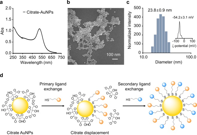
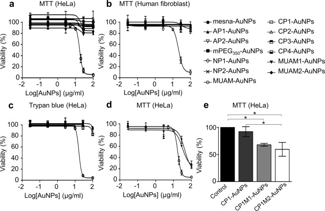
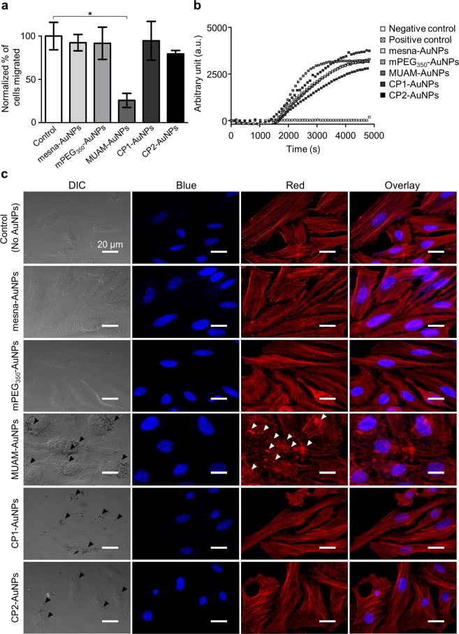
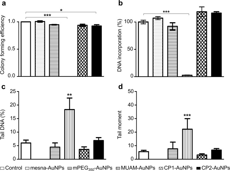
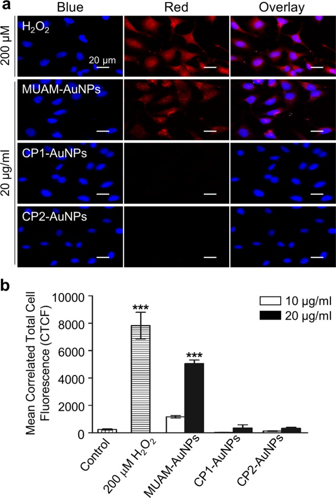
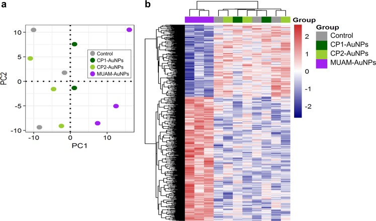
Gold nanoparticles (AuNPs) with diverse physicochemical properties are reported to affect biological systems differently, but the relationship between the physicochemical properties of AuNPs and their biological effects is not clearly understood. Here, we aimed to elucidate the molecular origins of AuNP-induced cytotoxicity and their mechanisms, focusing on the surface charge and structural properties of modified AuNPs. We prepared a library of well-tailored AuNPs exhibiting various functional groups and surface charges. Through this work, we revealed that the direction or the magnitude of surface charge is not an exclusive factor that determines the cytotoxicity of AuNPs. We, instead, suggested that toxic AuNPs share a common structural characteristics of a hydrophobic moiety neighbouring the positive charge, which can induce lytic interaction with plasma membrane. Mechanistic study showed that the toxic AuNPs interfered with the formation of cytoskeletal structure to slow cell migration, inhibited DNA replication and caused DNA damage via oxidative stress to hinder cell proliferation. Gene expression analysis showed that the toxic AuNPs down-regulated genes associated with cell cycle processes. We discovered structural characteristics that define the cytotoxic AuNPs and suggested the mechanisms of their cytotoxicity. These findings will help us to understand and to predict the biological effects of modified AuNPs based on their physicochemical properties.


Conflict of interest statement

The authors declare no competing interests.

Figures

Figure 1
Figure 1
Characterization of gold nanoparticles (AuNPs) and schematics of AuNP modification. AuNPs were synthesized and analysed using UV-Vis spectroscopy (a), Field emission scanning electron microscopy (FE-SEM) at 5 kV (b), Dynamic laser scattering (DLS) (c), and zeta-potential measurements (c, inset). AuNPs were modified via place-exchange reaction to introduce variously charged ligands (d).
Figure 2
Figure 2
Effect of modified gold nanoparticles (AuNPs) on cell viability. (a) The viability of AuNPs-treated HeLa cells were analysed using MTT assays. Only MUAM-AuNPs induced cell death with LD50 of 17.1 μg/ml. (b) The viability of AuNPs-treated human fibroblasts were analysed using MTT assays. Only MUAM-AuNPs induced cell death with LD50 of 20.5 μg/ml. (c) A trypan blue assay was performed on HeLa cells treated with modified AuNPs for 24 h. Only MUAM-AuNPs induced cell death with LD50 of 16.5 μg/ml. (d) MTT viability assay was performed on cells treated with three different MUAM-carrying AuNPs (MUAM-, MUAM1- and MUAM2-AuNPs). All three AuNPs showed comparable cytotoxicity regardless of ligand densities or the magnitude of positive charges. (e) MTT viability assay was performed on Cells treated with three different CP1-derived AuNPs (CP1-, CP1M1- and CP1M2-AuNPs). The introduction of hydrophobic chains increased cytotoxicity of CP1-derived AuNPs. The results are shown as mean ± standard error of mean (*p < 0.05, one-way ANOVA).
Figure 3
Figure 3
Effect of modified gold nanoparticles (AuNPs) on cell motility. (a) The motility of AuNPs-treated cells were monitored via a gap-filling assay. The migration rate decreased when cells were treated with MUAM-AuNPs (10 μg/ml) (*p < 0.05, one-way ANOVA). (b) In vitro actin polymerization assay was performed on HeLa cells treated with modified AuNPs (10 μg/ml). The rate of actin polymerization did not change noticeably when treated with AuNPs. (c) Cytoskeletal structures in AuNPs-treated cells were visualized using fluorescent phalloidin (DAPI-stained nucleus, blue; actin filaments, red). F-actins in MUAM-AuNPs treated cells were disassembled and fragmented (white arrows). Scale bar: 50 μm.
Figure 4
Figure 4
Effect of modified gold nanoparticles (AuNPs) on cell division and proliferation. (a) The Colony forming efficiency assay was performed on cell treated with modified AuNPs (10 μg/ml). MUAM-AuNPs treated cells did not form colonies over 50 cells. The number of colonies also decreased slightly in CP2-AuNP treated cells. (b) The effect of modified AuNPs on DNA replication was analysed. MUAM-AuNPs inhibited DNA replication near to completion. (c and d) AuNPs-induced DNA damage was monitored using the Comet Assay. Measurements of % tail DNA (c) and tail moment (d) suggest severe DNA damage in MUAM-AuNPs treated samples (*p < 0.05, **p < 0.01, ***p < 0.001, one-way ANOVA).
Figure 5
Figure 5
Reactive oxygen species generation induced by cationic gold nanoparticles (AuNPs). (a) The level of ROS generation in cationic AuNPs-treated cells were monitored. MUAM-AuNPs treated cells showed increased ROS level comparable to H2O2-treated control group, while other cationic AuNPs did not induce noticeable ROS generation. Scale bar: 20 μm. (b) The fluorescence intensity shows the amount of ROS in AuNPs-treated cells. The level of ROS increased as the concentration of MUAM-AuNPs increased (***p < 0.001, one-way ANOVA).
Figure 6
Figure 6
Gene expression profiles of HeLa cells treated with cationic gold nanoparticles (AuNPs) (a) Global gene expression profiles of the cationic AuNPs (MUAM-, CP1-, and CP2-AuNPs) treated cells were analysed by principal component analysis. The MUAM-AuNPs treated cells showed unique gene expression patterns, while the CP1- and CP2-AuNPs treated samples showed the patterns similar to the control. (b) Heat map analysis of gene expression pattern shows hierarchical clustering of 1,156 differentially expressed genes between MUAM-AuNPs treated samples and the group of three other samples. Red and blue colours indicate up- and down-expression levels, respectively.

References

    1. Oh Y-J, Kang M, Park M, Jeong K-H. Engineering hot spots on plasmonic nanopillar arrays for SERS: A review. BioChip J. 2016;10:297–309. doi: 10.1007/s13206-016-0406-2. - DOI
    1. Uehara N. Polymer-functionalized gold nanoparticles as versatile sensing materials. Anal Sci. 2010;26:1219–1228. doi: 10.2116/analsci.26.1219. - DOI - PubMed
    1. Huang X, El-Sayed MA. Gold nanoparticles: Optical properties and implementations in cancer diagnosis and photothermal therapy. J Adv Res. 2010;1:13–28. doi: 10.1016/j.jare.2010.02.002. - DOI
    1. Hutter E, Maysinger D. Gold nanoparticles and quantum dots for bioimaging. Microsc. Res. Tech. 2011;74:592–604. doi: 10.1002/jemt.20928. - DOI - PubMed
    1. Mahmoudi M, Serpooshan V, Laurent S. Engineered nanoparticles for biomolecular imaging. Nanoscale. 2011;3:3007–3026. doi: 10.1039/c1nr10326a. - DOI - PubMed

Publication types