Review
. 2019 May;20(5):282-297.
doi: 10.1038/s41583-019-0126-4.

Neuron-glia interactions in the pathophysiology of epilepsy

Affiliations
Review

Neuron-glia interactions in the pathophysiology of epilepsy

Dipan C Patel et al. Nat Rev Neurosci. 2019 May.

Abstract

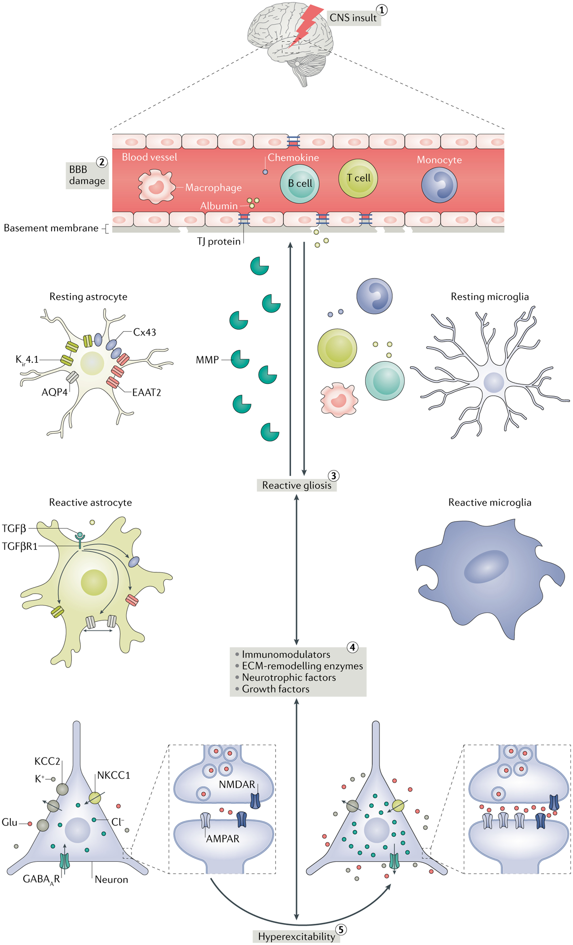
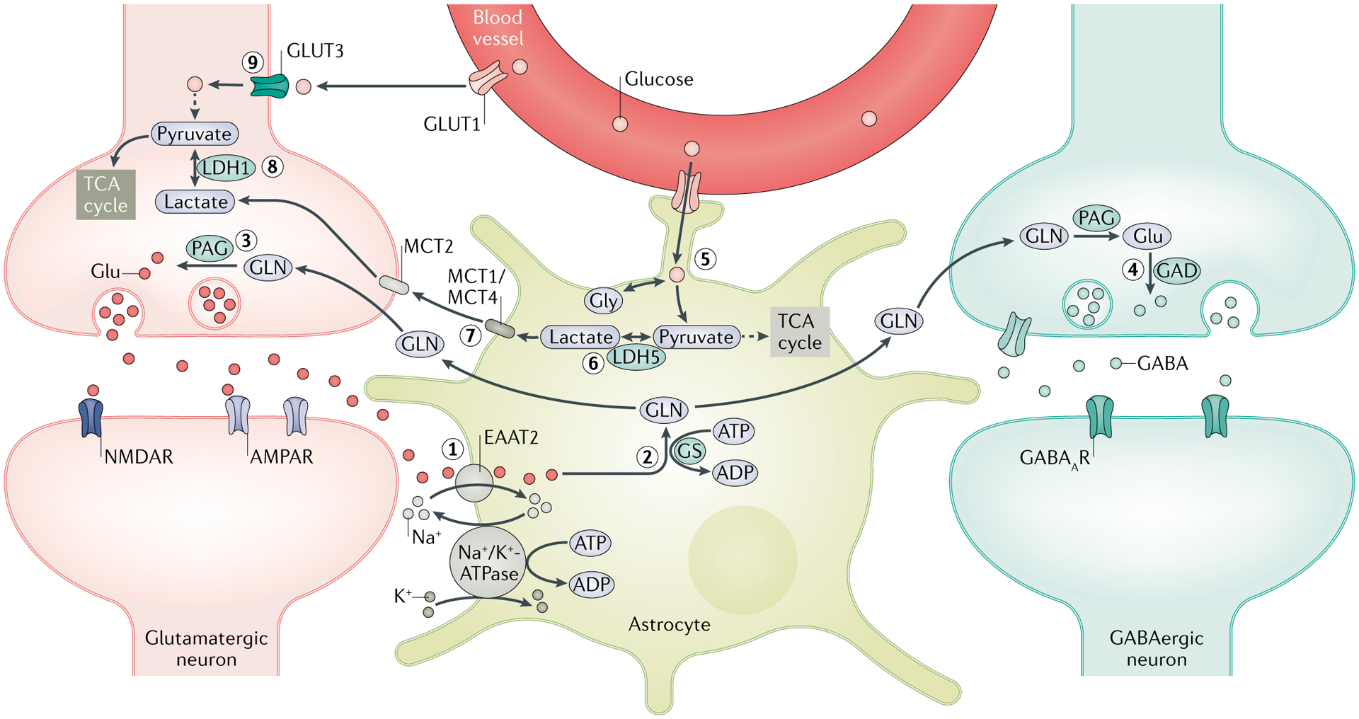
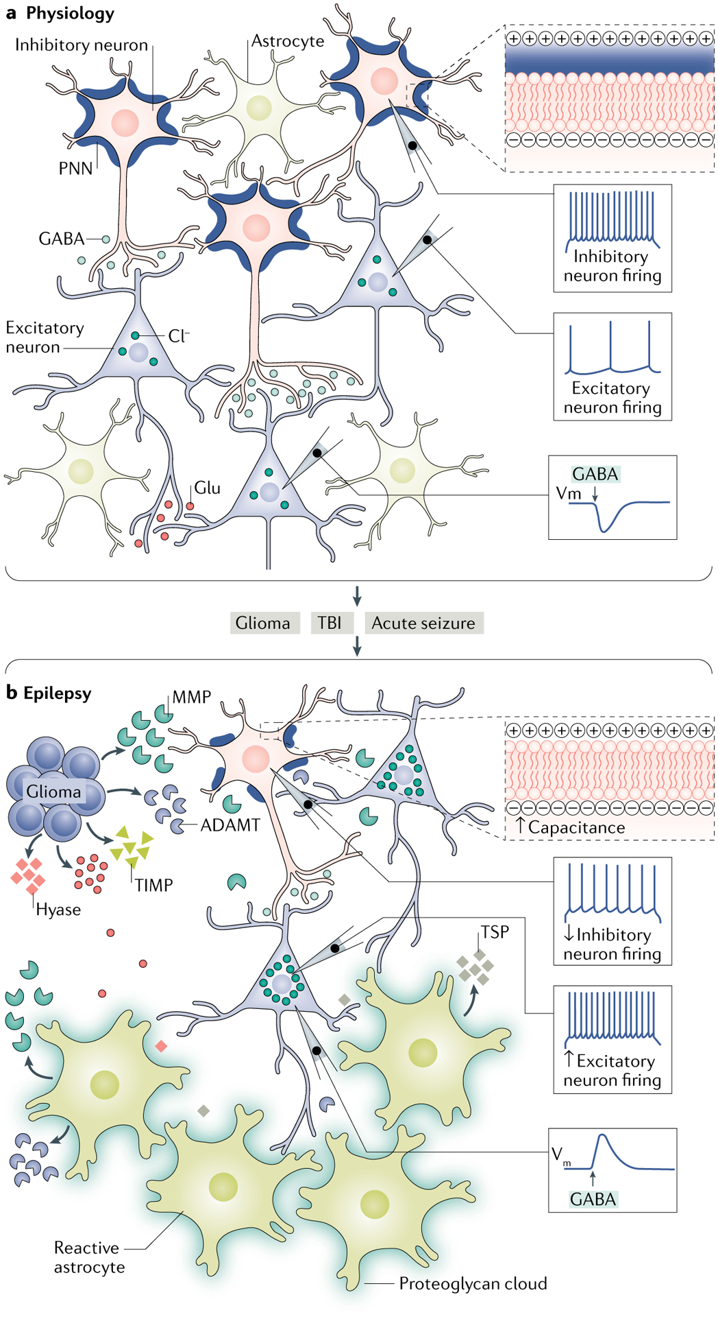
Epilepsy is a neurological disorder afflicting ~65 million people worldwide. It is caused by aberrant synchronized firing of populations of neurons primarily due to imbalance between excitatory and inhibitory neurotransmission. Hence, the historical focus of epilepsy research has been neurocentric. However, the past two decades have enjoyed an explosion of research into the role of glia in supporting and modulating neuronal activity, providing compelling evidence of glial involvement in the pathophysiology of epilepsy. The mechanisms by which glia, particularly astrocytes and microglia, may contribute to epilepsy and consequently could be harnessed therapeutically are discussed in this Review.


Conflict of interest statement

Competing interests

The authors declare no competing interests.

Figures

Fig. 1 |
Fig. 1 |. Interplay between CNS insult, reactive gliosis and seizures.
(1) Various CNS insults such as stroke, infection, trauma, tumours and hypoxia can cause blood–brain barrier (BBB) damage. (2) The resulting increased permeability of the BBB promotes extravasation of serum albumin, peripheral immune cells (macrophages, monocytes, B cells and T cells) and chemokines into the brain parenchyma, where, along with direct CNS insult, they induce reactive gliosis. (3) Reactive gliosis refers to morphological (hypertrophy of the cell bodies and processes) and physiological changes (changes in the expression and functions of many glial proteins) in glial cells in response to various CNS injuries. (4) Reactive glia release extracellular matrix (ECM)-remodelling enzymes such as matrix metalloproteinases (MMPs), neurotrophic factors such as brain-derived neurotrophic factor (BDNF), growth factors such as transforming growth factor-β (TGFβ) and immunomodulators such as cytokines that modulate the expression and function of receptors, transporters, enzymes and other molecules implicated in the functions of excitatory and inhibitory neurotransmission by a variety of mechanisms. MMPs can aggravate BBB impairment by damaging the tight junction (TJ) connections and promote the infiltration of serum proteins in the brain. In addition to TGFβ, serum albumin is known to activate TGFβ receptor 1 (TGFβR1) signalling in astrocytes by directly interacting with TGFβR1. Accordingly, the membrane expression of inwardly rectifying potassium channel 4.1 (Kir4.1), excitatory amino acid transporter 2 (EAAT2) and the gap junction protein connexin 43 (Cx43) are decreased. Activation of TGFβR1 also changes the trafficking and surface expression of aquaporin 4 (AQP4). As a result, extracellular concentrations of K+ and glutamate (Glu) increase, which promotes network hyperexcitability. Control of Cl concentration is also crucial for regulating hyperexcitability. The K+–Cl cotransporter (KCC2; a Cl exporter) and the Na+–K+–Cl cotransporter 1 (NKCC1; a Cl importer) play a crucial role in maintaining a low concentration of intracellular Cl, which is essential for maintaining the inhibitory activity of GABAergic neurotransmission. BDNF decreases the membrane expression of KCC2, causing net accumulation of Cl intracellularly and reversal of Cl flux through GABA type A receptor (GABAAR). Therefore, the activation of GABAAR paradoxically causes hyperexcitation. Cytokines (for example, tumour necrosis factor (TNF)) released from reactivated glia can increase the postsynaptic density of AMPA receptors (AMPARs) and excitatory neurotransmission. (5) As a consequence, network hyperexcitability and seizures occur owing to impairment in K+, Cl and glutamate buffering and imbalance in excitatory and inhibitory neurotransmission. Importantly, seizures can aggravate the gliotic condition by further increasing the release of neuromodulator molecules, setting the vicious cycle of gliosis and seizures. NMDAR, NMDA receptor.
Fig. 2 |
Fig. 2 |. Regulation of synaptic transmitter homeostasis and energy metabolites by astrocytes.
A crucial function of astrocytes is to maintain the optimal level of extracellular glutamate (Glu) through the Glu–glutamine (GLN) cycle. (1) Following synaptic transmission, excess extracellular Glu is absorbed by astrocytes through excitatory amino acid transporter 2 (EAAT2). (2) A large amount of Glu is converted by GLN synthetase (GS) into GLN, which is shuttled into the surrounding neurons. (3) GLN is then metabolized by phosphate-activated glutaminase (PAG) into Glu, which is packaged into synaptic vesicles in the glutamatergic neurons. (4) In GABAergic neurons, Glu is further converted by Glu decarboxylase (GAD) into GABA, which is released in the synapse during inhibitory neurotransmission. Rapid clearance of extracellular Glu following increased activity of synaptic transmission exerts an energetic burden on astrocytes owing to the energy-intensive nature of the Glu–GLN cycle, as both EAAT2 and GS require ATP. (5) The energy deficit that results stimulates the glycolytic oxidation of glucose imported from the blood through glucose transporter 1 (GLUT1) and is enzymatically derived from glycogen stores in astrocytes to generate energy metabolites — lactate and pyruvate. (6) Owing to the limited oxidative capacity of astrocytes, pyruvate is primarily converted into lactate via lactate dehydrogenase 5 (LDH5) and (7) shuttled into the surrounding neurons via monocarboxylate transporters (MCTs). (8) In neurons, LDH1 catalyses the conversion of lactate into pyruvate, which is subsequently used as an energy source through the tricarboxylic acid (TCA) cycle. This pathway is termed the astrocyte neuron lactate shuttle (ANLS) and is crucial in meeting the energy demands of neurons during high synaptic activity, such as during seizures. (9) Glucose is also taken up by neurons from the blood vessels through GLUT3; however, owing to the limited glycolytic capability of neurons and the lack of glycogen stores, neurons derive pyruvate primarily from astrocytes via the ANLS pathway. Disruptions in the expressions and functions of enzymes and transporters implicated in both the Glu–GLN cycle and the ANLS are associated with the development of neuronal hyperexcitability and seizures. AMPAR, AMPA receptor; GABAAR, GABA type A receptor; Gly, glycine; NMDAR, NMDA receptor.
Fig. 3 |
Fig. 3 |. Disintegration of perineuronal nets in glioma-associated epilepsy.
a,b | Immunohistochemical staining of the cerebral cortex from a glioma-implanted mouse (part a) shows 4′,6-diamidino-2-phenylindole (DAPI)-stained nuclei (blue), Wisteria floribunda agglutinin (WFA)-stained perineuronal nets (PNNs; yellow), parvalbumin-positive (PV+) interneurons (green) and NeuN-stained neurons (red) (scale = 50 μm). The presence of glioma is identified by densely packed DAPI-stained cells indicated in the upper part of the image demarcated by a white solid line (tumour border). The distance between the tumour border and each of the white dotted lines below is ~0.2 mm. Magnified individual images of PNNs, PV+ interneurons and NeuN+ neurons and their merged images corresponding to their distance from the tumour border are shown on the right (upper panels <0.2 mm, middle panels 0.2–0.4 mm and lower panels 0.4–0.6 mm; scale = 5 μm). c | Confocal images of PV+ interneurons (green) show disintegrated architecture of PNNs (yellow) in the peritumoural cortex (PTC) and are compared to sham-treated mouse cortex (scale = 2 μm). d | The WFA fluorescence intensity of a line drawn along the periphery of a PV+ interneuron in the peritumoural cortex (left panel of part c) and sham (right panel of part c) shows many high-intensity WFA peaks in sham compared with those in the peritumoural cortex. Upper and lower two-headed arrows within the two nearest WFA peaks indicate the size of a hole in the PNN from the sham and peritumoural cortex, respectively. au, arbitrary units. Adapted from REF., Springer Nature Limited.
Fig. 4 |
Fig. 4 |. Changes in the extracellular matrix as contributors to epilepsy.
a | Under physiological conditions, the extracellular space is filled with a dense amorphous interstitial matrix and highly organized perineuronal nets (PNNs) surrounding parvalbumin-positive (PV+) inhibitory interneurons. The presence of intact PNNs around the cell reduces the effective membrane capacitance of the cell by increasing the charge separation between intracellular and extracellular compartments (upper right panel). This enables interneurons to sustain high firing frequencies to maintain a steady-state inhibitory drive by releasing GABA onto the excitatory cells, which helps to maintain optimal firing of the excitatory neurons. Intact structure of the extracellular matrix (ECM) also helps to maintain a low concentration of intracellular chloride; therefore, activation of the GABA type A receptor (GABAAR) causes hyperpolarization owing to the chloride influx. b | Alterations in the ECM that occur during several pathological conditions such as glioma, traumatic brain injury (TBI) and acute seizures can contribute to the development of epilepsy. Glioma cells release glutamate (Glu) and ECM-remodelling molecules, including matrix metalloproteinases (MMPs), tissue inhibitors of metalloproteinases (TIMPs), hyaluronidase (hyase) and a disintegrin and metalloproteinase with thrombospondin motifs (ADAMTs). These ECM-remodelling molecules cause degradation of the PNNs around inhibitory interneurons and thinning of the interstitial matrix. The loss of PNNs increases the membrane capacitance of inhibitory interneurons by decreasing the charge separation between intracellular and extracellular compartments (upper right panel), thereby slowing their firing frequency and, as a consequence, reducing their GABA release and lowering inhibition of excitatory neurons. Hence, the firing of excitatory neurons increases, contributing to network hyperexcitability. Glioma cells also cause selective loss of PV+ inhibitory interneurons owing to Glu excitotoxicity further lowering the inhibitory drive, thereby leading to epileptiform activity. Impairment in the ECM structure also alters chloride homeostasis, which results in an increased intracellular concentration of chloride and a depolarizing shift in the activation of the GABAAR. In a manner similar to that of glioma, following TBI and acute seizures, reactive astrocytes become the primary source of the ECM-remodelling molecules (MMPs and ADAMTs), synaptogenic molecules (thrombospondins (TSPs)) and proteoglycans, which can contribute to epileptogenesis by degrading PNNs and the interstitial matrix, thinning the interstitial matrix and causing aberrant synaptogenesis. Vm, membrane potential.

References

    1. Sontheimer H Diseases of the Nervous System 61–95 (Elsevier, 2015).
    1. Fisher RS et al. ILAE official report: a practical clinical definition of epilepsy. Epilepsia 55, 475–482 (2014). - PubMed
    1. Kaplan DI, Isom LL & Petrou S Role of sodium channels in epilepsy. Cold Spring Harb. Perspect. Med 6, a022814 (2016). - PMC - PubMed
    1. Loscher W, Hirsch LJ & Schmidt D The enigma of the latent period in the development of symptomatic acquired epilepsy - traditional view versus new concepts. Epilepsy Behav.52, 78–92 (2015). - PubMed
    1. Sofroniew MV Astrogliosis. Cold Spring Harb. Perspect. Biol 7, a020420 (2014). - PMC - PubMed

Publication types