Review
. 2018;38(5):379-401.
doi: 10.1615/CritRevImmunol.2018026790.

Immunomodulatory Bonds of the Partnership between Dendritic Cells and T Cells

Affiliations
Review

Immunomodulatory Bonds of the Partnership between Dendritic Cells and T Cells

Jessica Bourque et al. Crit Rev Immunol. 2018.

Abstract

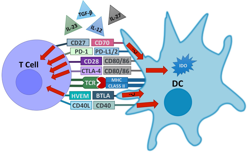
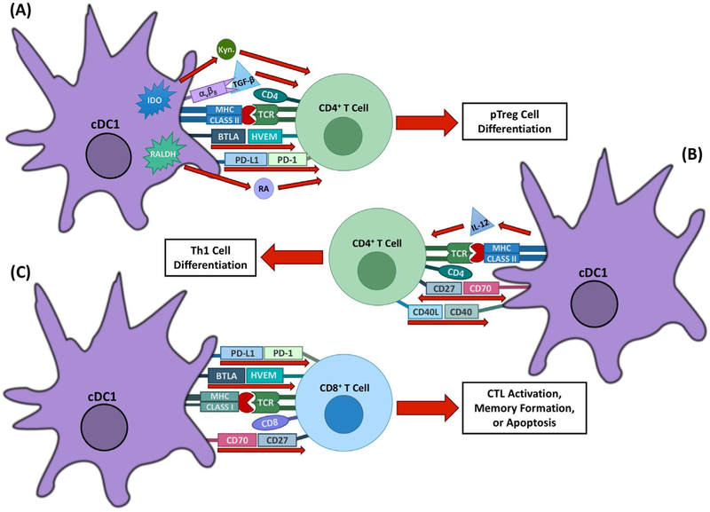
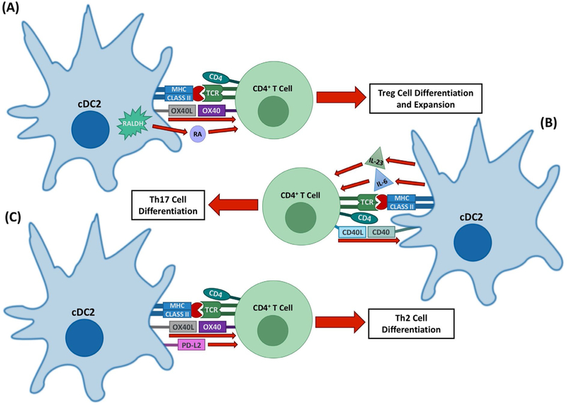
By acquiring, processing, and presenting both foreign and self-antigens, dendritic cells (DCs) initiate T cell activation that is shaped through the immunomodulatory functions of a variety of cell-membrane-bound molecules including BTLA-HVEM, CD40-CD40L, CTLA-4-CD80/CD86, CD70-CD27, ICOS-ICOS-L, OX40-OX40L, and PD-L1-PD-1, as well as several key cytokines and enzymes such as interleukin-6 (IL-6), IL-12, IL-23, IL-27, transforming growth factor-beta 1 (TGF-β1), retinaldehyde dehydrogenase (Raldh), and indoleamine 2,3-dioxygenase (IDO). Some of these distinct immunomodulatory signals are mediated by specific subsets of DCs, therefore contributing to the functional specialization of DCs in the priming and regulation of immune responses. In addition to responding to the DC-mediated signals, T cells can reciprocally modulate the immunomodulatory capacities of DCs, further refining immune responses. Here, we review recent studies, particularly in experimental mouse systems, that have delineated the integrated mechanisms of crucial immunomodulatory pathways that enable specific populations of DCs and T cells to work intimately together as single functional units that are indispensable for the maintenance of immune homeostasis.

Keywords: T cell; T cell differentiation; dendritic cell; effector T cell; immunoregulation; regulatory T cell.


Figures

FIG. 1:
FIG. 1:
DCs and T cells form a functional partnership. DCs and T cells express a variety of surface molecules that work together as ligands and receptors in order to control various aspects of the immune response. Some of these pathways, such as BTLA–HVEM and CD27–CD70, have been shown to induce bidirectional signaling, although the impact of such bidirectional interactions between DCs and T cells remains mostly unclear at this point. In addition, DCs secrete or activate a variety of cytokines that influence T cell differentiation and proliferation. DCs upregulate expression of the CD70 and IL-12 in response to CD40 signaling induced by T cell-expressed CD40L. DCs can also produce the tolerogenic tryptophan-catabolizing enzyme IDO in response to signals mediated by CTLA-4 in T cells.
FIG. 2:
FIG. 2:
Immunomodulatory mechanisms of cDC1s. cDC1s are important for the induction of Th1 and tolerogenic T cell responses through several mechanisms. (A) cDC1s mediate T cell tolerance through several mechanisms, including pTreg induction by BTLA–HVEM, PD-L1–PD-1, RA, and TGF-β signaling. Expression of the enzyme IDO in response to CTLA-4–CD80/86 signaling also promotes tolerance through tryptophan deprivation and the production of metabolites, including kynurenine. (B) cDC1-expressed IL-12 and CD70, which are upregulated in response to CD40 signaling, work together to promote Th1 differentiation from naive CD4+ T cells. (C) cDC1s can promote CD4+ T cell-dependent expansion and effectiveness of CD8+ T cell responses through CD70–CD27 interactions. BTLA–HVEM signaling additionally plays a role in memory formation of CD8+ T cells in response to both viral and bacterial infections. cDC1s can also induce apoptosis of CD8+ T cells through PD-L1–PD-1 signaling.
FIG. 3:
FIG. 3:
Immunomodulatory mechanisms of cDC2s. cDC2s are important for the induction of Th2 and Th17 responses as well as some aspects of tolerogenic T cell responses through several mechanisms. (A) cDC2s can promote the expansion of Treg cells, potentially through OX40 signaling. They also promote pTreg cell formation in some environments through their production of RA by Raldh2. (B) cDC2s are the main source of the Th17-polarizing cytokines IL-23 and IL-6, the expression of which can be upregulated by CD40 signaling. (C) cDC2 promote Th2 responses through OX40-L–OX40 signaling as well as through the production of Th2-polarizing cytokines. PD-L2 may also play a role in Th2 differentiation.

Similar articles

Cited by

References

    1. Iberg CA, Jones A, Hawiger D. Dendritic cells as inducers of peripheral tolerance. Trends Immunol 2017;38(11):793–804. - PMC - PubMed
    1. Akira S, Uematsu S, Takeuchi O. Pathogen recognition and innate immunity. Cell 2006;124(4):783–801. - PubMed
    1. Sancho D, Reis e Sousa C. Signaling by myeloid C-type lectin receptors in immunity and homeostasis. Annu Rev Immunol 2012;30:491–529. - PMC - PubMed
    1. Iwasaki A, Medzhitov R. Control of adaptive immunity by the innate immune system. Nat Immunol 2015;16(4):343–53. - PMC - PubMed
    1. Liu G, Wu C, Wu Y, Zhao Y. Phagocytosis of apoptotic cells and immune regulation. Scand J Immunol 2006;64(1):1–9. - PubMed

Publication types

LinkOut - more resources