Review
. 2019 Feb;7(1):3-12.
doi: 10.1093/gastro/goy052. Epub 2019 Feb 13.

Fight them or feed them: how the intestinal mucus layer manages the gut microbiota

Affiliations
Review

Fight them or feed them: how the intestinal mucus layer manages the gut microbiota

Bjoern O Schroeder. Gastroenterol Rep (Oxf). 2019 Feb.

Abstract

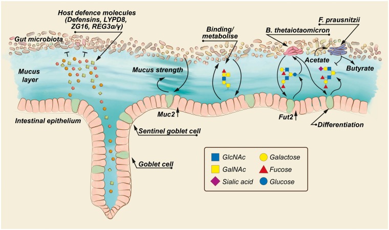
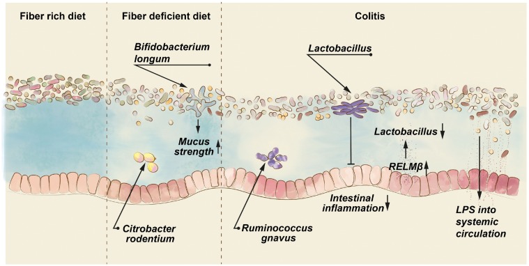
The intestinal tract is inhabited by a tremendous number of microorganisms, termed the gut microbiota. These microorganisms live in a mutualistic relationship with their host and assist in the degradation of complex carbohydrates. Although the gut microbiota is generally considered beneficial, the vast number of microbial cells also form a permanent threat to the host. Thus, the intestinal epithelium is covered with a dense layer of mucus to prevent translocation of the gut microbiota into underlying tissues. Intestinal mucus is an organized glycoprotein network with a host-specific glycan structure. While the mucus layer has long been considered a passive, host-designed barrier, recent studies showed that maturation and function of the mucus layer are strongly influenced by the gut microbiota. In return, the glycan repertoire of mucins can select for distinct mucosa-associated bacteria that are able to bind or degrade specific mucin glycans as a nutrient source. Because the intestinal mucus layer is at the crucial interface between host and microbes, its breakdown leads to gut bacterial encroachment that can eventually cause inflammation and infection. Accordingly, a dysfunctional mucus layer has been observed in colitis in mice and humans. Moreover, the increased consumption of a low-fiber Western-style diet in our modern society has recently been demonstrated to cause bacteria-mediated defects of the intestinal mucus layer. Here, I will review current knowledge on the interaction between gut bacteria and the intestinal mucus layer in health and disease. Understanding the molecular details of this host-microbe interaction may contribute to the development of novel treatment options for diseases involving a dysfunctional mucus layer, such as ulcerative colitis.

Keywords: Dietary fiber; gut microbiota; host–microbe interaction; inflammatory bowel disease; metabolic disease; mucin; mucosal barrier; mucus; probiotics; ulcerative colitis.


Figures

Figure 1.
Figure 1.
The structure of the mucus layer is affected by the gut microbiota. Gut bacteria are separated from the host epithelium by the intestinal mucus layer, which is fortified with host defense molecules, such as defensins, Ly6/PLAUR domain containing protein 8 (LYPD8), zymogen granulae protein 16 (ZG16), Regenerating islet-derived proteins 3 (REG3α/γ) and others. The presence of gut microbiota induces expression of the genes encoding mucin 2 (Muc2) and galactoside 2-alpha-L-fucosyltransferase 2 (Fut2), thereby affecting mucus strength and mucin glycan structure. This can in return affect gut-microbiota composition, as specific gut bacteria, such as Bacteroides thetaiotaomicron, can bind and metabolize mucin glycans as an energy source. Degradation of glycans leads to production of microbial metabolites, which not only affect goblet cell differentiation, but also via cross-feeding the abundance of other microbial species, for example Faecalibacterium prausnitzii. Accordingly, the composition of the gut microbiota affects mucus function through the availability of microbial metabolites.
Figure 2.
Figure 2.
Defects of the intestinal mucus layer exacerbate intestinal infection and inflammation. Diets lacking microbiota-accessible carbohydrates, as contained in dietary fiber, direct gut microbial species to degrade host glycans of the intestinal mucus layer (depicted by lighter mucus color), thereby deteriorating mucus strength. A defective mucus layer increases the risk for intestinal infections, for example by the mouse pathogen Citrobacter rodentium. The diet-mediated mucus defect can be prevented by specific gut bacteria, such as Bifidobacterium longum. During colitis (depicted by intense red epithelial cells), increased expression of resistin-like molecule β (Relmβ) induces production of the antimicrobial protein regenerating islet-derived protein 3β (REG3β), which reduces beneficial Lactobacillus species. Correspondingly, application of Lactobacillus can ameliorate colitis. In the mucus of inflammatory bowel disease patients, an increased number of the mucus-degrading bacterium Ruminococcus gnavus has been observed, while a defective mucus layer may lead to increased translocation of bacterial lipopolysaccharide, thereby contributing to metabolic diseases.

References

    1. Schroeder BO, Bäckhed F.. Signals from the gut microbiota to distant organs in physiology and disease. Nat Med 2016;22:1079–89. - PubMed
    1. Rodríguez-Piñeiro AM, Bergström JH, Ermund A. et al. Studies of mucus in mouse stomach, small intestine, and colon. II. Gastrointestinal mucus proteome reveals Muc2 and Muc5ac accompanied by a set of core proteins. Am J Physiol Gastrointest Liver Physiol 2013;305:G348–56. - PMC - PubMed
    1. Swidsinski A, Loening-Baucke V, Theissig F. et al. Comparative study of the intestinal mucus barrier in normal and inflamed colon. Gut 2007;56:343–50. - PMC - PubMed
    1. Johansson MEV, Gustafsson JK, Holmén-Larsson J. et al. Bacteria penetrate the normally impenetrable inner colon mucus layer in both murine colitis models and patients with ulcerative colitis. Gut 2014;63:281–91. - PMC - PubMed
    1. Chassaing B, Raja SM, Lewis JD. et al. Colonic microbiota encroachment correlates with dysglycemia in humans. Cell Mol Gastroenterol Hepatol 2017;4:205–21. - PMC - PubMed