

Protein Composition Reflects Extracellular Vesicle Heterogeneity
- PMID: 30793499
- PMCID: PMC7521840
- DOI: 10.1002/pmic.201800167
Protein Composition Reflects Extracellular Vesicle Heterogeneity
Abstract
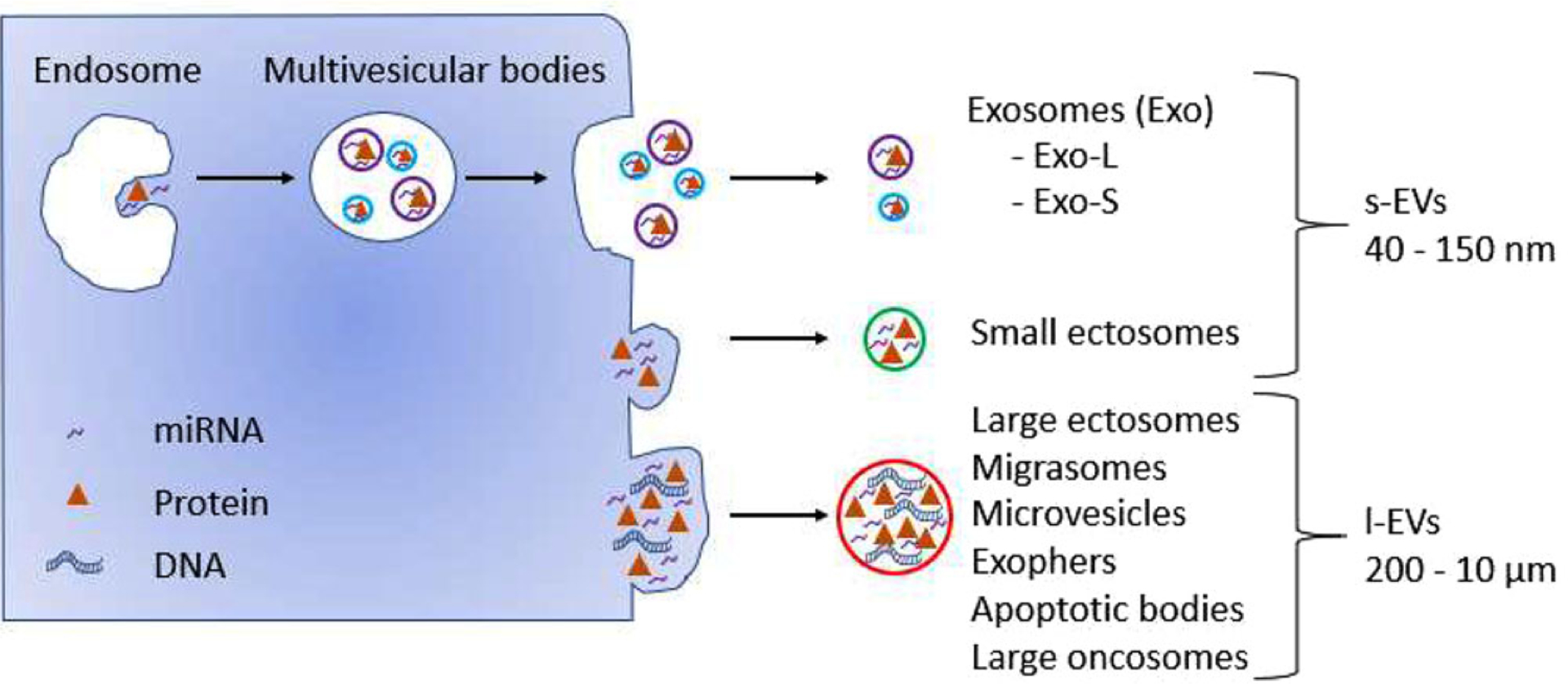
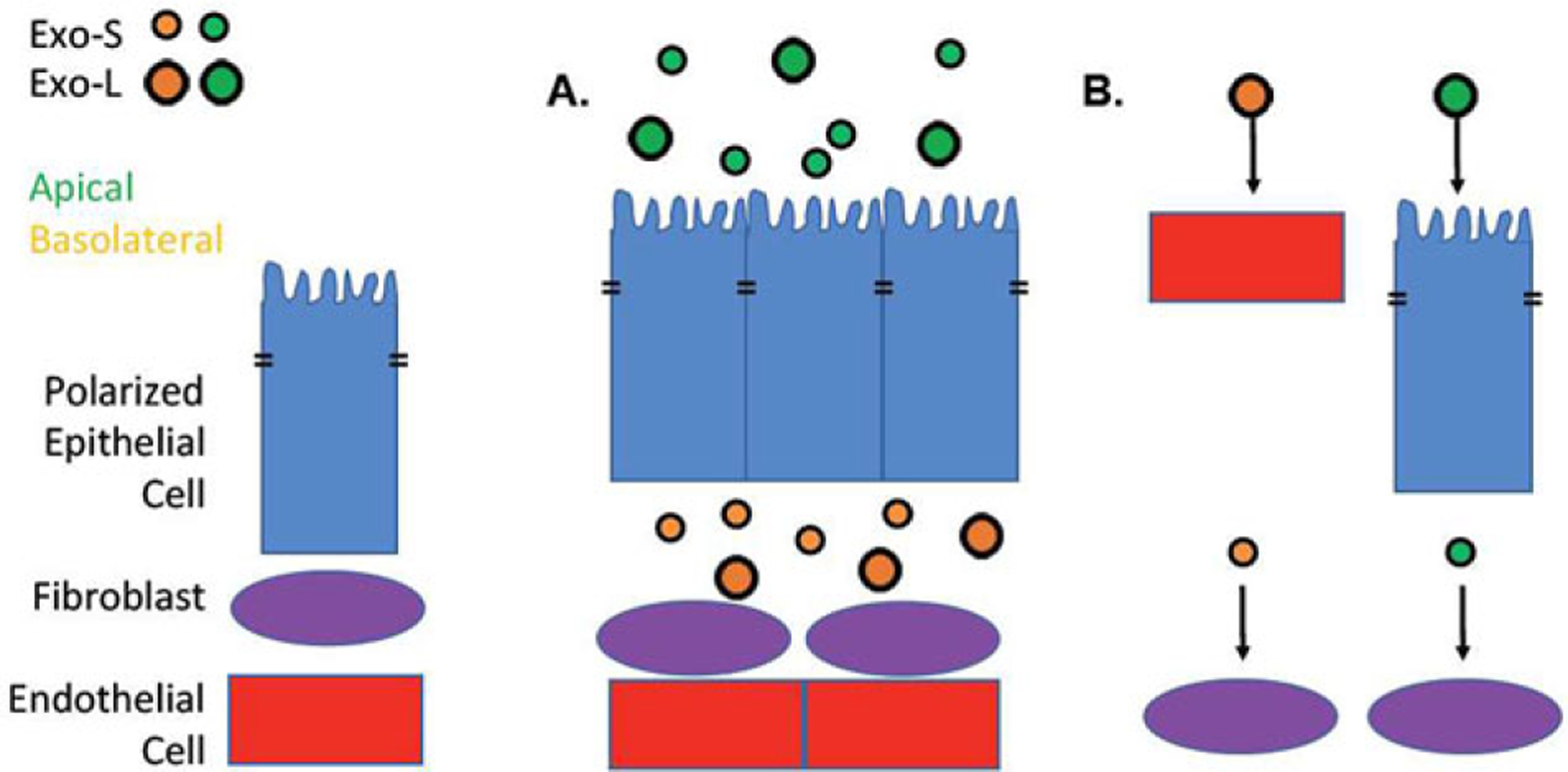
Extracellular vesicles (EVs) are membrane-enclosed particles that are released by virtually all cells from all living organisms. EVs shuttle biologically active cargo including protein, RNA, and DNA between cells. When shed by cancer cells, they function as potent intercellular messangers with important functional consequences. Cells produce a diverse spectrum of EVs, spanning from small vesicles of 40-150 nm in diameter, to large vesicles up to 10 μm in diameter. While this diversity was initially considered to be purely based on size, it is becoming evident that different classes of EVs, and different populations within one EV class may harbor distinct molecular cargo and play specific functions. Furthermore, there are considerable cell type-dependent differences in the cargo and function of shed EVs. This review focuses on the most recent proteomic studies that have attempted to capture the EV heterogeneity by directly comparing the protein composition of different EV classes and EV populations derived from the same cell source. Recent studies comparing protein composition of the same EV class(es) derived from different cell types are also summarized. Emerging approaches to study EV heterogeneity and their important implications for future studies are also discussed.
Keywords: biomarkers; cancer; extracellular vesicles; heterogeneity; protein profiling.
© 2019 WILEY-VCH Verlag GmbH & Co. KGaA, Weinheim.
Conflict of interest statement
Conflict of Interest
The authors declare no conflict of interest.
Figures
References
-
- Figueroa JM, Skog J, Akers J, Li H, Komotar R, Jensen R, Ringel F, Yang I, Kalkanis S, Thompson R, Loguidice L, Berghoff E, Parsa A, Liau L, Curry W, Cahill D, Bettegowda C, Lang FF, Chiocca EA, Henson J, Kim R, Breakefield X, Chen C, Messer K, Hochberg F, Carter BS, Neuro Oncol 2017, 19, 1494. - PMC - PubMed
-
- Fais S, O’Driscoll L, Borras FE, Buzas E, Camussi G, Cappello F, Carvalho J, Cordeiro Da Silva A, Del Portillo H, El Andaloussi S, Ficko Trček T, Furlan R, Hendrix A, Gursel I, Kralj-Iglic V, Kaeffer B, Kosanovic M, Lekka ME, Lipps G, Logozzi M, Marcilla A, Sammar M, Llorente A, Nazarenko I, Oliveira C, Pocsfalvi G, Rajendran L, Raposo G, Rohde E, Siljander P, Van Niel G, Vasconcelos MH, Yáñez-Mó M, Yliperttula ML, Zarovni N, Zavec AB, Giebel B, ACS Nano 2016, 10, 3886. - PubMed
Publication types
MeSH terms
Grants and funding
LinkOut - more resources
Full Text Sources
Other Literature Sources
