. 2019 May;13(5):812-822.
doi: 10.1002/term.2830. Epub 2019 Mar 21.

Scaffolding kidney organoids on silk

Affiliations

Scaffolding kidney organoids on silk

Ashwani Kumar Gupta et al. J Tissue Eng Regen Med. 2019 May.

Abstract

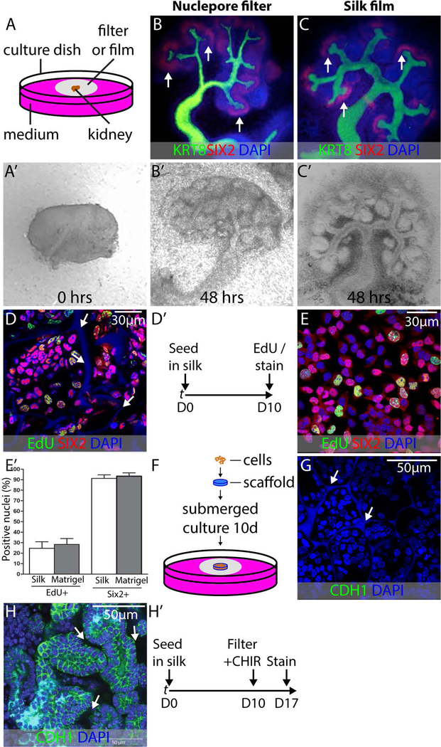
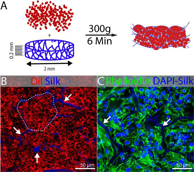
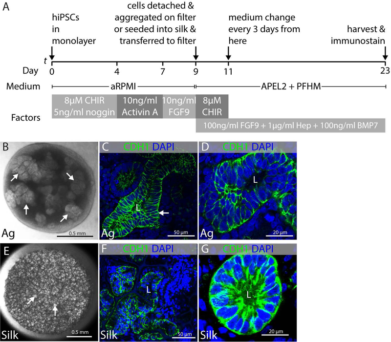
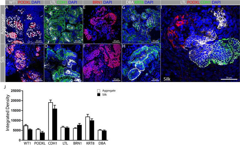
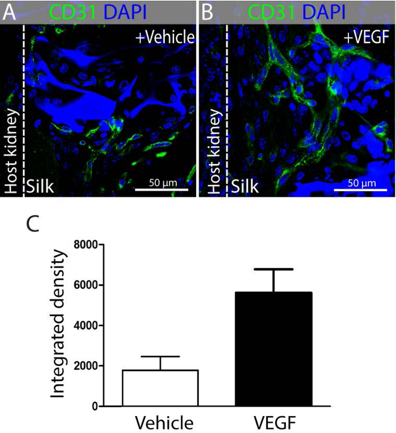
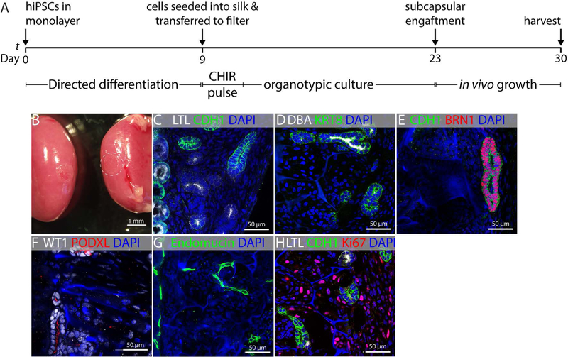
End stage kidney disease affects hundreds of thousands of patients in the United States. The therapy of choice is kidney replacement, but availability of organs is limited, and alternative sources of tissue are needed. Generation of new kidney tissue in the laboratory has been made possible through pluripotent cell reprogramming and directed differentiation. In current procedures, aggregates of cells known as organoids are grown either submerged or at the air-liquid interface. These studies have demonstrated that kidney tissue can be generated from pluripotent stem cells, but they also identify limitations. The first is that perfusion of cell aggregates is limited, restricting the size to which they can be grown. The second is that aggregates lack the structural integrity required for convenient engraftment and suturing or adhesion to regions of kidney injury. In this study, we evaluated the capacity of silk to serve as a support for the growth and differentiation of kidney tissue from primary cells and from human induced pluripotent stem cells. We find that cells can differentiate to epithelia characteristic of the developing kidney on this material and that these structures are maintained following engraftment under the capsule of the adult kidney. Blood vessel investment can be promoted by the addition of vascular endothelial growth factor to the scaffold, but the proliferation of stromal cells within the graft presents a challenge, which will require some readjustment of cell growth and differentiation conditions. In summary, we find that silk can be used to support growth of stem cell derived kidney tissue.

Keywords: directed differentiation; engraftment; fibroin.


Conflict of interest statement

Conflict of interest

The authors declare that they have no conflicts of interest.

Figures

FIGURE 1.
FIGURE 1.
Validation of silk biomaterial for kidney development applications. (A) Schematic illustration of embryonic kidney culture in organotypic conditions. All culture experiments were performed in biological triplicate, and representative examples are shown. (A’) E11.5 kidney imaged on a nuclepore filter at the fluid-air interface immediately after dissection. Note that there is a single epithelial tubule and that there is no branching of the collecting duct. (B,B’) E11.5 kidney after 48 hours culture in organotypic conditions on nuclepore filter. KRT8 labels collecting duct and SIX2 labels NPCs. Note extensive collecting duct branching and maintenance of NPCs at collecting duct tips (arrows). (C,C’) E11.5 kidney after 48 hours culture in organotypic conditions on silk film. Collecting duct branching and NPC maintenance are comparable to culture on nuclepore filter. (D) Purified primary NPCs seeded into silk scaffolds and grown in expansion medium for 10 days as shown in (D’). The SIX2 NPC marker is in red, the EdU proliferation marker is in green and the nuclear marked DAPI is in blue. Note the autofluorescence of the silk in the DAPI channel (arrows). (E) Purified primary NPCs seeded on Matrigel and grown in expansion medium for 10 days and stained with SIX2, EdU, and DAPI for comparison with silk-seeded NPCs. (E’) Quantification of EdU incorporation in NPCs after 10 days growth in silk scaffold versus growth on Matrigel. (F) Schematic summarizing the method for loading scaffolds with primary NPCs and subsequently culturing these constructs using organotypic conditions identical to those used for the embryonic kidney. (G) Immunostaining for CDH1 to detect epithelial organization in silk scaffolded NPCs on day 1 of the 7 day organotypic culture period. DAPI nuclear counterstain reveals nuclei and silk (arrows) (H) Expression of the epithelial marker CDH1 in NPCs expanded in silk scaffolds for 10 days, exposed to CHIR99021 and cultured in organotypic conditions for 7 days. Basolateral CDH1 expression is evident and cells are arranged in typical epithelial structures with lumens. The silk scaffold is within the unstained areas between presumptive tubules (arrows). (H’) Strategy for outgrowth of NPCs in silk scaffold and subsequent epithelialization.
FIGURE 2.
FIGURE 2.
Seeding of kidney progenitors in silk scaffold. Experiments were performed in triplicate and representative examples are shown. (A) Schematic illustration of spin-seeding in silk scaffold (0.2mm thick and 2mm diameter) by centrifugation at 300g for 6min. (B) Representative fluorescent image showing DiI labeled cells packed (dotted line) into the pockets of silk scaffold (white arrow) after spin-seeding. The DAPI channel was included in this image to show the localization of the autofluorescent silk, although DAPI staining was not performed. (C) Phalloidin 488 staining 3 days after seeding showing maintenance of cells packed into the pockets of silk scaffold. DAPI counterstain shows nuclei and the silk scaffold (arrows).
FIGURE 3.
FIGURE 3.
Epithelial differentiation of kidney progenitors. Experiments were performed in triplicate and data representative of a minimum of 3 independent experiments are shown. (A) Schematic illustration of protocol showing directed differentiation of hiPSCs into kidney progenitors on day 9 and subsequently into epithelial structures after making aggregates and seeding in silk scaffold. (B–D) Epithelial structures that differentiate in aggregates of cells derived from directed differentiation cultured in organotypic conditions. (B) Representative stereomicroscope image of kidney organoid generated from hiPSCs at air-liquid interface showing presumptive epithelial clusters (arrows). (C) Representative immunofluorescent image showing CDH1+ tubular epithelial structure with lumen (L) in aggregated cells from directed differentiation (arrow). (D) High magnification micrograph of epithelial tubule with lumen in aggregated cells from directed differentiation. (E–G) Epithelial structures that differentiate in cells from directed differentiation that have been seeded into silk scaffolds. (E) Representative stereomicroscope image of silk scaffold seeded with cells derived from directed differentiation showing clusters of cells within the pockets of the silk scaffold (arrows). (F) Representative immunofluorescent image showing CDH1+ epithelial tubules with lumens (L) within the pockets of the silk scaffold. (G) High magnification micrograph of CDH1+ epithelial tubule with lumen (L) within a pocket of the silk scaffold.
FIGURE 4.
FIGURE 4.
Representative fluorescent images of different nephron segments in aggregates and silk scaffolds. Experiments were performed in triplicate and data representative of a minimum of 3 independent experiments are shown. hiPSC-derived kidney progenitors were cultured either as aggregates (A, C, E, G) or cultured in silk scaffolds (B, D, F, H, I) as shown in Fig. 3A. Immunostaining was performed for: (A-B) Podocytes (PODXL+ WT1+); (C–D) Proximal tubule (CDH1 LTL+); (E–F) Distal tubule (BRN1+) and (G–H) Collecting duct (KRT8+, DBA+). (I) Representative fluorescent image showing nephron patterning in presumptive glomerulus (PODXL+), Proximal (LTL+), distal (CDH1+) and/or collecting duct (CDH1+). (J) Quantification of fluorescent intensity for each of the markers used reveals comparable levels of differentiation within the silk scaffold.
FIGURE 5.
FIGURE 5.
VEGF treatment of silk scaffolds promote vascularization after subcapsular engraftment. Images are representative from n=5 mice in each group. Representative immunofluorescent staining showing CD31+ vasculature in (A) vehicle and (B) VEGF treated silk scaffold, engrafted under kidney capsule of mice. (C) Quantification of CD31+ fluorescence signal in n=5 random high power fields (50µm) from vehicle-treated or VEGF-treated groups.
FIGURE 6.
FIGURE 6.
Maintenance of differentiated epithelial structures in vivo. Images are representative from both kidneys of n=3 mice. (A) Schematic illustration of work plan showing in vitro differentiation of hiPSC derived kidney progenitors after seeding in silk scaffold and their growth under mice kidney capsule. (B) Representative image showing engrafted silk scaffold with differentiated structures under kidney capsule (dotted circle). In vivo engraftment of differentiated renal epithelial structures showing maintenance of (C) Proximal tubule (CDH1 LTL+), (D) Collecting duct (KRT8+, DBA+), (E) Distal tubule (CDH1+ BRN1+) and (F) Podocytes (PODXL+ WT1+). Engrafted silk scaffold with renal epithelial structures were (G) vascularized (Endomucin+) and (H) proliferation (Ki67+) of mesenchymal cells was observed in the graft.

References

    1. Abbott A, Oxburgh L, Kaplan DL, Coburn JM, 2018. Avidin Adsorption to Silk Fibroin Films as a Facile Method for Functionalization. Biomacromolecules - PMC - PubMed
    1. Brown AC, Muthukrishnan SD, Oxburgh L, 2015. A synthetic niche for nephron progenitor cells. Developmental cell 34, 229–241. - PMC - PubMed
    1. Cairns DM, Chwalek K, Moore YE, Kelley MR, Abbott RD, Moss S, Kaplan DL, 2016. Expandable and Rapidly Differentiating Human Induced Neural Stem Cell Lines for Multiple Tissue Engineering Applications. Stem cell reports 7, 557–570. - PMC - PubMed
    1. Caralt M, Uzarski JS, Iacob S, Obergfell KP, Berg N, Bijonowski BM, Kiefer KM, Ward HH, Wandinger-Ness A, Miller WM, Zhang ZJ, Abecassis MM, Wertheim JA, 2015. Optimization and critical evaluation of decellularization strategies to develop renal extracellular matrix scaffolds as biological templates for organ engineering and transplantation. American journal of transplantation : official journal of the American Society of Transplantation and the American Society of Transplant Surgeons 15, 64–75. - PMC - PubMed
    1. Carroll TJ, Park JS, Hayashi S, Majumdar A, McMahon AP, 2005. Wnt9b plays a central role in the regulation of mesenchymal to epithelial transitions underlying organogenesis of the mammalian urogenital system. Developmental cell 9, 283–292. - PubMed

Publication types

LinkOut - more resources