. 2019 Mar 26;58(12):1660-1671.
doi: 10.1021/acs.biochem.9b00068. Epub 2019 Mar 7.

A Shared Mechanism for the Folding of Voltage-Gated K+ Channels

Affiliations

A Shared Mechanism for the Folding of Voltage-Gated K+ Channels

Sarah K McDonald et al. Biochemistry. .

Abstract

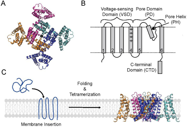
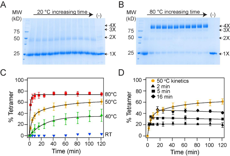
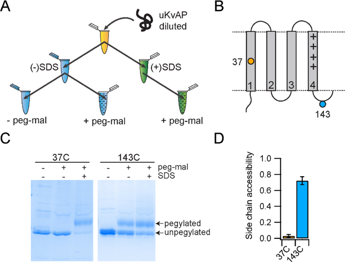
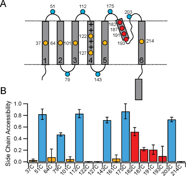
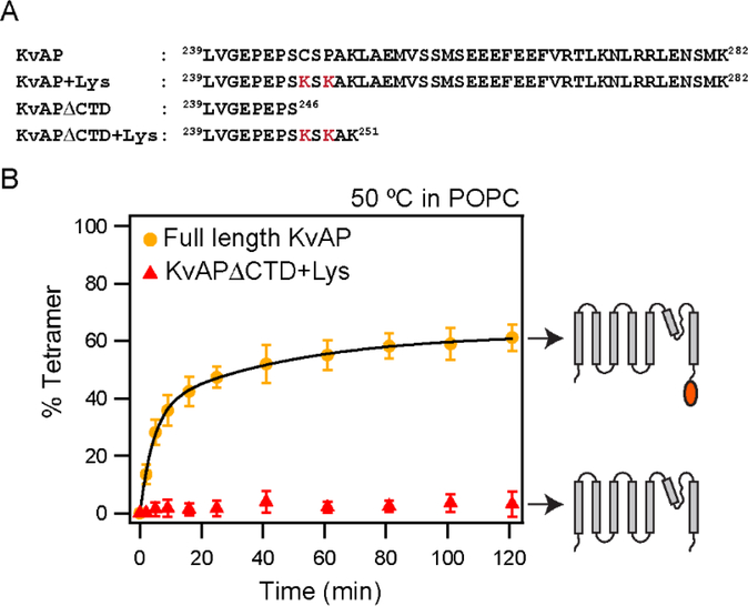
In this study, we probe the folding of KvAP, a voltage-gated K+ (Kv) channel. The KvAP channel, though of archaebacterial origin, is structurally and functionally similar to eukaryotic Kv channels. An advantage of the KvAP channel is that it can be folded in vitro from an extensively unfolded state and the folding can be controlled by temperature. We utilize these properties of the KvAP channel to separately study the membrane insertion and the tetramerization stages during folding. We use two quantitative assays: a Cys PEGylation assay to monitor membrane insertion and a cross-linking assay to monitor tetramerization. We show that during folding the KvAP polypeptide is rapidly inserted into the lipid bilayer with a "native-like" topology. We identify a segment at the C-terminus that is important for multimerization of the KvAP channel. We show that this C-terminal domain forms a dimer, which raises the possibility that the tetramerization of the KvAP channel proceeds through a dimer of dimers pathway. Our studies show that the in vitro folding of the KvAP channel mirrors aspects of the cellular assembly pathway for voltage-gated K+ channels and therefore suggest that evolutionarily distinct Kv channels share a common folding pathway. The pathway for the folding and assembly of a Kv channel is of central importance as defects in this pathway have been implicated in the etiology of several disease states. Our studies indicate that the KvAP channel provides an experimentally tractable system for elucidating the folding mechanism of Kv channels.


Figures

Figure 1.
Figure 1.. Structure and folding of the KvAP channel.
(A) Top view of the tetrameric KvAP channel. The structural model of the KvAP channel previously described is shown. The structure illustrates the domain swap between the pore and voltage sensor domains of adjacent subunits in the KvAP channel. (B) Topology map illustrating the domains and the structural features of a KvAP channel subunit. Dashed lines are membrane boundaries. (C) The folding pathway of the KvAP channel involves the stages of membrane insertion, secondary/tertiary folding and tetramerization. Structure figures were generated with VMD.
Figure 2.
Figure 2.. In vitro folding of the KvAP channel is dependent on temperature.
(A, B) Time course of the in vitro folding of the KvAP channel at 20 °C (A) and at 80 °C (B). Representative SDS-PAGE gels showing glutaraldehyde crosslinking of the in vitro folding reaction at various time points over 2 hours is shown. The last lane is a time point at 2 hours without glutaraldehyde added (−). The oligomeric nature of the crosslinked band is indicated. (C) Tetramerization kinetics of the KvAP channel at different temperatures are plotted. Data have been fitted with either a single (40 °C and 80 °C) or a double exponential (50 °C). Data at 20 °C could not be fitted to an exponential. (D) Temperature downshift arrests folding of KvAP. The in vitro folding reaction for the KvAP channel was carried out at 50 °C. After incubation for 2, 5 or 16 minutes, the samples were transferred from 50 °C to 20 °C and the tetramerization kinetics were followed. No additional tetramerization was observed at 20 °C. The data points following the downshift are fitted with a straight line. Error bars shown in C and D are standard deviations for n =3 using at least 2 independent protein preparations.
Figure 3.
Figure 3.. The Cys-PEGylation assay used to assess the membrane insertion of KvAP.
(A) The reaction scheme used in Cys-PEGylation assay is shown. (B) Topology map of the VSD of the KvAP channel illustrating the cysteines used to demonstrate the methodology. 37C is transmembrane and 143C is in a loop outside of the membrane. (C) SDS-PAGE gels showing the results of the Cys-PEGylation for 37C and 143C. (D) PEGylation results for 37C and D143C were quantified and plotted as bar graphs. Error bars are standard deviations from n=3 experiments using at least 2 independent protein preparations.
Figure 4.
Figure 4.. Membrane insertion of KvAP during folding.
(A) Topology map of the KvAP channel showing the location of the Cys substitutions used for the PEGylation assays. (B) Bar graph showing the PEGylation results for the Cys substitutions. Sites colored in gold are transmembrane, blue are in loops outside of the membrane and red are sites in the pore helix. Error bars are standard deviations from n=3 experiments using at least 2 independent protein preparations. Some error bars are zero because the measured reactivity was zero.
Figure 5.
Figure 5.. Deletion of the CTD abolishes tetramerization of KvAP in POPC lipids.
(A) The C-terminal sequences of the KvAP channel constructs used are shown. All constructs contain the wild type KvAP channel residues 1 – 238. The S6 transmembrane segment in the KvAP channel ends at residue 237. The Lys residues introduced to enhance glutaraldehyde crosslinking are indicated in red. (B) Time course of in vitro folding of the KvAPΔCTD+ Lys channel and the full length control in POPC lipid vesicles at 50 °C. Shown to the right are schematic illustrations of the constructs used. Data for the full-length KvAP channel is from Figure 2. Tetramerization kinetics were quantitated from n=3 experiments with at least 2 protein preparations. Error bars are standard deviations. Tetramerization kinetics for KvAPΔCTD+Lys could not be fitted with an exponential equation.
Figure 6.
Figure 6.. The CTD of KvAP is a dimer.
(A) Schematic illustration of the SUMO-GCN4 and SUMO-KvAP CTD constructs used. (B) SEC of the SUMO-KvAP-CTD, SUMO-GCN4 and SUMO proteins show that the SUMO- KvAP-CTD is a dimer, SUMO-GCN4 is a tetramer while the SUMO protein migrates as a monomer. (C) SDS-PAGE gel showing the glutaraldehyde cross-linking of the SUMO- KvAP-CTD and SUMO-GCN4 proteins confirms that the SUMO- KvAP-CTD is a dimer while SUMO-GCN4 is a tetramer.
Figure 7.
Figure 7.. The CTD inhibits in vitro folding of KvAP.
(A) The reaction scheme used in the competition assay is shown. The competitors used are the S6-CTD or the S6 peptide. The schematic representation of the proteins used in the assay are shown. (B) SDS-PAGE gel showing the glutaraldehyde crosslinking of the KvAP folding reaction carried out in the presence of the S6+CTD or the S6 peptide. The identity of the protein bands observed on glutaraldehyde crosslinking are indicated. The protein bands indicated by the asterisk potentially corresponds to a KvAP+S6-CTD dimer and a S6-CTD tetramer based on their molecular weights. Control lanes where the competitor was added after completion of folding are marked with a C. A five-fold molar ratio of competitor to unfolded KvAP is used in all lanes. Monomers of S6-CTD and the S6 peptide are not visible on this gel because of their low molecular weight. A decrease in the density of the KvAP tetramer band is seen in the case of folding in the presence of S6-CTD but not in the presence of the S6 peptide. (C) Inhibition of the in vitro folding reaction. Bar graph showing the % of tetramer inhibition observed in the presence of the S6+CTD and the S6 peptide. Error bars correspond to the standard deviation for n = 3 from at least 2 protein preparations.
Figure 8:
Figure 8:. Dimerization of the CTD is not dependent on temperature.
(A) The reaction scheme used for the CTD dimerization assay. The SUMO-CTD construct is used in this assay for easy visualization on an SDS-PAGE gel. (B) SDS-PAGE gel showing the extent of dimerization of SUMO-CTD as determined by glutaraldehyde crosslinking. The dimeric SUMO-CTD is dissociated by treatment with 0.35 % SDS, re-associated at the indicated temperature and the extent of re-association is determined by glutaraldehyde crosslinking. Glutaraldehyde crosslinking of the native SUMO-CTD before (−) and after treatment (+) with 0.35% SDS is also shown. Glutaraldehyde crosslinking of the monomeric and the dimeric SUMO-CTD proteins results in an additional band that is indicated by an asterisk. (C) The extent of SUMO-CTD dimerization at the different temperatures were quantified and plotted as a bar graph. The error bars are standard deviations from three experiments with two different protein preparations.

Similar articles

Cited by

References

    1. Hille B (2001) Ion Channels of Excitable Membranes, Sinauer; Sunderland, MA.
    1. Kuo MM, Haynes WJ, Loukin SH, Kung C, and Saimi Y (2005) Prokaryotic K(+) channels: from crystal structures to diversity, FEMS Microbiol Rev 29, 961–985. - PubMed
    1. Long SB, Tao X, Campbell EB, and MacKinnon R (2007) Atomic structure of a voltage-dependent K+ channel in a lipid membrane-like environment, Nature 450, 376–382. - PubMed
    1. Kim DM, and Nimigean CM (2016) Voltage-Gated Potassium Channels: A Structural Examination of Selectivity and Gating, Cold Spring Harb Perspect Biol 8. - PMC - PubMed
    1. Pongs O, and Schwarz JR (2010) Ancillary subunits associated with voltage-dependent K+ channels, Physiol Rev 90, 755–796. - PubMed

Publication types

MeSH terms