

Early post-surgical recurrence of metastatic vertebral neuro-endocrine tumour treated effectively with chemo-radiotherapy
- PMID: 30794152
- PMCID: PMC6385615
- DOI: 10.1051/bmdcn/2019090105
Early post-surgical recurrence of metastatic vertebral neuro-endocrine tumour treated effectively with chemo-radiotherapy
Abstract
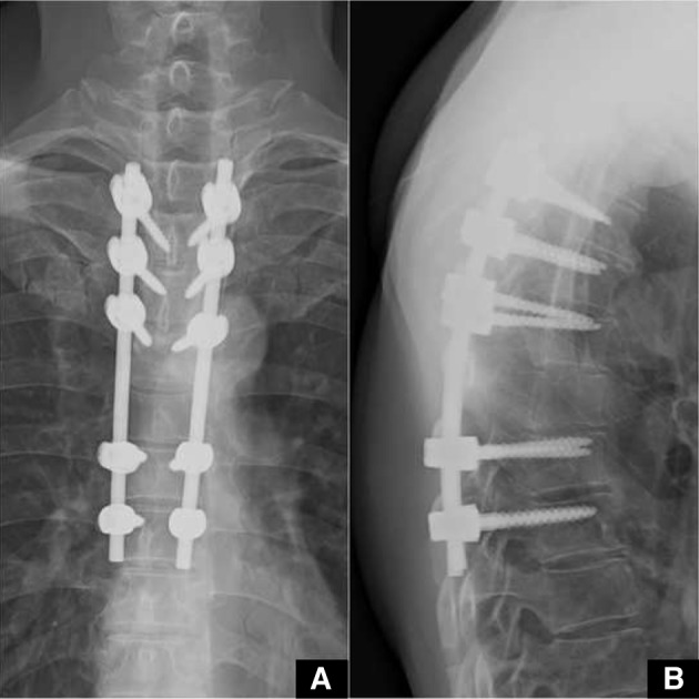
Spinal metastasis of neuro-endocrine tumours (NETs) usually arise from a primary in the lung. We encountered such a patient with NET metastasis to T6 vertebra causing severe cord compression. Considering the neurological status, immediate decompression surgery along with T3-T8 posterior stabilization was done. Early recurrence of the tumour causing near total obliteration of the spinal canal leading to significant neurological compromise was noted within one month of surgery. A second surgery at this stage was avoided due to the risk involved and concurrent chemo-radiotherapy was initiated. The tumour was sensitive to chemo-radiotherapy and rapid resolution was noted on subsequent follow-up visits. With appropriate rehabilitation, patient regained full power to become ambulant with support. This case report highlights a rare, early and aggressive recurrence of metastatic vertebral NET following index surgery which was effectively managed with chemo-radiotherapy.
© Author(s) 2019. This article is published with open access by China Medical University.
Figures


Similar articles
-
Intraoperative radiotherapy combined with posterior decompression and stabilization for non-ambulant paralytic patients due to spinal metastasis.Spine (Phila Pa 1976). 2008 Aug 1;33(17):1898-904. doi: 10.1097/BRS.0b013e31817c0410. Spine (Phila Pa 1976). 2008. PMID: 18670344
-
Complete percutaneous treatment of vertebral body tumors causing spinal canal compromise using a transpedicular cavitation, cement augmentation, and radiosurgical technique.Neurosurg Focus. 2009 Dec;27(6):E9. doi: 10.3171/2009.9.FOCUS09184. Neurosurg Focus. 2009. PMID: 19951062
-
Solitary vertebral metastasis of primary clear cell carcinoma of the liver: a case report and review of literature.J Spine Surg. 2017 Jun;3(2):287-293. doi: 10.21037/jss.2017.06.06. J Spine Surg. 2017. PMID: 28744515 Free PMC article.
-
Intraoperative vertebroplasty during surgical decompression and instrumentation for aggressive vertebral hemangiomas: a retrospective study of 39 patients and review of the literature.Spine J. 2018 Jul;18(7):1128-1135. doi: 10.1016/j.spinee.2017.11.003. Epub 2017 Nov 14. Spine J. 2018. PMID: 29154998 Review.
-
Spinal intradural primary germ cell tumour--review of literature and case report.Acta Neurochir (Wien). 2009 Mar;151(3):277-84. doi: 10.1007/s00701-009-0200-1. Epub 2009 Feb 25. Acta Neurochir (Wien). 2009. PMID: 19240975 Review.
Cited by
-
Thoracic dumbbell spinal metastasis secondary to neuroendocrine tumor of unknown origin: Case report and literature review.Surg Neurol Int. 2022 May 13;13:199. doi: 10.25259/SNI_341_2022. eCollection 2022. Surg Neurol Int. 2022. PMID: 35673674 Free PMC article.
References
LinkOut - more resources
Full Text Sources