. 2019 Apr;18(2):e12850.
doi: 10.1111/acel.12850. Epub 2019 Feb 22.

Knockout of receptor for advanced glycation end-products attenuates age-related renal lesions

Affiliations

Knockout of receptor for advanced glycation end-products attenuates age-related renal lesions

Thibault Teissier et al. Aging Cell. 2019 Apr.

Abstract

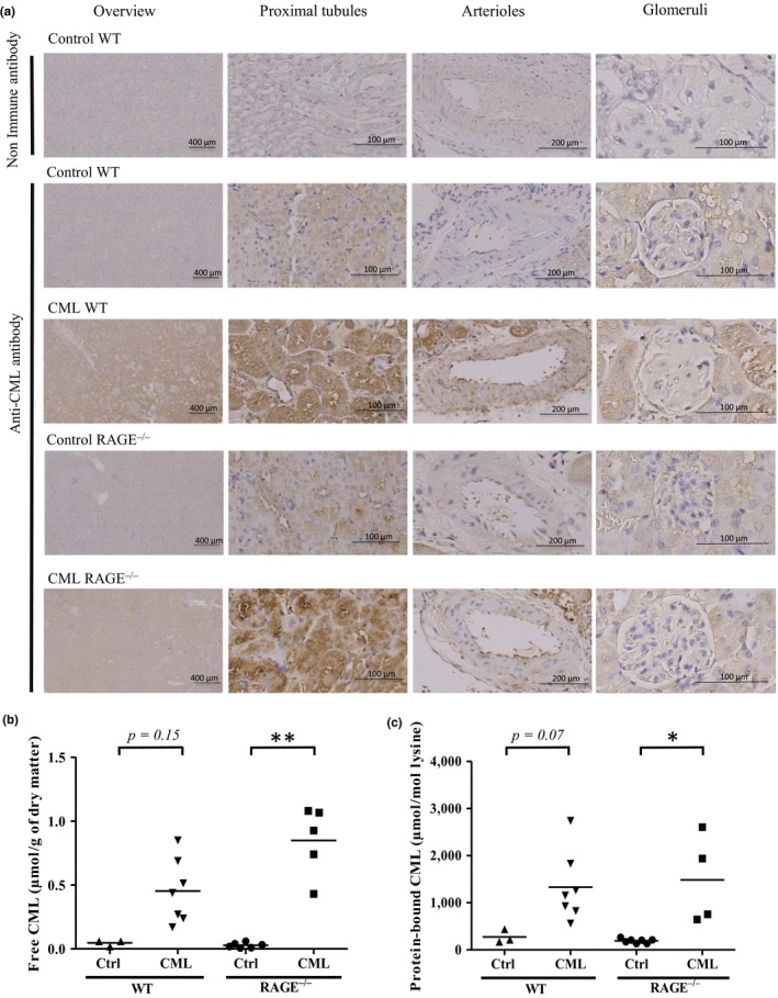
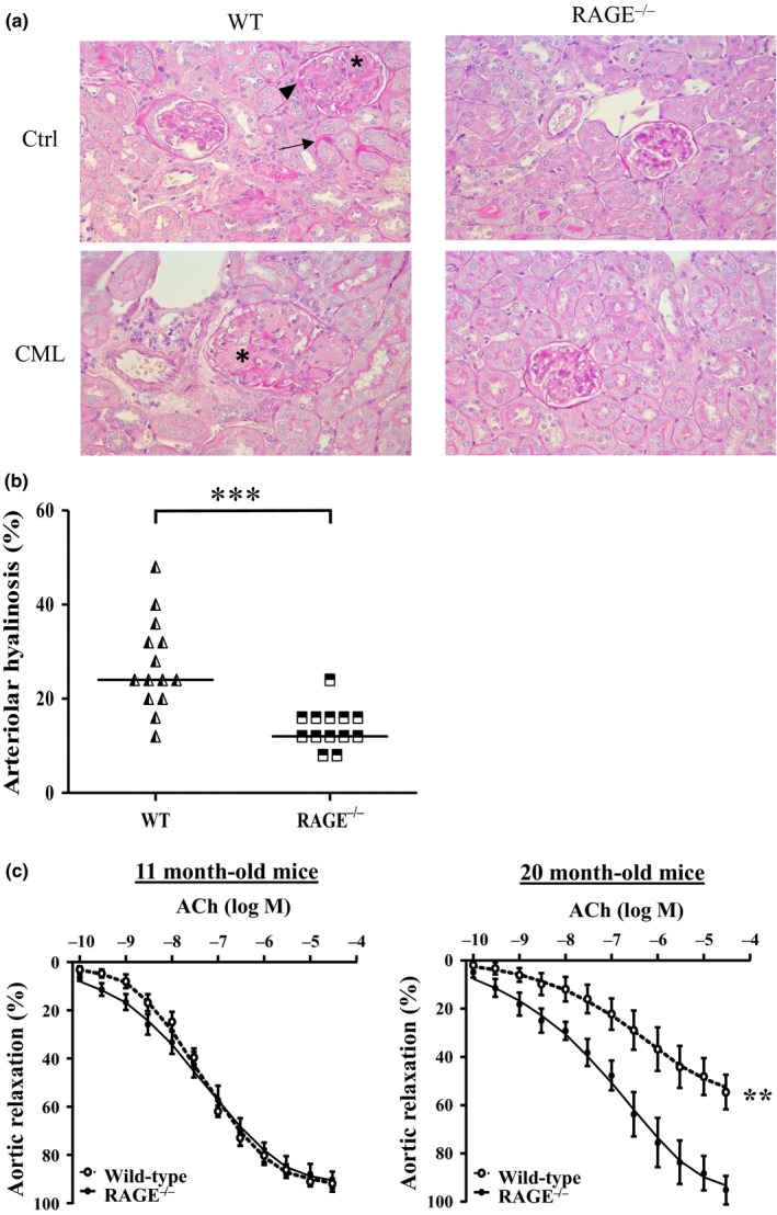
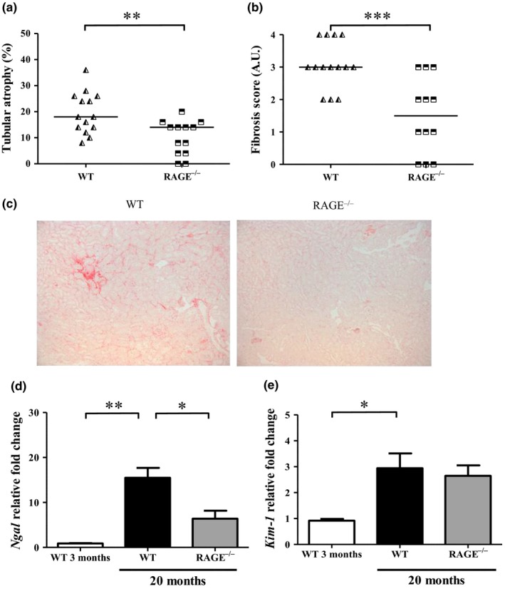
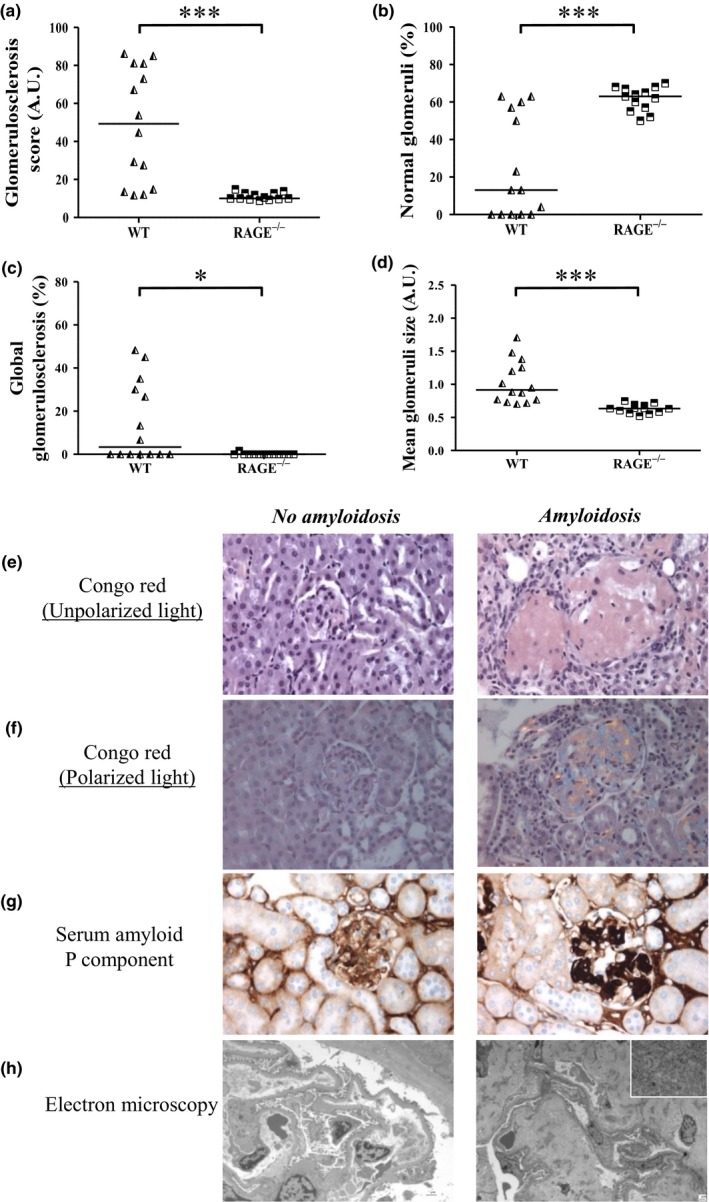
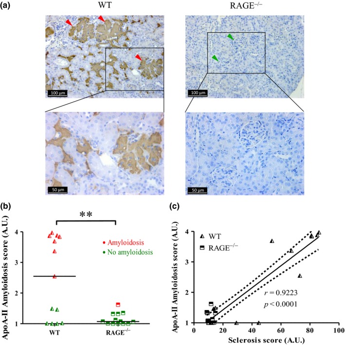
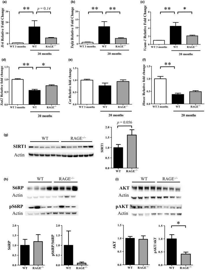
Pro-aging effects of endogenous advanced glycation end-products (AGEs) have been reported, and there is increasing interest in the pro-inflammatory and -fibrotic effects of their binding to RAGE (the main AGE receptor). The role of dietary AGEs in aging remains ill-defined, but the predominantly renal accumulation of dietary carboxymethyllysine (CML) suggests the kidneys may be particularly affected. We studied the impact of RAGE invalidation and a CML-enriched diet on renal aging. Two-month-old male, wild-type (WT) and RAGE-/- C57Bl/6 mice were fed a control or a CML-enriched diet (200 μg CML/gfood ) for 18 months. Compared to controls, we observed higher CML levels in the kidneys of both CML WT and CML RAGE-/- mice, with a predominantly tubular localization. The CML-rich diet had no significant impact on the studied renal parameters, whereby only a trend to worsening glomerular sclerosis was detected. Irrespective of diet, RAGE-/- mice were significantly protected against nephrosclerosis lesions (hyalinosis, tubular atrophy, fibrosis and glomerular sclerosis) and renal senile apolipoprotein A-II (ApoA-II) amyloidosis (p < 0.001). A positive linear correlation between sclerosis score and ApoA-II amyloidosis score (r = 0.92) was observed. Compared with old WT mice, old RAGE-/- mice exhibited lower expression of inflammation markers and activation of AKT, and greater expression of Sod2 and SIRT1. Overall, nephrosclerosis lesions and senile amyloidosis were significantly reduced in RAGE-/- mice, indicating a protective effect of RAGE deletion with respect to renal aging. This could be due to reduced inflammation and oxidative stress in RAGE-/- mice, suggesting RAGE is an important receptor in so-called inflamm-aging.

Keywords: advanced glycation end-products; amyloidosis; chronic kidney disease; nephrosclerosis; receptor for AGEs; renal aging.


Conflict of interest statement

The authors declare no conflict of interest.

Figures

Figure 1
Figure 1
CML accumulation in the kidneys of WT and RAGE−/−mice was diet‐dependant. (a) Representative localization of protein‐bound CML studied by IHC on kidney sections showed that mice fed a CML‐enriched diet exhibited predominantly tubular staining. From left to right: low magnification, high magnification on proximal tubules, arterioles and glomeruli. (b‐c) Quantification by HPLC‐MS/MS of (b) free and (c) protein‐bound CML in kidneys. *p < 0.05, **p < 0.01, Kruskal–Wallis test
Figure 2
Figure 2
Vascular aging of renal arterioles was reduced in RAGE−/− mice. (a) Representative PAS staining of paraffin‐embedded kidney sections of WT (left panels) and RAGE−/− mice (right panels) fed a control (upper panels) or CML‐enriched diet (lower panels) revealed the presence of hyalinosis (arrowheads), tubules undergoing tubular atrophy (arrows) and glomerulosclerosis (asterisks) (x400). (b) Kidney arteriolar hyalinosis determined by PAS staining in WT and RAGE−/− mice and (c) alteration in endothelium‐dependant relaxation in 11‐month‐old and 20‐month‐old WT and RAGE−/− mice fed a control diet. Aortic relaxation is expressed as mean ±SEM (n = 5–6 mice in duplicate). **p < 0.01, ***p < 0.001, unpaired t test on area under the curve
Figure 3
Figure 3
RAGE−/− mice were protected against tubulo‐interstitial aging. Quantification of (a) tubular atrophy was determined by PAS staining and (b) interstitial fibrosis by Sirius red staining. (c) Representative Sirius red staining of control and CML WT and RAGE−/− mice at low magnification (x 100). (d‐e) Expression of kidney injury markers Ngal (d) and Kim‐1 (e) in renal tissue from 3‐ or 20‐month‐old WT and RAGE−/− mice (mean ±SEM, n = 3 for 3‐month‐old mice and n = 10 for 20‐month‐old mice). *p < 0.05, **p < 0.01, ***p < 0.001, unpaired t test or Kruskal–Wallis test for multiple comparisons
Figure 4
Figure 4
Amyloidosis‐linked glomerulosclerosis was largely prevented in RAGE−/− mice. Quantification of GS in paraffin‐embedded kidney sections indicated (a) the GS score (60 glomeruli/mouse) which was used to determine (b) the percentage of normal glomeruli (glomeruli with GS score of 0) and of (c) global GS (glomeruli with a GS score of 100). (d) Mean glomeruli size (30 glomeruli/mouse). Determination of amyloidosis‐positive lesions in paraffin‐embedded kidney sections after (e) Congo red staining under nonpolarized light (x 400) and (f) polarized light showing birefringence (x400), (g) Immunohistochemistry of Serum Amyloid P component (SAP, x 400) and (h) electron microscopy showing the absence in control WT mouse (left panel, x 1,000) and the presence in CML WT mouse (right panel, x 1670 low magnification, x 21,560 high magnification in the frame) of amyloid fibrils. *p < 0.05, ***p <0.001, Mann–Whitney test, representative images
Figure 5
Figure 5
AApoA‐II was largely absent in RAGE−/− mice. AApoA‐II in kidney tissue sections from 20‐month‐old WT mice (left panel) and RAGE−/− mice (right panel) were immunostained using an anti‐ApoA‐II antibody (a) green arrowheads, no deposits; red arrowheads, extensive deposits (representative image). (b) Scoring of ApoA‐II amyloid deposits obtained from IHC and calculated from the grading of 100 glomeruli per mouse, where 1 = no deposit, 2 = small, 3 = moderate and 4 = extensive deposits. Red dots, amyloidosis‐positive mice; green dots, amyloidosis‐negative mice. (c) Linear regression analysis between AApoA‐II and sclerosis scores obtained for all four conditions, y = 0.036 x + 0.70, r = 0.9223, dotted line: 95% confidence intervals (C). **p < 0.01, unpaired t test
Figure 6
Figure 6
Inflammation and oxidation markers were decreased in RAGE−/− mice. Inflammation markers (a) Il‐6, (b) Tnfα and (c) Vcam‐1 and oxidation markers (d) Sod2, (e) Cat and (f) HmoxmRNA expression were measured in renal tissue from 3‐ or 20‐month‐old WT and RAGE−/− mice (mean ±SEM, n = 3 for 3‐month‐old mice and n = 10 for 20‐month‐old mice). Representative western blot and quantification of protein levels of (g) SIRT1 (mean ±SEM, n = 10), (h) pS6RP (Ser235/236)/S6RP and (i) pAKT (Ser473)/AKT in kidney extracts of 22‐ to 26‐month‐old mice (mean ±SEM, n = 4). *p < 0.05, **p < 0.01, Mann–Whitney test or Kruskal–Wallis test for multiple comparisons

References

    1. Alamir, I. , Niquet‐Leridon, C. , Jacolot, P. , Rodriguez, C. , Orosco, M. , Anton, P. M. , & Tessier, F. J. (2013). Digestibility of extruded proteins and metabolic transit of N ε ‐carboxymethyllysine in rats. Amino Acids, 44(6), 1441–1449. 10.1007/s00726-012-1427-3 - DOI - PubMed
    1. Alderson, H. V. , Ritchie, J. P. , Pagano, S. , Middleton, R. J. , Pruijm, M. , Vuilleumier, N. , & Kalra, P. A. (2016). The associations of blood kidney injury molecule‐1 and neutrophil gelatinase‐associated lipocalin with progression from CKD to ESRD. Clinical Journal of the American Society of Nephrology, 11(12), 2141–2149. 10.2215/CJN.02670316 - DOI - PMC - PubMed
    1. Anantharamaiah, G. M. , Hughes, T. A. , Iqbal, M. , Gawish, A. , Neame, P. J. , Medley, M. F. , & Segrest, J. P. (1988). Effect of oxidation on the properties of apolipoproteins A‐I and A‐II. Journal of Lipid Research, 29(3), 309–318. - PubMed
    1. Bolignano, D. , Mattace‐Raso, F. , Sijbrands, E. J. G. , & Zoccali, C. (2014). The aging kidney revisited: A systematic review. Ageing Research Reviews, 14, 65–80. 10.1016/j.arr.2014.02.003 - DOI - PubMed
    1. Calve, S. , Witten, A. J. , Ocken, A. R. , & Kinzer‐Ursem, T. L. (2016). Incorporation of non‐canonical amino acids into the developing murine proteome. Scientific Reports, 6, 32377 10.1038/srep32377 - DOI - PMC - PubMed

Publication types

Substances

LinkOut - more resources