Comparative Study
. 2019 May 1;316(5):R463-R471.
doi: 10.1152/ajpregu.00305.2018. Epub 2019 Feb 22.

Relation between resting sympathetic outflow and vasoconstrictor responses to sympathetic nerve bursts: sex differences in healthy young adults

Affiliations
Comparative Study

Relation between resting sympathetic outflow and vasoconstrictor responses to sympathetic nerve bursts: sex differences in healthy young adults

Austin T Robinson et al. Am J Physiol Regul Integr Comp Physiol. .

Abstract

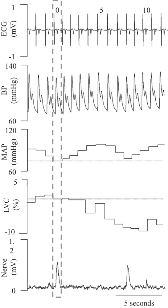
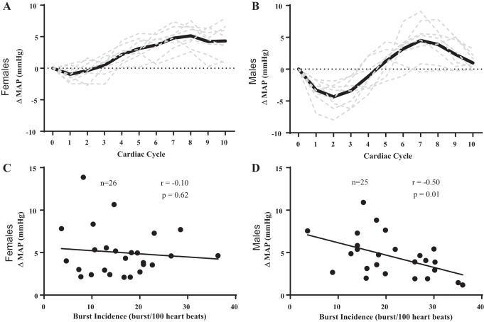
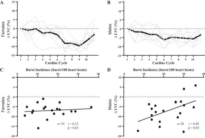
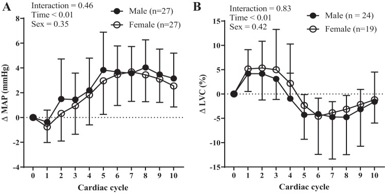
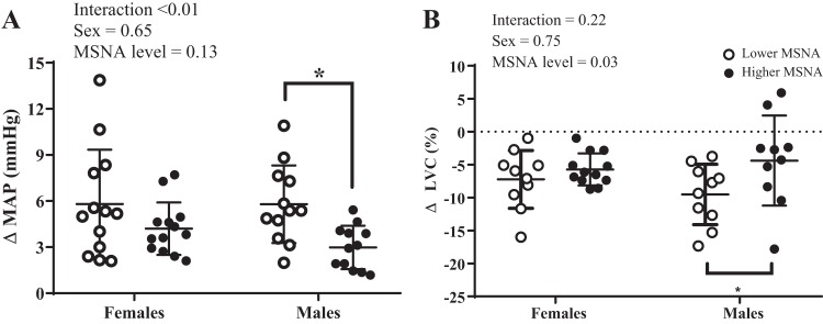
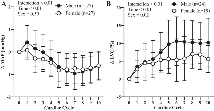
Previous studies have demonstrated an inverse relation between resting muscle sympathetic nerve activity (MSNA) and vasoconstrictor responsiveness (i.e., sympathetic transduction), such that those with high resting MSNA have low vascular responsiveness, and vice versa. The purpose of this investigation was to determine whether biological sex influences the balance between resting MSNA and beat-to-beat sympathetic transduction. We measured blood pressure (BP) and MSNA during supine rest in 54 healthy young adults (27 females: 23 ± 4 yr, 107 ± 8/63 ± 8 mmHg; 27 males: 25 ± 3 yr, 115 ± 11/64 ± 7 mmHg; means ± SD). We quantified beat-to-beat fluctuations in mean arterial pressure (MAP, mmHg) and limb vascular conductance (LVC, %) for 10 cardiac cycles after each MSNA burst using signal averaging, an index of sympathetic vascular transduction. In females, there was no correlation between resting MSNA (burst incidence; burst/100 heartbeats) and peak ΔMAP (r = -0.10, P = 0.62) or peak ΔLVC (r = -0.12, P = 0.63). In males, MSNA was related to peak ΔMAP (r = -0.50, P = 0.01) and peak ΔLVC (r = 0.49, P = 0.03); those with higher resting MSNA had blunted increases in MAP and reductions in LVC in response to a burst of MSNA. In a sub-analysis, we performed a median split between high- versus low-MSNA status on ΔMAP and ΔLVC within each sex and found that only males demonstrated a significant difference in ΔMAP and ΔLVC between high- versus low-MSNA groups. These findings support an inverse relation between resting MSNA and sympathetic vascular transduction in males only and advance our understanding on the influence of biological sex on sympathetic nervous system-mediated alterations in beat-to-beat BP regulation.

Keywords: blood pressure; muscle sympathetic nerve activity; sex differences; vascular physiology; vasoconstriction.


Conflict of interest statement

No conflicts of interest, financial or otherwise, are declared by the authors.

Figures

Fig. 1.
Fig. 1.
Illustration depicting original electrocardiogram (ECG), microneurographic, and blood pressure (BP) traces, along with a calculated vascular conductance curve in a male participant. MAP, mean arterial pressure; LVC, limb vascular conductance.
Fig. 2.
Fig. 2.
Influence of resting sympathetic outflow on peak changes in beat-to-beat mean arterial pressure (MAP) in females vs. males. Examples of the signal averaging for a female participant (A) and a male participant (B) illustrate the changes in MAP following spontaneous bursts of muscle sympathetic nerve activity. There was no relation between peak change in MAP and burst incidence in female participants (C). However, there was a significant negative correlation between peak change in MAP and burst incidence in male participants (D). Sample size (n) is provided within each graph.
Fig. 3.
Fig. 3.
Influence of resting sympathetic outflow on peak changes in beat-to-beat limb vascular conductance (LVC) in females vs. males. Examples of the signal averaging for a female participant (A) and a male participant (B) illustrate the changes in LVC following spontaneous bursts of muscle sympathetic nerve activity. There was no relation between peak change in LVC and burst incidence in female participants (C). There was a significant positive correlation between peak change in LVC and burst incidence in male participants (D). Sample size (n) is provided within each graph.
Fig. 4.
Fig. 4.
Resting sympathetic vascular transduction in female vs. male participants. There was no effect of sex on transduction when quantified as either mean arterial pressure (MAP) (A) or limb vascular conductance (LVC) (B). Sample size (n) is provided within each graph. Data are means ± SD.
Fig. 5.
Fig. 5.
The sex-specific effects of high vs. low muscle sympathetic nerve activity (MSNA) on peak changes in mean arterial pressure (MAP) and limb vascular conductance (LVC). There was a significant interaction effect (sex × MSNA level) on ΔMAP (A). Post hoc analyses indicated male participants with lower resting MSNA demonstrated greater changes in ΔMAP compared with male participants with higher resting MSNA (n values for MAP: low MSNA females = 12, high MSNA females = 13, low MSNA males = 12, high MSNA males = 12). There was a significant main effect of resting MSNA level on ΔLVC (B). Post hoc analyses indicated male participants with lower MSNA demonstrated greater changes in ΔLVC compared with male participants with higher resting MSNA, but this was not the case in female participants (n values for LVC: low MSNA females = 10, high MSNA females = 9, low MSNA males = 10, high MSNA males = 10). Data are means ± SD. We performed a median split to determine high vs. low MSNA. *P < 0.05, high vs. low MSNA.
Fig. 6.
Fig. 6.
Vascular hemodynamics following non-bursts in female vs. male participants. There was no effect of sex on changes in hemodynamics following nonbursts when quantified as mean arterial pressure (MAP) (A). However, female participants demonstrated smaller changes in limb vascular conductance (LVC) (B). Sample size (n) is provided within each graph. Data are means ± SD.

References

    1. Baker SE, Limberg JK, Ranadive SM, Joyner MJ. Neurovascular control of blood pressure is influenced by aging, sex, and sex hormones. Am J Physiol Regul Integr Comp Physiol 311: R1271–R1275, 2016. doi:10.1152/ajpregu.00288.2016. - DOI - PMC - PubMed
    1. Boggia J, Asayama K, Li Y, Hansen TW, Mena L, Schutte R. Cardiovascular risk stratification and blood pressure variability on ambulatory and home blood pressure measurement. Curr Hypertens Rep 16: 470, 2014. doi:10.1007/s11906-014-0470-8. - DOI - PubMed
    1. Brian MS, Matthews EL, Watso JC, Babcock MC, Wenner MM, Rose WC, Stocker SD, Farquhar WB. The influence of acute elevations in plasma osmolality and serum sodium on sympathetic outflow and blood pressure responses to exercise. J Neurophysiol 119: 1257–1265, 2018. doi:10.1152/jn.00559.2017. - DOI - PMC - PubMed
    1. Charkoudian N, Joyner MJ, Johnson CP, Eisenach JH, Dietz NM, Wallin BG. Balance between cardiac output and sympathetic nerve activity in resting humans: role in arterial pressure regulation. J Physiol 568: 315–321, 2005. doi:10.1113/jphysiol.2005.090076. - DOI - PMC - PubMed
    1. Charkoudian N, Joyner MJ, Sokolnicki LA, Johnson CP, Eisenach JH, Dietz NM, Curry TB, Wallin BG. Vascular adrenergic responsiveness is inversely related to tonic activity of sympathetic vasoconstrictor nerves in humans. J Physiol 572: 821–827, 2006. doi:10.1113/jphysiol.2005.104075. - DOI - PMC - PubMed

Publication types

LinkOut - more resources