. 2019 May;33(5):6226-6238.
doi: 10.1096/fj.201801682R. Epub 2019 Feb 22.

Genome-wide effect of pulmonary airway epithelial cell-specific Bmal1 deletion

Affiliations

Genome-wide effect of pulmonary airway epithelial cell-specific Bmal1 deletion

Zhenguang Zhang et al. FASEB J. 2019 May.

Abstract

Pulmonary airway epithelial cells (AECs) form a critical interface between host and environment. We investigated the role of the circadian clock using mice bearing targeted deletion of the circadian gene brain and muscle ARNT-like 1 (Bmal1) in AECs. Pulmonary neutrophil infiltration, biomechanical function, and responses to influenza infection were all disrupted. A circadian time-series RNA sequencing study of laser-captured AECs revealed widespread disruption in genes of the core circadian clock and output pathways regulating cell metabolism (lipids and xenobiotics), extracellular matrix, and chemokine signaling, but strikingly also the gain of a novel rhythmic transcriptome in Bmal1-targeted cells. Many of the rhythmic components were replicated in primary AECs cultured in air-liquid interface, indicating significant cell autonomy for control of pulmonary circadian physiology. Finally, we found that metabolic cues dictate phasing of the pulmonary clock and circadian responses to immunologic challenges. Thus, the local circadian clock in AECs is vital in lung health by coordinating major cell processes such as metabolism and immunity.-Zhang, Z. Hunter, L., Wu, G., Maidstone, R., Mizoro, Y., Vonslow, R., Fife, M., Hopwood, T., Begley, N., Saer, B., Wang, P., Cunningham, P., Baxter, M., Durrington, H., Blaikley, J. F., Hussell, T., Rattray, M., Hogenesch, J. B., Gibbs, J., Ray, D. W., Loudon, A. S. I. Genome-wide effect of pulmonary airway epithelial cell-specific Bmal1 deletion.

Keywords: circadian clock; circadian lung function; food entrainment; influenza infection; metabolic entrainment.


Conflict of interest statement

The authors thank Ian Donaldson and Andy Hayes (Bioinformatics and Genomic Technologies Core Facilities, University of Manchester) for providing support with regard to chromatin immunoprecipitation sequencint (ChIP) and RNA-seq analysis and sequencing, Dr. Halina Dobrzynski and Garry Ashton (University of Manchester) for access to LCM machines, Prof. Colin Bingle’s group (University of Sheffield, Sheffield, England) in the training of primary AEC ALI culture, Dr. Emma Rawlins (University of Cambridge, Cambridge, United Kingdom) for communications of the Cre virus transfection method, Dr. Christoph Ballestrem, Rachel Lennon, and Patrick Caswell (all from the University of Manchester) for discussions of epithelial cell biology, the Genomics Center for RNA-seq Service, the Histology Laboratory, and Bioimaging Facility, University of Manchester for tissue section studies, and members of the laboratories of A.S.I.L. and D.W.R. for general discussions. The work is supported by Biotechnology and Biological Sciences Research Council (BBSRC) grants awarded to A.S.I.L. and D.W.R. (BB/L000954/1 and BB/K003097/1). D.W.R. and A.S.I.L. are Wellcome Investigators (Wellcome Trust; 107849/Z/15/Z). J.B.H. is supported by the U.S. National Institutes of Health, National Institute of Neurological Disorders and Stroke (2R01NS054794 to J.B.H. and A.S.I.L.). The authors declare no conflicts of interest.

Figures
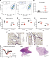
Figure 1
Figure 1
Altered pulmonary homeostasis and functions in CCSP-Bmal1−/− mice. BAL samples were collected in the morning from unchallenged young CCSP-Bmal1−/− and Bmal1flox/flox littermate controls for flow cytometry study. A) Flow cytometry plots for BAL macrophages and neutrophils from Bmal1flox/flox and CCSP-Bmal1−/− mice. B) BAL neutrophilia in Bmal1flox/flox and CCSP-Bmal1−/− mice. Data shown are means ± sd; Student’s t test (n = 5–6/group). *P < 0.05. C–E) Measurement of lung biomechanical function in 4-mo-old and 12-mo-old Bmal1flox/flox and CCSP-Bmal1−/− mice assessed at ZT4–6. Rrs, lung resistance; Crs, lung compliance; and Ers, lung. Data shown are means ± sd; Student’s t test between genotypes in the same age group (n = 5–7 in the 4-mo group, n = 3–4 in the 12-mo group). *P < 0.05, **P < 0.01. F) Examples of picrosirius red staining of larger airways in aged CCSP-Bmal1−/− and Bmal1flox/flox mice. G) Body weight changes over 21 d following influenza infection. Data shown are means ± sd; 2-way ANOVA with post hoc test (n = 5–6/group). **P < 0.01, ***P < 0.001, ****P < 0.0001. H) Example of lung hematoxylin and eosin staining 11 d postinfection.
Figure 2
Figure 2
Disrupted core clock gene expression in CCSP-Bmal1−/− mice. Time-series (48 h) RNA-seq studies were carried out in LCM distal small AECs and RNA reads were normalized by DEseq2 method. Transcripts of core clock genes from Bmal1flox/flox (black solid circle) and CCSP-Bmal1−/− (red solid square) mice are shown. Y-axis labels DESeq2-normalized reads count, and x-axis labels CT.
Figure 3
Figure 3
Circadian transcription in LCM distal AECs over 2 cycles. A) Group 1: rhythmically expressed transcripts in Bmal1flox/flox mice (left) compared with the same genes plotted for CCSP-Bmal1−/− mice (right). Group 2: common rhythmic genes in both genotypes. Group 3: newly emergent rhythmic transcripts in CCSP-Bmal1−/− mice (right) and expression of the same transcripts in Bmal1flox/flox group (left). B) Phase distribution of rhythmic genes of groups 1–3, which centers at CT5 and CT15 in groups 1 and 2, and at CT9 and CT20 in group 3. C) KEGG pathway enrichment analysis of genes in groups 1–3, ranked by significance, with most significant pathways as propionate metabolism in group 1, circadian rhythm in group 2, and antigen processing and presentation in group 3. D) Examples of enriched DNA motifs of rhythmic genes shown above in order of significance of fit.
Figure 4
Figure 4
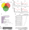
Analysis of metabolic-related DE gene sets in Bmal1-targeted cells. A) Overlap in expression of circadian gene sets from Fig. 3 with DE gene sets. B) Examples of 4 DE genes from Bmal1flox/flox (black solid circle) and CCSP-Bmal1−/− (red solid square). C) KEGG pathway enrichment analysis of DE genes, ranked in order of significance. D) Plots of phase and log2 fold changes of a subset of DE genes defined in metabolic pathways, which were rhythmic in Bmal1flox/flox group. E) HMGCS2 Western blots in whole-lung tissues from CCSP-Bmal1−/− (left) and global Bmal1−/− (right) mice. Samples were taken at ZT4. Each lane contained a single sample.
Figure 5
Figure 5
Cell-autonomous Bmal1 function in ALI culture of primary mouse tracheal cells. ALI cultures were established using primary tracheal epithelial cells from WT and global Bmal1−/− mice, with 7 d under submerged condition and another 10 d exposed to air in the apical side of cells. A, B) Electron microscopy images for cilia in ALI primary tracheal cells from WT and global Bmal1−/− mice, respectively. C, D) Immunofluorescence staining of cilia markers (acetylated tubulin) and epithelial cell markers (E-cadherin) in ALI primary tracheal cells from WT and global Bmal1−/− mice, respectively. E, F) Cxcl5 and Cxcl15 gene expression measurement by qPCR in ALI primary tracheal cells from WT and global Bmal1−/− mice. Data shown are means ± sd; Student’s t test (n = 3/genotype). *P < 0.05, **P < 0.01. G) Expression of Bmal1 exon 8 in primary tracheal cells 2 d after infection. Data shown are means ± sd; Student’s t test (n = 3/genotype). ***P < 0.001. H) Bioluminescence traces of Per2-luc in control and Adv-Cre–transfected ALI primary tracheal cells 21 d in culture. I) Schematic description of viral Cre infection in primary tracheal cells from Bmal1flox/flox mice. J) Overlap of expression of rhythmic genes during ALI culture and LCM studies. K) KEGG pathway enrichment of DE genes in cell culture study. Pathways are ranked by significance value.
Figure 6
Figure 6
Reverse feeding resets time of day variation in pulmonary LPS response. A) Schematic description of experiment design. The food-reversal experiment was undertaken using 2 separate cohorts. BD) Food reversal without LPS treatment was performed in cohort 1. EG) Aerosolized LPS exposure experiment at ZT0 vs. ZT12 was performed in cohort 2 and total BAL cells, neutrophils, and CXCL5 concentrations measured. Gene expression was determined in cohort 2. Data analyzed by 2-way ANOVA with post hoc test to examine time of day difference within genotypes (n = 6–8). *P < 0.05, **P < 0.01, ***P < 0.001, ****P < 0.0001 (significant time of day difference within genotypes).
Figure 7
Figure 7
Illustration of key findings. Feeding sets the time of lung in vivo, driving rhythmic expression of core clock proteins like BMAL1 and more widespread rhythmic genes in club cells, involving mainly metabolic- and chemokine-signaling pathways. Deletion of Bmal1 in club cells abolishes these rhythmically expressed genes and uncovers a different set of newly rhythmic genes. This may be due to other rhythmic signals in vivo. CCSP-Bmal1−/− mice show altered lung mechanic functions and more severe influenza infection. In vitro, primary AECs were able to generate rhythmic gene expression under temperature entrainment, and deletion of Bmal1 disrupts genome-wide rhythmic gene expression without generating newly rhythmic gene expression. Flu stands for influenza in the picture.

References

    1. Takahashi J. S. (2017) Transcriptional architecture of the mammalian circadian clock. Nat. Rev. Genet. 18, 164–179 - PMC - PubMed
    1. Kondratov R. V., Kondratova A. A., Gorbacheva V. Y., Vykhovanets O. V., Antoch M. P. (2006) Early aging and age-related pathologies in mice deficient in BMAL1, the core componentof the circadian clock. Genes Dev. 20, 1868–1873 - PMC - PubMed
    1. Bunger M. K., Wilsbacher L. D., Moran S. M., Clendenin C., Radcliffe L. A., Hogenesch J. B., Simon M. C., Takahashi J. S., Bradfield C. A. (2000) Mop3 is an essential component of the master circadian pacemaker in mammals. Cell 103, 1009–1017 - PMC - PubMed
    1. Bass J., Lazar M. A. (2016) Circadian time signatures of fitness and disease. Science 354, 994–999 - PubMed
    1. Papagiannakopoulos T., Bauer M. R., Davidson S. M., Heimann M., Subbaraj L., Bhutkar A., Bartlebaugh J., Vander Heiden M. G., Jacks T. (2016) Circadian rhythm disruption promotes lung tumorigenesis. Cell Metab. 24, 324–331 - PMC - PubMed

Publication types

LinkOut - more resources