. 2019 Feb 22;15(2):e1007992.
doi: 10.1371/journal.pgen.1007992. eCollection 2019 Feb.

A DNA repair protein and histone methyltransferase interact to promote genome stability in the Caenorhabditis elegans germ line

Affiliations

A DNA repair protein and histone methyltransferase interact to promote genome stability in the Caenorhabditis elegans germ line

Bing Yang et al. PLoS Genet. .

Abstract

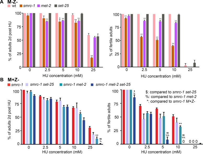
Histone modifications regulate gene expression and chromosomal events, yet how histone-modifying enzymes are targeted is poorly understood. Here we report that a conserved DNA repair protein, SMRC-1, associates with MET-2, the C. elegans histone methyltransferase responsible for H3K9me1 and me2 deposition. We used molecular, genetic, and biochemical methods to investigate the biological role of SMRC-1 and to explore its relationship with MET-2. SMRC-1, like its mammalian ortholog SMARCAL1, provides protection from DNA replication stress. SMRC-1 limits accumulation of DNA damage and promotes germline and embryonic viability. MET-2 and SMRC-1 localize to mitotic and meiotic germline nuclei, and SMRC-1 promotes an increase in MET-2 abundance in mitotic germline nuclei upon replication stress. In the absence of SMRC-1, germline H3K9me2 generally decreases after multiple generations at high culture temperature. Genetic data are consistent with MET-2 and SMRC-1 functioning together to limit replication stress in the germ line and in parallel to promote other germline processes. We hypothesize that loss of SMRC-1 activity causes chronic replication stress, in part because of insufficient recruitment of MET-2 to nuclei.


Conflict of interest statement

The authors have declared that no competing interests exist.

Figures

Fig 1
Fig 1. SMRC-1 reduces replication stress-induced sterility and lethality.
(A) Impact of HU treatment on viability (left) and fertility (right) of smrc-1 M-Z- F2 mutants. wt, met-2, and set-25 were tested in parallel. (B) Impact of HU treatment on viability (left) and fertility (right) of M+Z- F1 generation mutants. smrc-1 F1 M+Z- mutants, smrc-1 set-25 M+Z- double, smrc-1 met-2 M+Z- double, and met-2 smrc-1 set-25 M+Z- triple mutants were tested in parallel. (A, B) Animals were exposed to HU for 16 hr at 25°C as L1 larvae, transferred to regular NGM plates, and maintained at 25°C for 48 hr. Error bars indicate standard error of the mean. smrc-1(om136) was used for smrc-1 single and smrc-1 set-25 double mutant strains; smrc-1(om138) was used in smrc-1 met-2 and smrc-1 met-2 set-25 strains. (A) Mutant and wildtype data were compared using two-tailed t-test on arcsine transformed data. * indicates P <0.05, ** indicates P <0.01. (B) Pair-wise comparison were performed as indicated in the legend. $, %, * indicate P <0.05; $ $, %%, ** indicate P <0.01. ≥ 3 biological replicates were performed with 40 L1 larvae/replicate.
Fig 2
Fig 2. smrc-1 mutants maintained long-term at high culture temperature have variably reduced brood sizes.
16 smrc-1(ea8) lines were passaged for 33 generations in parallel at 25°C. The first larva to reach L4 was passaged in each case; if sterile, a fertile sibling was chosen to rescue the line. The second generation where the oldest L4 was sterile, the line is indicated as sterile in the graph. Brood sizes were binned, as indicated. See text and Supplemental Materials and methods.
Fig 3
Fig 3. SMRC-1 promotes genome integrity.
(A) Apoptotic germ cells are identified by CED-1::GFP expression in control and representative fertile and sterile smrc-1;ced-1::gfp adult gonads. Each gonad is oriented with the loop region to the right. Plots show the number of CED-1::GFP positive cells per gonad arm in wildtype, met-2, and fertile smrc-1 mutants; box represents the middle 50% of values, line represents the 50th percentile (median) value, and bars indicate the full range of values. CED-1::GFP is observed throughout sterile smrc-1 and smrc-1 met-2 gonads; these individuals were not quantified because it was difficult to reliably distinguish individual cells. Elevated apoptosis is CEP-1-dependent. ***P<0.001, Student’s t-test. N.s., not significant. See S2C Fig for more smrc-1 met-2 images. (B) Reversion of the unc-58(e665) phenotype is enhanced by smrc-1 loss-of-function at 20° and 25°C. Total = number of individuals assayed. (C) The dog-1 poly G/C deletion phenotype is elevated in smrc-1 and met-2 mutants. Data represent the deletion frequency within vab-1 exon 5. Total = number of individuals assayed. Deletion = number of deletions identified. Note that smrc-1 met-2 double mutants were assayed in the M+Z- generation. (D) Antibody S9.6 immunolabeling detects elevated RNA:DNA hybrids throughout the smrc-1 germ line. The proportion of nuclei with S9.6 foci (the S9.6 index) was calculated independently for proliferative, leptotene/ zygotene (transition), and pachytene regions of 25°C wildtype and smrc-1 M-Z- hermaphrodite germ lines. *P<0.02, **P<0.002. (E) smrc-1 mutations alter bivalent formation. Images show representative diakinesis-stage nuclei in the -1 oocyte. Wildtype image contains 6 larger DAPI-bright bodies, representing 6 bivalents, and spo-11(ok79) image contains 12 smaller DAPI-bright bodies, representing 12 univalents. Righthand panel shows the number of DAPI-stained bodies per diakinesis nucleus in strains raised at 25°C. smrc-1(om138) and smrc-1(ea8) oocytes contain 4–7 distinct DAPI-bright bodies of variable size; larger bodies often appear to contain chromosomes held together by DNA bridging (arrows). DNA linkages were verified by rotation of confocal microscopy Z-stacks. smrc-1(om138);spo-11(ok79) oocytes contain 5–12 DAPI-bright bodies of variable size.
Fig 4
Fig 4. SMRC-1 associates with MET-2.
(A) Protein blot containing total lysate (left two lanes) and immunoprecipitated material (right two lanes) was probed with anti-MET-2 antibody. Nuclear protein extracts were prepared from him-8(e1489) (control) and met-2(n4256) mutants, and IP was performed with anti-MET-2 antibody. *, Cross-reacting polypeptide routinely observed on protein blots but not recovered in IP under our conditions. Images are from a single immunoblot with uninformative lanes cropped out. (B) Protein blots containing total nuclear and cytoplasmic lysates and material immunoprecipitated with anti-FLAG antibody were probed with anti-MET-2. Extracts were prepared from wildtype and 3xflag::smrc-1 adults, as indicated.
Fig 5
Fig 5. SMRC-1 localizes to germ cell nuclei.
Images show dissected adult gonads labeled with anti-FLAG antibody (green), counterstained with DAPI (red), and visualized with epifluorescence microscopy. (A) A complete XO male gonad is shown above; lower panels correspond to the boxed images. (B) An XX hermaphrodite gonad is shown above; lower panels correspond to the boxed regions. Nuclear 3xFLAG::SMRC-1 is detected throughout the (A) male and (B) hermaphrodite germ lines. Scale bars: 16 μm.
Fig 6
Fig 6. Nuclear MET-2 is detected throughout the XO and XX germ line.
Images show dissected adult gonads labeled with anti-FLAG antibody (green) and counterstained with DAPI (red). (A) A complete male gonad is shown above; lower panels correspond to the boxed images presented in distal to proximal order. (B) A hermaphrodite gonad is shown above; lower panels correspond to the boxed regions. Scale bar: 16 μm. (C) Pachytene germ cells co-labeled for 3xMYC::MET-2 and 3xFLAG::SMRC-1. Images show a portion of a dissected adult germ line immunolabeled with anti-MYC and anti-FLAG antibodies, counterstained with DAPI, and visualized with confocal microscopy. Single-label images are shown in grey scale. Merged image: MET-2 (red), SMRC-1 (green). Scale bar: 5 μm. Arrows indicate example of regions with co-labeling. See S4 Fig for additional images.
Fig 7
Fig 7. Nuclear SMRC-1 and MET-2 abundance in distal germline nuclei is elevated upon hydroxyurea treatment.
(A) 3xFLAG::SMRC-1 abundance in distal germline nuclei with/without HU treatment. Images show anti-FLAG immunolabeled gonads from control and HU-treated adults. Gonad arms are oriented with the distal end to the left (A-D). L4 larvae were exposed to 25mM HU for 24 hours at room temperature (22°C) prior to immunolabeling. Box and whisker plots show normalized 3xFLAG::SMRC-1 signals with/without HU treatment; box represents the middle 50% of values, line represents the 50th percentile (median) value, and bars indicate the full range of values. SMRC-1 signal is shown normalized to mean DAPI fluorescence intensity (left) and mCherry::HIS-58 fluorescence intensity (right). For each germ line, 5–7 mitotic nuclei in a similar state of chromatin condensation and a common focal plane were measured; nuclei were random with respect to size. 6–8 germ lines were measured per treatment and per replicate (see Supplemental materials and methods). See S5 Fig for normalization to total histone H3. N = 3 biological replicates. (B, C) 3xFLAG::MET-2 abundance in distal germline nuclei with/without HU treatment in (B) control and (C) smrc-1 mutant germ lines. HU treatment and quantification were performed as in (A). N = 2 biological replicates. (D) H3K9me2 abundance in distal nuclei is higher following HU treatment. **, P<0.01.
Fig 8
Fig 8. smrc-1 germ cells are resistant to mitotic arrest.
(A) In wildtype and smrc-1 mutants, HU treatment significantly reduced the number of distal germ cell nuclei and increased their size. However, the response in smrc-1 was significantly milder than in wt germ cells. Plots show number of nuclei in the mitotic region (above) and average nuclear area (below); box represents the middle 50% of values, line represents the 50th percentile (median) value, and bars indicate the full range of values. (B) wt and smrc-1 have the same mitotic index under standard growth conditions. Mitotic germ cells were identified by anti-H3S10phos labeling (pH3). Plot shows the mitotic index (number of H3S10phos-positive nuclei distal to meiotic entry/ total number of nuclei distal to meiotic entry); box represents the middle 50% of values, line represents the 50th percentile (median) value, and bars indicate the full range of values. Upon HU treatment, there is a significant reduction in the mitotic index in wildtype but not in smrc-1. **, Mitotic index of HU-treated wt and smrc-1 germ cells is significantly different, P<0.05. N = 14–20 germ lines assayed per treatment.
Fig 9
Fig 9. H3K9me2 labeling is reduced in serially passaged smrc-1 mutants.
(A) H3K9me2 distribution as detected by immunolabeling in wildtype and smrc-1(om136) F2 worms raised at 25°C. Gonads were dissected at 18 hr post-L4 stage and labeled with anti-H3K9me2 antibody and counterstained with DAPI to visualize DNA. Panel show sets of pachytene nuclei. (B) H3K9me2 distribution as detected in wildtype and smrc-1(ea8) animals passaged for 30 generations at 25°C. Gonads were dissected at 18 hr post-L4 stage and labeled with anti-H3K9me2 antibody and counterstained with DAPI to visualize DNA. Each panel shows a set of pachytene nuclei from four different individuals with different staining intensities and/or patterns. Histograms indicate the corrected total nuclear fluorescence (CTNF, see Materials and methods) for a set of nuclei within randomly selected wildtype and smrc-1 F30 germ lines; asterisks indicate the germ lines included in the images. Number of gonads assayed: 4 wt and 21 smrc-1 males; 4 wt and 20 smrc-1 hermaphrodites. 6 germline nuclei assayed/gonad.
Fig 10
Fig 10. Alternative models for the relationship between MET-2 and SMRC-1 activity.
Association of MET-2 with SMRC-1 may promote deposition of H3K9 methylation (A) on nascent chromatin following DNA replication and/or (B) at sites of DNA damage (star). This second alternative may occur during DNA replication and/or at other times in the cell cycle (as depicted). In this scenario, SMRC-1 is recruited to the damaged site to facilitate the repair of a DNA lesion. MET-2 associates with SMRC-1 and methylates H3K9. Deposition of H3K9me2 and establishment of heterochromatin near a DNA break site stabilizes the exposed break until repair occurs.

References

    1. Gursoy-Yuzugullu O, Ayrapetov MK, Price BD. Histone chaperone Anp32e removes H2A.Z from DNA double-strand breaks and promotes nucleosome reorganization and DNA repair. 2015;112(24):7507–12. 10.1073/pnas.1504868112 - DOI - PMC - PubMed
    1. Caridi PC, Delabaere L, Zapotoczny G, Chiolo I. And yet, it moves: nuclear and chromatin dynamics of a heterochromatic double-strand break. Philosophical transactions of the Royal Society of London Series B, Biological sciences. 2017;372(1731):20160291 10.1098/rstb.2016.0291 - DOI - PMC - PubMed
    1. Ayrapetov MK, Gursoy-Yuzugullu O, Xu C, Xu Y, Price BD. DNA double-strand breaks promote methylation of histone H3 on lysine 9 and transient formation of repressive chromatin. 2014;111(25):9169–74. 10.1073/pnas.1403565111 - DOI - PMC - PubMed
    1. Madireddy A, Gerhardt J. Replication Through Repetitive DNA Elements and Their Role in Human Diseases In: Masai H, Foiani M, editors. DNA Replication: From Old Principles to New Discoveries. Singapore: Springer Singapore; 2017. p. 549–81. - PubMed
    1. Bhatia V, Herrera-Moyano E, Aguilera A, Gómez-González B. The Role of Replication-Associated Repair Factors on R-Loops. Genes. 2017;8(7):171. - PMC - PubMed

Publication types