. 2019 Feb 22;14(2):e0212005.
doi: 10.1371/journal.pone.0212005. eCollection 2019.

Secreted frizzled related protein is a target of PaxB and plays a role in aquiferous system development in the freshwater sponge, Ephydatia muelleri

Affiliations

Secreted frizzled related protein is a target of PaxB and plays a role in aquiferous system development in the freshwater sponge, Ephydatia muelleri

Chelsea Hall et al. PLoS One. .

Abstract

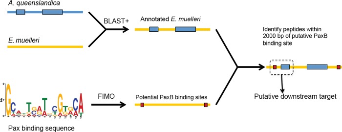
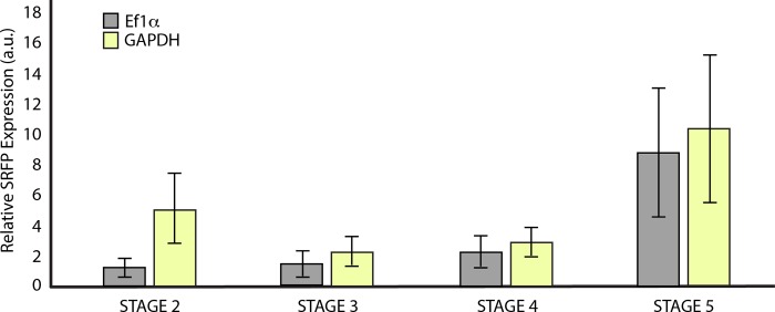
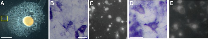
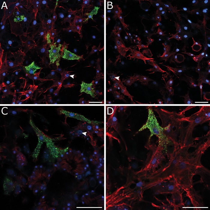
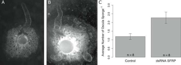
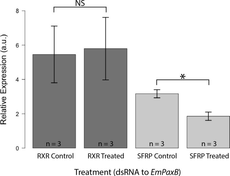
Canonical and non-canonical Wnt signaling, as well as the Pax/Six gene network, are involved in patterning the freshwater sponge aquiferous system. Using computational approaches to identify transcription factor binding motifs in a freshwater sponge genome, we located putative PaxB binding sites near a Secreted Frizzled Related Protein (SFRP) gene in Ephydatia muelleri. EmSFRP is expressed throughout development, but with highest levels in juvenile sponges. In situ hybridization and antibody staining show EmSFRP expression throughout the pinacoderm and choanoderm in a subpopulation of amoeboid cells that may be differentiating archeocytes. Knockdown of EmSFRP leads to ectopic oscula formation during development, suggesting that EmSFRP acts as an antagonist of Wnt signaling in E. muelleri. Our findings support a hypothesis that regulation of the Wnt pathway by the Pax/Six network as well as the role of Wnt signaling in body plan morphogenesis was established before sponges diverged from the rest of the metazoans.


Conflict of interest statement

The authors have declared that no competing interests exist.

Figures

Fig 1
Fig 1. Schematic of computational approach used to identify potential downstream targets of EmPaxB.
An Ephydatia muelleri low quality draft genome was “roughly annotated” against Amphimedon queenslandica predicted peptides database. Separately, potential sponge-specific binding sites of EmPaxB were identified in the E. muelleri genome using Find Individual Motif Occurrences (FIMO).
Fig 2
Fig 2. EmSFRP phylogenetic analysis.
Tree of SFRP and Frizzled relationships from sponges and other metazoans based on PhyML. The two monophyletic Frizzled groups are shown with branches in blue. A monophyletic group of SFRPs (some with netrin domains) are designated with purple branches, including Ephydatia muelleri SFRP (denoted EPHMU SFRP in the phylogeny) and other sponge SFRPs in light purple. Additional SFRPs and other frizzled domain proteins without netrin domains are in green. Node support was run using aLRT and 90–100% supported nodes are designated with a red dot, 76–89% with an orange dot, 65–75% with yellow, and nodes with under 65% are not shown. Pfam domains are given in the key such that Frizzled domains with a predicted glycosylation site are dark green, Frizzled domains without a glycosylation site are light green, netrin domains are purple, and other domains are designated by beige/brown/brick red.
Fig 3
Fig 3. Relative expression levels of EmSFRP across asexual developmental stages of gemmulating E. muelleri.
Relative EmSFRP expression (a.u. = arbitrary units) levels were normalized to Ef1α and GAPDH, averages (± SEM) are shown. A one-way ANOVA indicated no significant differences among stages (Ef1α: F3,4 = 5.012, p = 0.077; GAPDH: F3,4 = 3.33; p = 0.138). Developmental stage diagrams and pictures reprinted from [28] under a CCC license, with permission from Elsevier, original Copyright, 2016.
Fig 4
Fig 4. EmSFRP expression in amoeboid cells with filipodia in mesohyl at periphery of stage 5 sponge.
A) Stage 5 juvenile sponge with functional aquiferous system. Scale: 1 mm. B&D) In situ hybridization of EmSFRP with staining in a subset of amoeboid cells with filipodia in the mesohyl of the pinacoderm. Multiple morphologies are shown, but all EmSFRP positive cells are amoeboid in shape. Region of sponge where photos were taken is shown in yellow box in A. C&E) Dapi staining of sections shown in B and D show locations of staining and not staining cells. Scales: 20 μM.
Fig 5
Fig 5. EmSFRP protein localization in amoeboid cells in mesohyl of pinacoderm and choanoderm.
A) EmSFRP found in subset of amoeboid cells with filipodia in mesohyl. Other amoeboid cells containing nucleolated nuclei (white arrowhead) are shown. B) Antibody control shows no background staining. C) Closer magnification showing EmSFRP localized in amoeboid cell abutting choanocyte chamber (top center) and another next to amoeboid cells containing nucleolated nuclei (white arrowhead; likely archeocytes; top right). D) EmSFRP localization in amoeboid cell in basal pinacoderm next to other amoeboid and connective cells. Images show DNA in blue, anti-EmSFRP in green, and F-actin in red. Scales: 20 μm.
Fig 6
Fig 6. EmSFRP knockdown with dsRNA causes multiple oscula.
A) Representative control sponge phenotype with a single primary osculum. B) Representative EmSFRP knockdown sponge exhibiting multiple oscula. C) Eight repeated trials show average oscula number (± SEM) per sponge in treated and control sponges. Welch’s two sample t-test indicated that the treatment group had significantly more oscula than the control (t11.6 = -2.403, p = 0.034).
Fig 7
Fig 7. EmSFRP expression is decreased in sponges treated with dsRNA to EmPaxB.
Relative expression (a.u. = arbitrary units) levels of EmRXR and EmSFRP were normalized to Ef1α, averages (± SEM) are shown. 48-hour control and treated sponges showed no significant difference in EmRXR expression when treated with dsRNA to EmPaxB (t3.96 = -0.138, p = 0.897). Whereas, control and treated sponges showed significant differences in EmSFRP expression when treated with dsRNA to EmPaxB (t3.99 = 3.9, p = 0.018).

Similar articles

Cited by

References

    1. Moroz LL, Kocot KM, Citarella MR, Dosung S, Norekian TP, Povolotskaya IS, et al. The ctenophore genome and the evolutionary origins of neural systems. Nature. 2014;510: 109–114. 10.1038/nature13400 - DOI - PMC - PubMed
    1. Pisani D, Pett W, Dohrmann M, Feuda R, Rota-Stabelli O, Philippe H, Lartillot N, Wörheide G. Genomic data do not support comb jellies as the sister group to all other animals. P Natl Acad Sci USA. 2015;112: 15402–15407. - PMC - PubMed
    1. Whelan NV, Kocot KM, Moroz LL, Halanych KM. Error, signal, and the placement of Ctenophora sister to all other animals. P Natl Acad Sci USA. 2015;112: 5773–5778. - PMC - PubMed
    1. Telford MJ, Moroz LL, Halanych KM. A sisterly dispute. Nature. 2016;529: 286–287. P Natl Acad Sci USA. 2016;113: E948-E949. 10.1038/529286a - DOI - PubMed
    1. Halanych KM, Whelan NV, Kocot DM, Kohn AB, Moroz LL. Miscues misplace sponges. P Natl Acad Sci USA. 2016;113: E946–E947. - PMC - PubMed

MeSH terms

Substances