. 2019 Feb 22;14(2):e0212538.
doi: 10.1371/journal.pone.0212538. eCollection 2019.

Autophagic flux modulation by Wnt/β-catenin pathway inhibition in hepatocellular carcinoma

Affiliations

Autophagic flux modulation by Wnt/β-catenin pathway inhibition in hepatocellular carcinoma

Lilia Turcios et al. PLoS One. .

Abstract

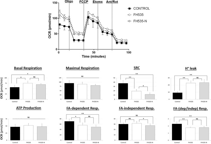
Autophagy targets cellular components for lysosomal-dependent degradation in which the products of degradation may be recycled for protein synthesis and utilized for energy production. Autophagy also plays a critical role in cell homeostasis and the regulation of many physiological and pathological processes and prompts this investigation of new agents to effect abnormal autophagy in hepatocellular carcinoma (HCC). 2,5-Dichloro-N-(2-methyl-4-nitrophenyl) benzenesulfonamide (FH535) is a synthetic inhibitor of the Wnt/β-catenin pathway that exhibits anti-proliferative and anti-angiogenic effects on different types of cancer cells. The combination of FH535 with sorafenib promotes a synergistic inhibition of HCC and liver cancer stem cell proliferation, mediated in part by the simultaneous disruption of mitochondrial respiration and glycolysis. We demonstrated that FH535 decreased HCC tumor progression in a mouse xenograft model. For the first time, we showed the inhibitory effect of an FH535 derivative, FH535-N, alone and in combination with sorafenib on HCC cell proliferation. Our study revealed the contributing effect of Wnt/β-catenin pathway inhibition by FH535 and its derivative (FH535-N) through disruption of the autophagic flux in HCC cells.


Conflict of interest statement

CL and DSW have partial ownership in a private venture, Epionc, Inc., incorporated to develop small-molecule inhibitors for cancer treatment through a licensing agreement with the University of Kentucky. In accord with University of Kentucky policies, CL and DSW disclosed prior work to the University of Kentucky’s Intellectual Property Committee and complied with stipulations of the University’s Conflict of Interest Oversight Committee. Epionc, Inc., has no license for compounds described in this paper. This does not alter our adherence to PLOS ONE policies on sharing data and materials.

Figures

Fig 1
Fig 1. Structure of FH535 (2,5-dichloro-N-(2-methyl-4-nitrophenyl)benzenesulfonamide) and FH535-N (2,5-dichloro-N-(4-nitronaphthalen-1-yl)benzenesulfonamide).
Fig 2
Fig 2. FH535 effect in vivo.
(A) Mice body weight after FH535 treatment. C57BL/6 mice (n = 5) were treated by intraperitoneal injection with 15 mg/Kg of FH535 or DMSO vehicle control every 4 days for 6 weeks. Mice were monitored before injections for signs of body weight loss, impaired mobility, labored breathing and body score based on the Ullman-Culleré MH, Foltz CJ method [27]. (B-D) FH535 reduces tumor growth in vivo in a xenograft tumor model. Huh7 cell were injected subcutaneously on the right flank of athymic nude mice. FH535 (15 mg/Kg) or vehicle (DMSO) were administrated by intraperitoneal injection every other day when tumor size reached 100 mm3. (B) Tumor growth was monitored every other day until day 10 of starting treatments when mice were euthanized according to the AVMA guidelines, *p< 0.05 (n = 5, each group); (C) Tumor weight of excised tumors after 10 day treatment with FH535 reduced the tumor weight in 42 ± 8% compared to vehicle treatment, **p< 0.001 (n = 4, each group). (D) H&E and ki67 staining from one representative tumor of each group treatment. Pictures were taken at 400X magnification. H&E staining showed poorly differentiated carcinoma comprised of sheets of epithelioid cells with increased N/C ratio, enlarged nuclei with prominent nucleoli, high mitotic activity and tumor necrosis (lower right corner of the picture for FH535, and left upper corner and left mid area of the picture for control group). The Ki-67 immunohistochemical staining highlights very high mitotic index with nuclear staining in more than 95% of the viable neoplastic cells for both groups.
Fig 3
Fig 3. FH535 regulates autophagic activity in HCC cells.
Western blot analysis of Huh7 cell after 40 h treatment with FH535 at indicated concentrations in absence (-CQ) or presence (+CQ) of 50 μM chloroquine for 8h. LC3B (A) and p62 (B) were used as autophagy markers for western blot analysis. Band intensity were estimated using ImageJ software. Autophagic flux was determined by subtracting the band intensity of LC3B II western blot in presence of CQ and the corresponding treatment in absence of CQ which is referred as ΔLC3II (LC3II (+CQ)—LC3II (-CQ)) (A, right panel). mRNA p62 expression levels were assessed by RT-qPCR in absence of CQ (C).
Fig 4
Fig 4. FH535 regulates autophagic flux in Huh7 cells.
Autophagic activity of Huh7 cells after 40 h FH535 treatment in absence (-CQ) or presence (+CQ) of 50 μM CQ (8h) was determined by flow cytometry analysis using the Cyto-ID autophagy detection reagent. Results are shown as GeoMean ± SD from viables cells (Zombie negative population). Autophagic flux was determined by the difference in GeoMean between cells treated with CQ and corresponding treatment in absence of CQ also referred as ΔGeoMean (GeoMean (+CQ)—GeoMean (-CQ)) (right panel). *: p ≤ 0.05.
Fig 5
Fig 5. Effect of FH535-N on HCC cells proliferation.
Cell proliferation was measured on Huh7, PLC/PRF/5 and Hep3B cells using 3H-thymidine incorporation after 72 h treatment with FH535 or FH535-N alone or in combination with sorafenib at the concentrations indicated. Results are represented as mean ± SD, n = 4. *: p ≤ 0.05, **p ≤ 0.001.
Fig 6
Fig 6. Effect FH535-N on inhibition of Wnt/β-catenin pathway.
(A) Effect of FH535-N on TOPFlash activation. Huh7 cells were co-transfected with Top-Flash and phRL-TK plasmid. After 5 h of transfection, cells were treated with vehicle, FH535 or FH535-N at the concentration indicated in presence of 10 mM LiCl. Vehicle in absence of LiCl was used as control for basal levels of Wnt/β-catenin activity. Results are represented as mean ± SD, n = 3. #: p ≤ 0.05, *: p ≤ 0.001. (B-C). Effect of FH535-N on expression of β-catenin targets. Protein expression levels of downstream β-catenin targets from Huh7 cells treated with FH535-N for 36 h were determined by western blot analysis (B, left pannel). Densitometry analysis was performed using ImageJ software (B, right pannel). mRNA expression of downstream β-catenin targets from Huh7 cells treated with FH535-N for 36 h were determined by RT-qPCR (D).
Fig 7
Fig 7. Effect of FH535-N alone or in combination with sorafenib on apoptosis of HCC cells.
Analysis of apoptosis by Annexin V-APC/propidium iodide (PI) double staining of HuH7 and PLC/PRF/5 cells after 48 h treatment at the concentration of FH535, FH535-N and sorafenib indicated. (A) Two-color flow cytometry dot plots show the percentages of living cells as negative for both annexin V and PI; early-stage apoptotic cells as the populations testing Annexin V positive and PI negative, and late-stage apoptotic/necrotic cells as double-positive cells. Results are represented in (B) as mean ± SD, n = 3. *: p ≤ 0.05, **: p ≤ 0.001.
Fig 8
Fig 8. Mitochondrial respiration changes induced after 24 h-treatment of Huh7 cells with FH535, FH535-N alone or in combination with sorafenib.
Representative OCR profiles of Huh7 cells shown as percentage change with respect to the OCR levels after addition of the ATP-synthase inhibitor Oligomycin (O). The parameters of ATP turnover, proton leak, and spare respiratory capacity were calculated as area under the curve (AUC) values as described in Materials and Methods section. Data are shown as mean ± SEM, n = 6–8.*: p < 0.05 and **: p<0.001. Statistical comparisons were performed using one-way ANOVA and Dunnett’s multiple comparisons test and pairwise comparisons with Student’s t test. (NS = non-significant; p≥0.05).
Fig 9
Fig 9. FH535-N regulates autophagic activity in HCC cells.
Western blot analysis of Huh7 cell after 40 h treatment with FH535-N at indicated concentrations in absence (-CQ) or presence (+CQ) of 50 μM chloroquine for 8h. (A) Protein expression levels of LC3BII. Autophagic flux was determined by subtracting the band intensity of LC3B II western blot in presence of CQ and the corresponding treatment in absence of CQ which is referred as ΔLC3II (LC3II (+CQ)—LC3II (-CQ)). (B) Protein expression levels of p62 in absence of CQ. Band intensity from Western blots were estimated using ImageJ software.
Fig 10
Fig 10. FH535-N regulates autophagic flux in Huh7 cells.
Autophagic activity of Huh7 cells after 40 h FH535 treatment in absence (-CQ) or presence (+CQ) of 50 μM CQ (8h) was determined by flow cytometry analysis using the Cyto-ID autophagy detection reagent. Results are shown as GeoMean ± SD from viables cells (Zombie negative population). Autophagic flux was determined by the difference in GeoMean between cells treated with CQ and corresponding treatment in absence of CQ also referred as ΔGeoMean (GeoMean (+CQ)—GeoMean (-CQ)) (right panel). *: p ≤ 0.05.
Fig 11
Fig 11. FH535-N regulates autophagic flux in Huh7 and PLC/PRF/5 cells.
Autophagic activity of Huh7 cells after 40 h FH535 treatment in absence (-CQ) or presence (+CQ) of 50 μM CQ (8h) was determined by flow cytometry analysis using the Cyto-ID autophagy detection reagent. Results are shown as GeoMean ± SD from viables cells (Zombie negative population). Autophagic flux was determined by the difference in GeoMean between cells treated with CQ and corresponding treatment in absence of CQ also referred as ΔGeoMean (GeoMean (+CQ)—GeoMean (-CQ)) (right panel).*: p < 0.05 and **: p<0.001. (NS = non-significant; p≥0.05).

References

    1. Mittal S, El-Serag HB. Epidemiology of hepatocellular carcinoma: consider the population. Journal of clinical gastroenterology. 2013;47 Suppl:S2–6. Epub 2013/05/02. 10.1097/MCG.0b013e3182872f29 - DOI - PMC - PubMed
    1. Society AC. Facts & Figures 2018 Atlanta, Ga.2018 [cited 2018 Sept 9]. Available from: https://www.cancer.org/cancer/liver-cancer/about/what-is-key-statistics.....
    1. Altekruse SF, McGlynn KA, Reichman ME. Hepatocellular carcinoma incidence, mortality, and survival trends in the United States from 1975 to 2005. Journal of clinical oncology: official journal of the American Society of Clinical Oncology. 2009;27(9):1485–91. Epub 2009/02/20. 10.1200/jco.2008.20.7753 - DOI - PMC - PubMed
    1. Dimitroulis D, Damaskos C, Valsami S, Davakis S, Garmpis N, Spartalis E, et al. From diagnosis to treatment of hepatocellular carcinoma: An epidemic problem for both developed and developing world. World journal of gastroenterology: WJG. 2017;23(29):5282–94. Epub 2017/08/26. 10.3748/wjg.v23.i29.5282 - DOI - PMC - PubMed
    1. Lee HC, Kim M, Wands JR. Wnt/Frizzled signaling in hepatocellular carcinoma. Frontiers in bioscience: a journal and virtual library. 2006;11:1901–15. Epub 2005/12/22. . - PubMed

Publication types

MeSH terms