. 2019 Feb 22;14(2):e0212589.
doi: 10.1371/journal.pone.0212589. eCollection 2019.

Stimulated hepatic stellate cell promotes progression of hepatocellular carcinoma due to protein kinase R activation

Affiliations

Stimulated hepatic stellate cell promotes progression of hepatocellular carcinoma due to protein kinase R activation

Yusuke Imai et al. PLoS One. .

Abstract

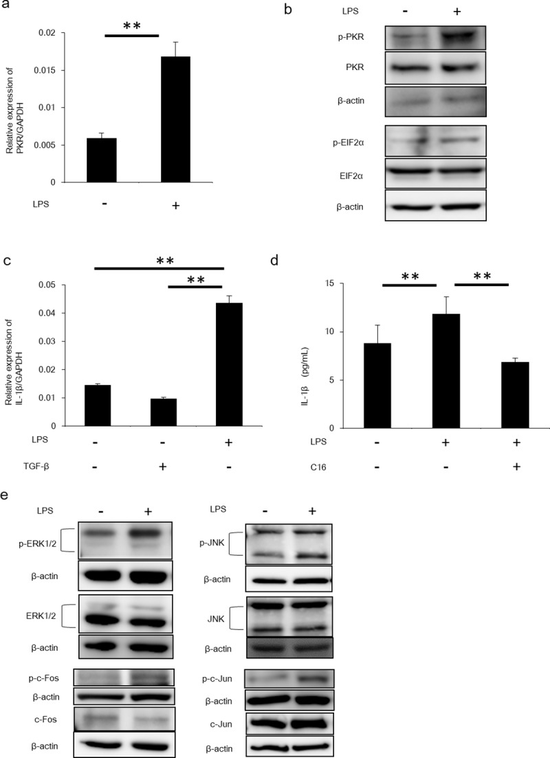
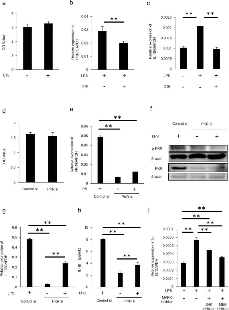
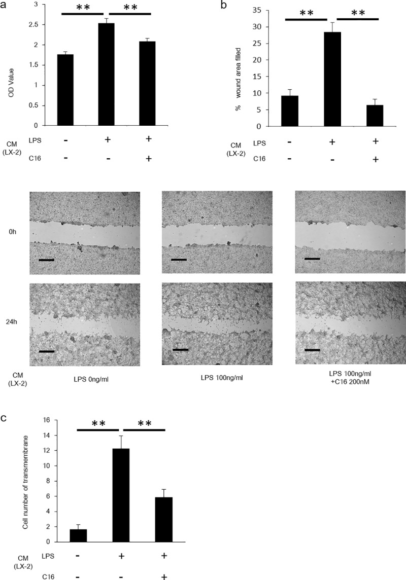
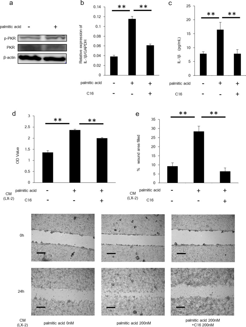
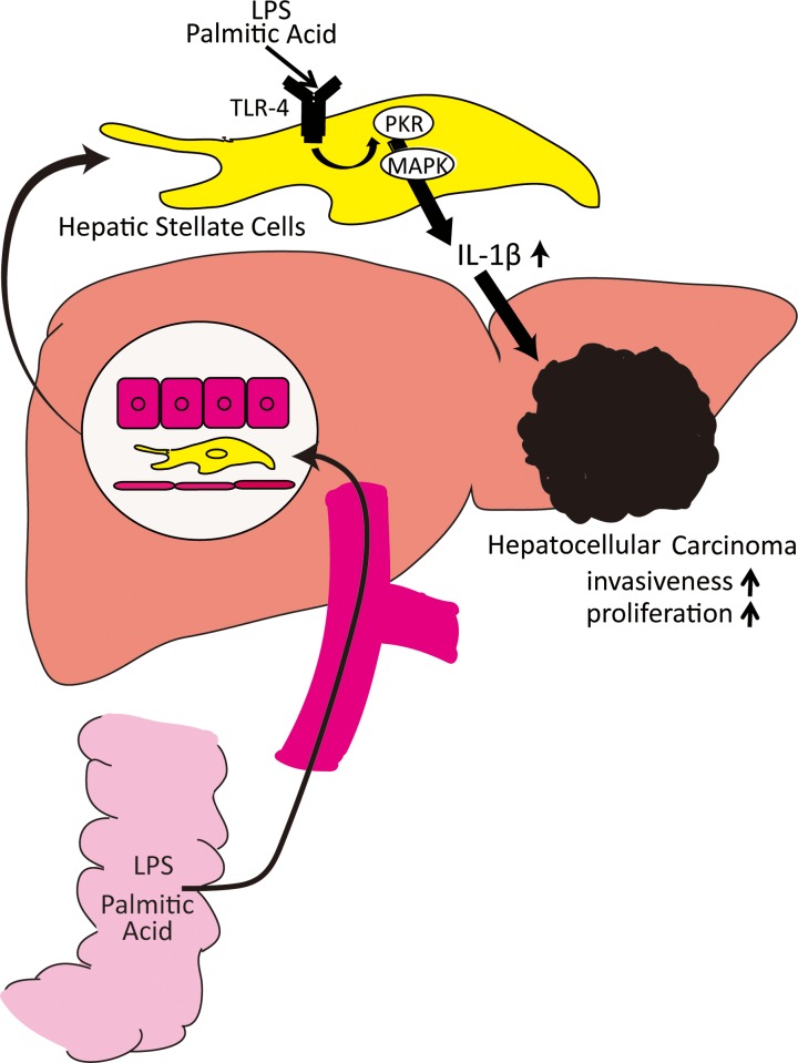
Hepatic stellate cells (HSCs) were reported to promote the progression of hepatocellular carcinoma (HCC), however its mechanism is uncertain. We previously reported that protein kinase R (PKR) in hepatocytes regulated HCC proliferation. In this study, we focused on the role of PKR in HSCs, and clarified the mechanism of its association with HCC progression. We confirmed the activation of PKR in a human HSC cell line (LX-2 cell). IL-1β is produced from HSCs stimulated by lipopolysaccharide (LPS) or palmitic acid which are likely activators of PKR in non-alcoholic steatohepatitis (NASH). Production was assessed by real-time PCR and ELISA. C16 and small interfering RNA (siRNA) were used to inhibit PKR in HSCs. The HCC cell line (HepG2 cell) was cultured with HSC conditioning medium to assess HCC progression, which was evaluated by proliferation and scratch assays. Expression of PKR was increased and activated in stimulated HSCs, and IL-1β production was also increased molecular. Key molecules of the mitogen-activated protein kinase pathway were also upregulated and activated by LPS. Otherwise, PKR inhibition by C16 and PKR siRNA decreased IL-1β production. HCC progression was promoted by HSC-stimulated conditioning medium although it was reduced by the conditioning medium from PKR-inhibited HSCs. Moreover, palmitic acid also upregulated IL-1β expression in HSCs, and conditioning medium from palmitic acid-stimulated HSCs promoted HCC proliferation. Stimulated HSCs by activators of PKR in NASH could play a role in promoting HCC progression through the production of IL-1β, via a mechanism that seems to be dependent on PKR activation.


Conflict of interest statement

The authors have declared that no competing interests exist.

Figures

Fig 1
Fig 1. LPS promoted IL-1β production from HSCs via PKR signalling.
(A) PKR mRNA in HSCs stimulated by LPS was measured by RT-PCR. Mean ± SEM of four replicates. **P<0.05. (B) Protein expression of PKR and EIF2α and each phosphorylated form was determined by Western blotting. (C) LX-2 cells were stimulated by LPS or TGF-β, following which mRNA levels of IL-1β were quantified by RT-PCR. Mean ± SEM of four replicates. **P<0.05. (D) The amount of IL-1β in LX-2 cells stimulated by LPS and inhibition by C16 was measured by ELISA. Mean ± SEM of three replicates. **P<0.05. (E) Upregulation of PKR mRNA by LPS stimulation was measured by RT-PCR. Mean ± SEM of four replicates. **P<0.05. Protein expression of JNK, ERK1/2, c-Jun and c-Fos and each phosphorylated form were determined by Western blotting.
Fig 2
Fig 2
IL-1β was downregulated by inhibition of PKR. (A) Cytotoxicity assay was performed for assessing cell viability of C16-treated LX-2 cells. Mean ± SEM of eight replicates. **P<0.05. (B) Following LX-2 cells stimulated by LPS and inhibition by C16, PKR mRNA was quantified by RT-PCR. Mean ± SEM of four replicates. **P<0.05. (C) IL-1β mRNA in LX-2 cell stimulation by LPS and inhibition by C16 was measured by RT-PCR. Mean ± SEM of four replicates. **P<0.05. (D) Cytotoxicity assay was performed for assessing cell viability of siRNA-treated LX-2 cells. Mean ± SEM of eight replicates. **P<0.05. (E) LX-2 cells were transfected with PKR siRNA or control siRNA and stimulated by LPS. PKR mRNA was measured by RT-PCR. Mean ± SEM of three replicates. **P<0.05. (F) Protein expression of phosphorylated forms of PKR were determined by Western blotting. (G) IL-1β mRNA in LX-2 transfected with PKR siRNA was measured by RT-PCR. Mean ± SEM of three replicates. **P<0.05. (H) The amount of IL-1β in LX-2 transfected with PKR siRNA was measured by ELISA. Mean ± SEM of three replicates. **P<0.05. (I) Following LX-2 cell stimulation by LPS and inhibition by JNK or MEK inhibitor, IL-1β mRNA was quantified by RT-PCR. Upregulation of PKR mRNA was inhibited by MAPK inhibitors. Mean ± SEM of four replicates. **P<0.05.
Fig 3
Fig 3. LPS-stimulated HSCs increased proliferation and invasiveness of HepG2 cells.
(A) Proliferation assay was performed for assessing proliferation of HepG2 cells incubated by conditioning medium from LPS-stimulated or C16-treated LX-2 cells. Mean ± SEM of eight replicates. **P<0.05. (B) Scratch assay was performed for assessing proliferation of HepG2 cells incubated by conditioning medium from LPS-stimulated or C16-treated LX-2 cells. Scale bars represent 500 μm. Mean ± SEM of four replicates. **P<0.05. (C) Invasion assay was performed for assessing invasiveness of HepG2 was performed for assessing proliferation of HepG2 cells incubated by conditioning medium from LPS-stimulated or C16-treated LX-2 cells. Mean ± SEM of four replicates. **P<0.05.
Fig 4
Fig 4. Palmitic acid-stimulated HSCs demonstrated increased IL-1β expression and enhanced the proliferation of HepG2 cells.
(A) Protein expression of PKR and its phosphorylated form was determined by Western blotting. (B) Following stimulation of the LX-2 cells by palmitic acid, IL-1β mRNA levels were quantified by RT-PCR. Mean ± SEM of four replicates. **P<0.05. (C) The amount of IL-1β in LX-2 cells was measured by ELISA. Mean ± SEM of three replicates. **P<0.05. (D) Proliferation assay was performed for assessing proliferation of HepG2 cells incubated by conditioning medium from palmitic acid-stimulated or C16-treated LX-2 cells. Mean ± SEM of six replicates. **P<0.05. (E) Scratch assay was performed for assessing proliferation of HepG2 cells incubated by conditioning medium from palmitic acid-stimulated or C16-treated LX-2 cells. Scale bars represent 500 μm. Mean ± SEM of four replicates. **P<0.05.
Fig 5
Fig 5. Proposed model of interplay between hepatic stellate cells and hepatocellular carcinoma.
In nonalcoholic steatohepatitis cases, LPS and palmitic acid from the intestine stimulate HSCs and upregulate PKR expression. Upregulation of PKR increases IL-1β production via MAPK pathways. The secreted IL-1β enhances the progression of hepatocellular carcinoma.

Similar articles

Cited by

References

    1. Balogh J, Victor D 3rd, Asham EH, Burroughs SG, Boktour M, Saharia A, et al. Hepatocellular carcinoma: a review. J Hepatocell Carcinoma. 2016;3:41–53. 10.2147/JHC.S61146 - DOI - PMC - PubMed
    1. Vernon G, Baranova A, Younossi ZM. Systematic review: the epidemiology and natural history of non-alcoholic fatty liver disease and non-alcoholic steatohepatitis in adults. Aliment Pharmacol Ther. 2011;34:274–85. 10.1111/j.1365-2036.2011.04724.x - DOI - PubMed
    1. Tateishi R, Okanoue T, Fujiwara N, Okita K, Omata M, Kumada H, et al. Clinical characteristics, treatment, and prognosis of non-B, non-C hepatocellular carcinoma: a large retrospective multicenter cohort study. J Gastroenterol. 2015;50:350–360. 10.1007/s00535-014-0973-8 - DOI - PMC - PubMed
    1. Saraswat VA, Pandey G, Shetty S. Treatment algorithms for managing hepatocellular carcinoma. J Clin Exp Hepatol. 2014;4:S80–89. 10.1016/j.jceh.2014.05.004 - DOI - PMC - PubMed
    1. Siegel AB, Olsen SK, Magun A, Brown RS. Sorafenib: where do we go from here? Hepatology. 2010;52:360–369. 10.1002/hep.23633 - DOI - PMC - PubMed

Publication types

MeSH terms