. 2019 Feb 22;14(2):e0212540.
doi: 10.1371/journal.pone.0212540. eCollection 2019.

Identification and characterization of the first pectin methylesterase gene discovered in the root lesion nematode Pratylenchus penetrans

Affiliations

Identification and characterization of the first pectin methylesterase gene discovered in the root lesion nematode Pratylenchus penetrans

Cláudia S L Vicente et al. PLoS One. .

Erratum in

Abstract

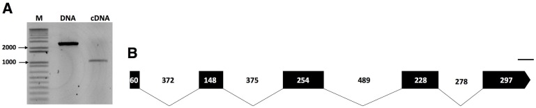
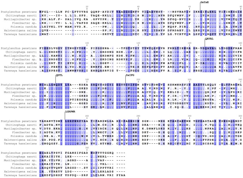
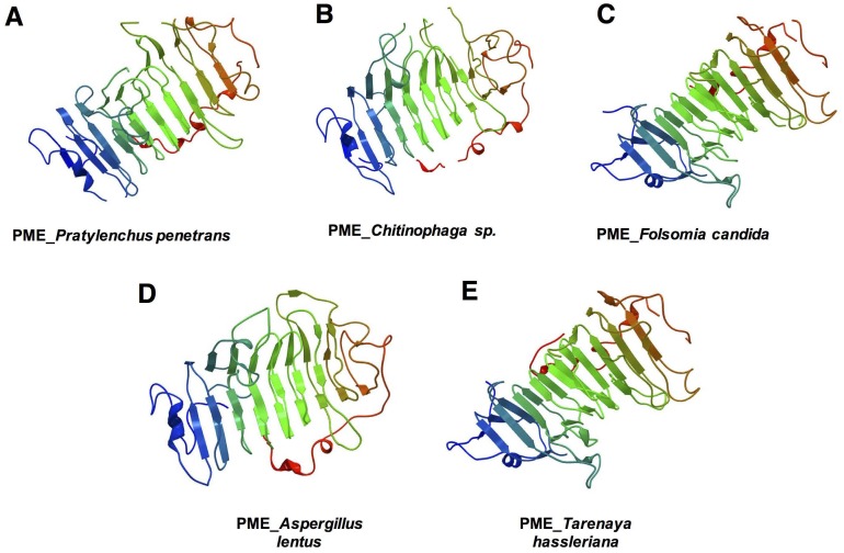
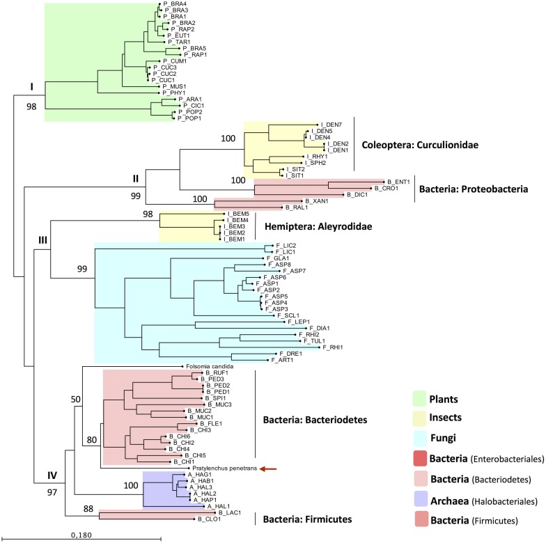
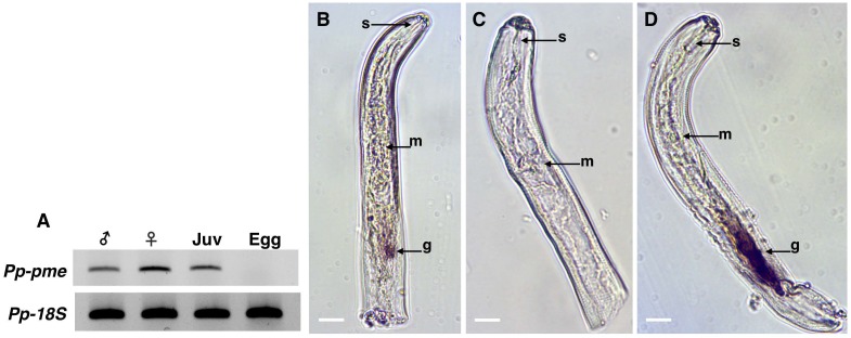
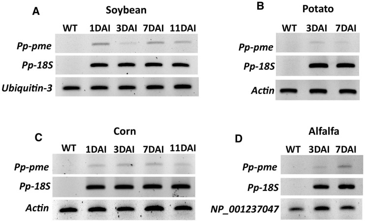
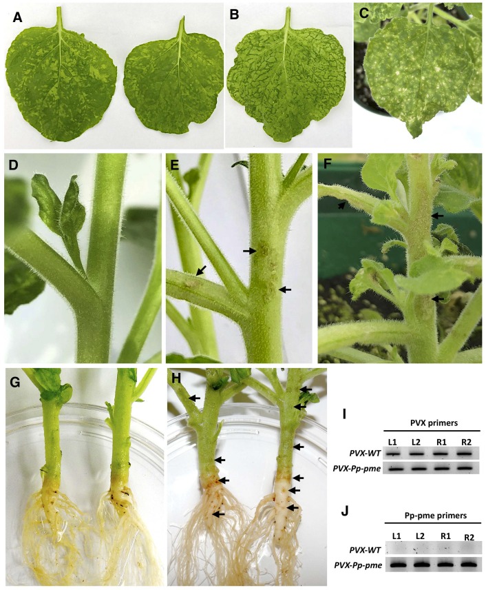
Similar to other plant-parasitic nematodes, root lesion nematodes possess an array of enzymes that are involved in the degradation of the plant cell wall. Here we report the identification of a gene encoding a cell wall-degrading enzyme, pectin methylesterase PME (EC 3.1.1.11), in the root lesion nematode Pratylenchus penetrans. Both genomic and coding sequences of the gene were cloned for this species, that included the presence of four introns which eliminated a possible contamination from bacteria. Expression of the Pp-pme gene was localized in the esophageal glands of P. penetrans as determined by in situ hybridization. Temporal expression of Pp-pme in planta was validated at early time points of infection. The possible function and activity of the gene were assessed by transient expression of Pp-pme in plants of Nicotiana benthamiana plants via a Potato virus X-based vector. To our knowledge, this is the first report on identification and characterization of a PME gene within the phylum Nematoda.


Conflict of interest statement

The authors have declared that no competing interests exist.

Figures

Fig 1
Fig 1. Molecular characterization of the pectin methylesterase (Pp-pme) of Pratylenchus penetrans.
(A) Amplicons of both genomic (2,501 bp) and cDNA coding (987 bp) sequences of Pp-pme, respectively. (B) Schematic representation of the Pp-pme gene structure. Relative positions and respective sizes of the exons are indicated by dark boxes and introns by lines.
Fig 2
Fig 2. Multiple sequence alignment of the predicted Pp-PME protein of Pratylenchus penetrans with PMEs of other organisms.
Representative species of bacteria (Chitinophaga sancti, Mucilaginibacter sp., Flexibacter sp.), collembolan (Folsomia candida), Archaea (Haloterrigena salina) and plants (Tarenaya hassleriana) were used. The five conserved sequence segments typical of PME proteins are numbered according to their positions in the corresponding Pp-PME predicted protein (64_GxYxE, 146_QAVAL, 168_QDTL, 195_DxIFG, and 251_LGRPW). Conserved residues among species are indicated by dark blue shading and dots, whereas similar residues are represented in light blue using a threshold for shading of 50% similarity. The accession numbers corresponding to each species are presented in S3 Table.
Fig 3
Fig 3. Three-dimensional model predicted for the pectin methylesterase of different organisms.
(A) Pratylenchus penetrans, (B) bacteria (Chitinophaga sp.), (C) collembolan (Folsomia candida), (D) fungi (Aspergillus lentus), and (E) plants (Tarenaya hassleriana). The models of P. penetrans and Chitinophaga sp. were based on the three-dimensional model of the Erwinia chrysanthemi (Phyre2 fold library ID: d1gq8a), F. candida and T. hassleriana were based on the model of Daucus carota (Phyre2 fold library ID: d1qjva), and A. lentus on the model of A. niger (Phyre2 fold library ID: c5c1cA), respectively. The N-terminal is indicated in blue and the C-terminal is shown in red.
Fig 4
Fig 4. Phylogenetic tree based on the closest homology to the catalytic domain of Pp-PME.
The PME sequences across different taxa were chosen based on the top BLAST hits against the Pp-PME predicted pectinesterase domain. The corresponding species names and range of e-values are presented in S3 Table. The red arrow indicates the position of P. penetrans PME. The phylogenetic tree was deduced by Maximum Likelihood with the “Whelan and Goldman” (WAG) model with discrete gamma distribution and 1000 bootstrap replicates.
Fig 5
Fig 5. Expression and localization of Pratylenchus penetrans Pp-pme transcripts.
(A) Determination of Pp-pme expression in different nematode developmental stages of P. penetrans by semi-quantitative RT-PCR. As a positive control, all cDNA templates were amplified with primers derived from the 18S gene of P. penetrans. The nematode stages were separated as males, females, juveniles (J2-J4), and eggs. (B-C) Detection of the Pp-pme transcripts by in situ hybridization. Nematode sections were hybridized with antisense (B), or sense (C) Pp-pme digoxigenin-labeled cDNA probes. (D) As a positive control, in situ hybridization was performed with the antisense probe designed for the CWDE (Pp-eng-1) specifically localized within the esophageal glands g: esophageal glands; m: metacorpus; s: stylet.
Fig 6
Fig 6. Semi-quantitative RT-PCR showing the transcript levels of Pp-pme in different host plants at different time points after infection.
Total RNA extracted from nematode infected roots of economically important host plants (A) soybean, (B) potato, (C) corn, and (D) alfalfa was used to validate the relative expression of Pp-pme at different days after nematode infection (DAI). The nematode 18S rDNA gene was used as internal control to validate the presence of P. penetrans within the infected roots, while specific plant reference genes were used for each specific host plant. Wild-type (WT) correspond to non-infected plants.
Fig 7
Fig 7. Phenotypic changes in Nicotiana benthamiana plants infected with the recombinant PVX-Pp-pme virus.
All photos were taken 14 days after inoculation. (A) characteristic mosaic-like symptom developed in N. benthamiana plants infected with transcripts generated from the empty PVX vector. (B-C) Distinct leaf chlorosis symptoms (B) and lesion-like spots (C) developed in N. benthamiana plants infected with PVX-Pp-pme transcripts, respectively. (D) Stem and branches of the N. benthamiana plant infected with transcripts generated from the empty PVX vector displayed no symptoms. (E-F) Lesion-like symptoms observed on stems and branches of N. benthamiana plants infected with PVX-Pp-pme transcripts. (G) Stem and roots of N. benthamiana plant infected with empty PVX vector displayed no symptoms. (H) Browning in the area of the stem-root joint and lesion-like spots observed on the roots of N. benthamiana plants infected with PVX-Pp-pme transcripts. (I-J) Detection of PVX-WT and PVX-Pp-pme transcripts in inoculated N. benthamiana plants by semi-quantitative RT-PCR. L1, L2: leaves from two independently-inoculated plants; R1, R2: roots from two independently-inoculated plants.

Similar articles

Cited by

References

    1. Cosgrove DJ. Growth of the plant cell wall. Nat Rev Mol Cell Biol. 2005;6: 850–861. 10.1038/nrm1746 - DOI - PubMed
    1. Keegstra K. Plant Cell Walls. Plant Physiol. 2010;154: 483–486. 10.1104/pp.110.161240 - DOI - PMC - PubMed
    1. Sørensen I, Domozych D, Willats WGT. How have plant cell walls evolved? Plant Physiol. 2010;153: 366–372 10.1104/pp.110.154427 - DOI - PMC - PubMed
    1. Jordan DB, Bowman MJ, Braker JD, Dien BS, Hector RE, Lee CC, et al. Plant cell walls to ethanol. Biochem J. 2012;442: 241–252. 10.1042/BJ20111922 - DOI - PubMed
    1. Mohnen D. Pectin structure and biosynthesis. Curr Opin Plant Biol. 2008;11: 266–277. 10.1016/j.pbi.2008.03.006 - DOI - PubMed

Publication types

MeSH terms