Comparative Study
. 2019 Feb 22;14(2):e0212800.
doi: 10.1371/journal.pone.0212800. eCollection 2019.

Comparative purification and characterization of hepatitis B virus-like particles produced by recombinant vaccinia viruses in human hepatoma cells and human primary hepatocytes

Affiliations
Comparative Study

Comparative purification and characterization of hepatitis B virus-like particles produced by recombinant vaccinia viruses in human hepatoma cells and human primary hepatocytes

Edith Reuschel et al. PLoS One. .

Abstract

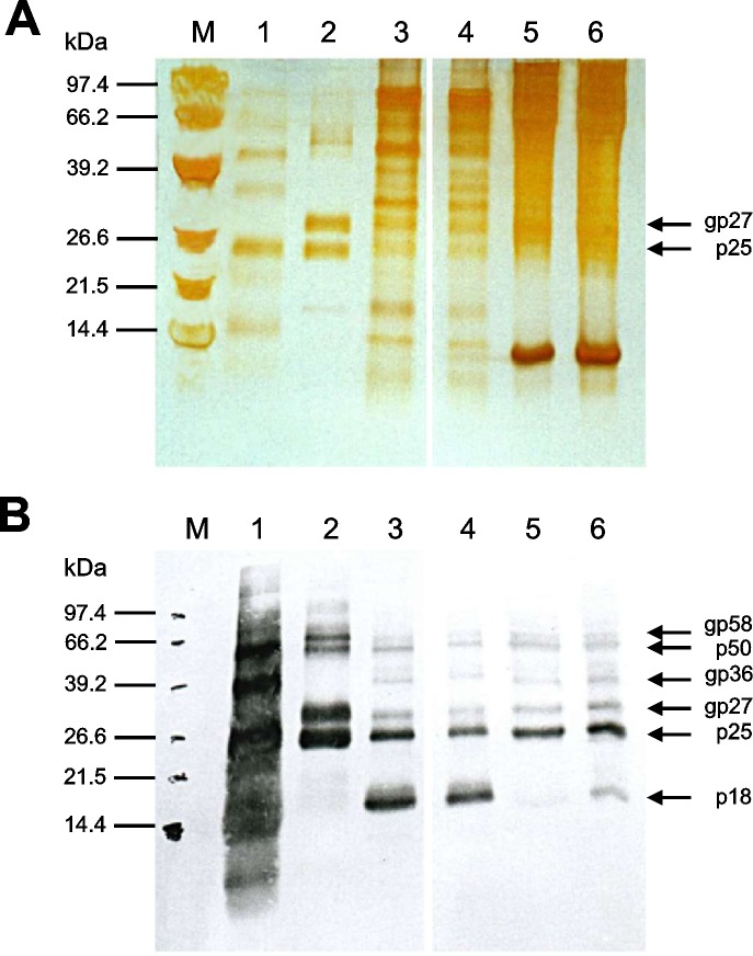
This study describes the comparative expression and purification of hepatitis B surface antigen (HBsAg) particles produced upon infection of human primary hepatocytes and human hepatoma cell lines (HuH-7 and HepG2) with recombinant vaccinia viruses. The highest levels of HBsAg expression were found in HuH-7 hepatoma cells following infection with recombinant vaccinia viruses, which contain the S gene under control of a 7.5 k-promoter. Four different methods for purification of the HBsAg particles were examined: isopycnic ultracentrifugation, sucrose cushion sedimentation, isocratic column gel filtration, and binding to anti-HBs-coated microparticles. The highest degree of purity of HBsAg particles was reached by the method based on anti-HBs-coated microparticles. The resulting product was >98% pure. Biochemical analysis and characterization of purified HBsAg particles were performed by sodium dodecyl sulfate polyacrylamide gel electrophoresis (SDS-PAGE), western blotting, and electron microscopy. The HBsAg, purified from human hepatoma cell lines and from human primary hepatocytes, consisted of both the non-glycosylated (p25) and the glycosylated (gp27) form and assembled into typical 22-nm particles, and thus may be of great interest and importance for research, diagnostics, and medical treatments.


Conflict of interest statement

The authors have declared that no competing interests exist.

Figures

Fig 1
Fig 1. Expression of HBsAg particles by recombinant vaccinia viruses in different cell lines.
(A) The hepatoma cell line HuH-7 was infected with various M.O.I. [multiplicity of infection] of HBsAg-recombinant vaccinia viruses. The expression level of HBsAg was measured from the cell culture supernatant (S/N-value) by an IMx microparticle enzyme immunoassay at indicated time points: 24, 48, 72, and 96 h after infection. (B) HuH-7 hepatoma cells, primary hepatocytes, and HepG2 were comparatively infected with recombinant vaccinia viruses at the indicated M.O.I´s. Four days after infection, cells were harvested, and the expression level of HBsAg was measured in the supernatant by an IMx microparticle enzyme immunoassay.
Fig 2
Fig 2. Long-term HBsAg-expression by human primary hepatocytes following infection with recombinant vaccinia viruses.
Human primary liver cells were isolated by EGTA/collagenase-perfusion and cultivated between 2 collagen layers. After infection with HBsAg-recombinant vaccinia viruses, the HBsAg concentration was determined from the supernatant over a period of more than 40 days using an IMx MEIA test.
Fig 3
Fig 3. Purification of HBsAg particles by isopycnic kalium bromide (KBr) gradient centrifugation.
(A) Cell culture supernatants of primary hepatocytes, which were infected with recombinant vaccinia viruses, were pre-cleared, and proteins were concentrated by trichloroacetic acid (TCA) precipitation prior to separation by isopycnic ultracentrifugation with KBr. Eleven fractions were collected, and the HBsAg in each fraction was detected by an IMx microparticle enzyme immunoassay. (B) Peak fraction was loaded on SDS-PAGE and immunostained with HBsAg-specific goat polyclonal antibodies: M: Premixed protein low range molecular weight standard marker (Boehringer); Lane 1: KBr gradient peak fraction 8. Sizes of reactive proteins are indicated in kilodaltons (KDa).
Fig 4
Fig 4. Isocratic column gel filtration.
(A) HepG3 protein separation was performed by preparative gel filtration on a Superdex 200 prep grade column. Flow rate was 0.5 ml at a total volume of 120 ml. Flow buffer was PBS (pH 7.5). Sixty fractions of 3 ml each were collected, and the total protein concentration was determined by spectrophotometry at a wavelength of 280 nm. (B) The HBsAg content in collected fractions was determined by a commercial IMx microparticle enzyme immunoassay. (C) Silver-stained (SDS-PAGE) and (D) western blot analysis of selected fractions: Lane 1: Positive control (microparticle purified HBsAg). Lane 2: Fraction 12 as negative control. Lane 3: Fraction 17; Lane 4: Fraction 23; Lane 5: Fraction 25. The highest concentration of HBsAg was found in fraction 23 (Lane 4).
Fig 5
Fig 5. Comparison between HBsAg purified by anti-HBs-coated microparticles and sucrose-cushion purified HBsAg from differently cultured hepatoma-cell-lines.
Constitutively HBsAg expressing HepG3-cells were cultured in serum-free and serum-supplemented medium. After 5 days of cultivation HBsAg was concentrated, subjected to 15% SDS-PAGE and analyzed by silver staining (A) or immunoblotting with HBsAg-specific antibodies (B). M: Molecular weight standard; Lane 1: Commercially available human plasma reactive for HBsAg as positive control (Abbott IMx HBsAg Control No. 2228–10); Lane 2: HBsAg purified from recombinant vaccinia virus-infected HuH-7 cells by anti-HBs-coated microparticles (Abbott IMx HBsAg (V2) Reagent Pack No. 2228–21). Lane 3 and 4: Two fractions of HBsAg expressed by HepG3-cells under serum-free conditions. Lane 5 and 6: Two fractions of HBsAg expressed by HepG3-cells in serum-supplemented media. Protein samples were loaded on one gel. Two non-relevant lanes were cut out of the silver-stain (A) and Western-blot (B) images, as indicated. The positions of anti-HBsAg antibody reactive proteins are depicted on the right.
Fig 6
Fig 6. Electron microscopy of microparticle-purified HBsAg particles derived from recombinant vaccinia virus-infected primary hepatocytes.
HBsAg particles (22 nm) were negatively stained with 2% aqueous phosphotungstic acid (pH 7.3) and examined by electron microscopy.

Similar articles

Cited by

References

    1. Mormile R. Hepatitis B vaccine non response: A predictor of latent autoimmunity? Med Hypotheses. 2017;104: 45–47. 10.1016/j.mehy.2017.05.020 - DOI - PubMed
    1. Hadiji-Abbes N, Mihoubi W, Martin M, Karakasyan-Dia C, Frikha F, Gergely C, et al. Characterization of C69R variant HBsAg: effect on binding to anti-HBs and the structure of virus-like particles. Arch Virol. 2015;160: 2427–2433. 10.1007/s00705-015-2515-y - DOI - PubMed
    1. World Health Organization, World Health Organization, Global Hepatitis Programme. Global hepatitis report, 2017 [Internet]. 2017. Available: http://apps.who.int/iris/bitstream/10665/255016/1/9789241565455-eng.pdf?...
    1. Stasi C, Silvestri C, Voller F. Emerging Trends in Epidemiology of Hepatitis B Virus Infection. J Clin Transl Hepatol. 2017;5: 272–276. 10.14218/JCTH.2017.00010 - DOI - PMC - PubMed
    1. Shepard CW, Simard EP, Finelli L, Fiore AE, Bell BP. Hepatitis B virus infection: epidemiology and vaccination. Epidemiol Rev. 2006;28: 112–125. 10.1093/epirev/mxj009 - DOI - PubMed

Publication types

MeSH terms

Substances