

Comment
doi: 10.1016/j.cell.2019.01.053.
A Watershed Finding for Heart Regeneration
Affiliations
- PMID: 30794778
- PMCID: PMC8240015
- DOI: 10.1016/j.cell.2019.01.053
Item in Clipboard
Comment
A Watershed Finding for Heart Regeneration
Cell.
.
Abstract
The adult mammalian heart is minimally regenerative after injury, whereas neonatal hearts fully recover even after major damage. New work from the Red-Horse and Woo labs (Das et al., 2019) shows that collateral artery formation is a key mechanism contributing to successful regeneration in newborn mice and provides insights into how collateral arteries form.
Copyright © 2019 Elsevier Inc. All rights reserved.
Figures
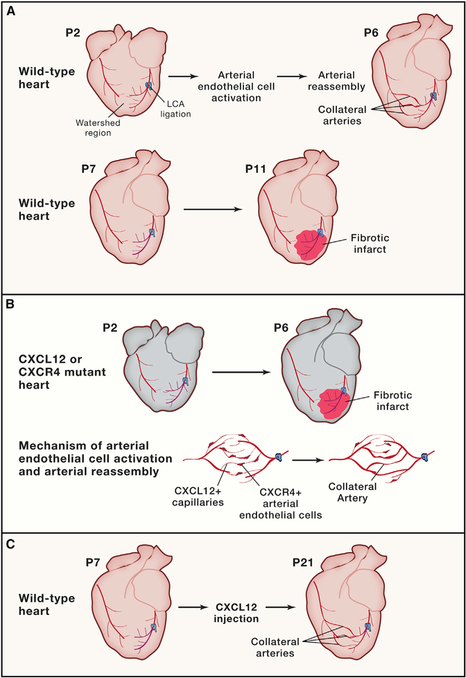
(A) Neonatal (P2) hearts subjected to left coronary artery ligation are able to restore blood flow to the injured region through the formation of collateral arteries by arterial reassembly. After the regenerative window (P7) hearts subjected to left coronary artery (LCA) ligation do not form collateral arteries. (B) Neonatal hearts lacking Cxcr4 in the arterial endothelial cells or Cxcl12 in the capillary bed are unable to recruit arterial endothelial cells (AECs) for arterial reassembly and form fibrotic scars after LCA ligation. Collateral artery reassembly is triggered by CXCL12-expressing capillaries, which activate and recruit CXCR4-expressing AECs to migrate, proliferate, and form new collateral arteries. (C)The injection of a high dose of CXCL12 rescues the formation of collateral arteries in P7 hearts.
Comment on
-
A Unique Collateral Artery Development Program Promotes Neonatal Heart Regeneration.Cell. 2019 Feb 21;176(5):1128-1142.e18. doi: 10.1016/j.cell.2018.12.023. Epub 2019 Jan 24. Cell. 2019. PMID: 30686582 Free PMC article.
References
-
- Habib GB, Heibig J, Forman SA, Brown BG, Roberts R, Terrin ML, and Bolli R; The TIMI Investigators (1991). Influence of coronary collateral vessels on myocardial infarct size in humans. Results of phase I thrombolysis in myocardial infarction (TIMI) trial. Circulation 83, 739–746. - PubMed
Publication types
MeSH terms
Grants and funding
LinkOut - more resources
Full Text Sources
