. 2019 May;189(5):999-1014.
doi: 10.1016/j.ajpath.2019.01.014. Epub 2019 Feb 19.

Genetic Strain and Sex Differences in a Hyperoxia-Induced Mouse Model of Varying Severity of Bronchopulmonary Dysplasia

Affiliations

Genetic Strain and Sex Differences in a Hyperoxia-Induced Mouse Model of Varying Severity of Bronchopulmonary Dysplasia

Sean Leary et al. Am J Pathol. 2019 May.

Abstract

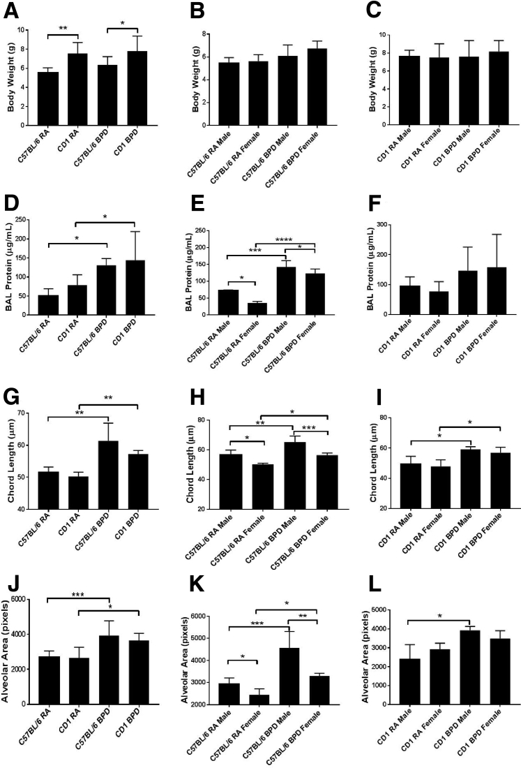
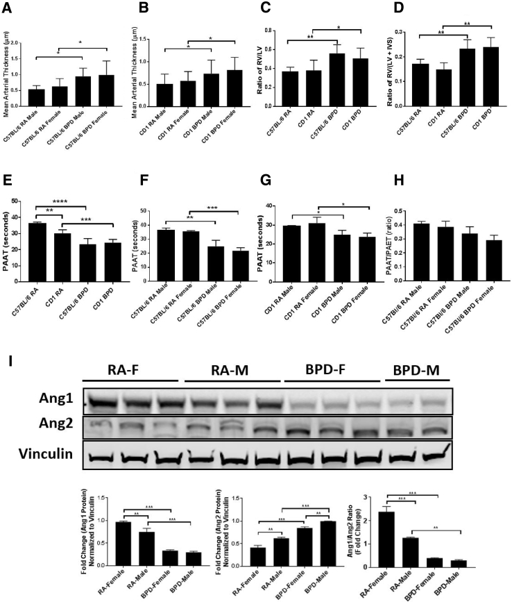
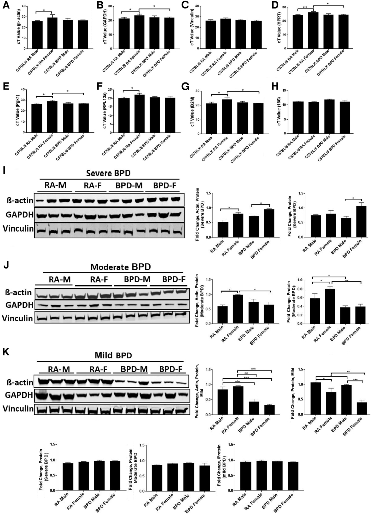
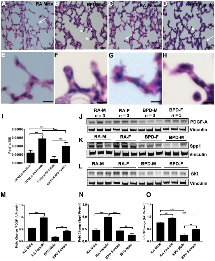
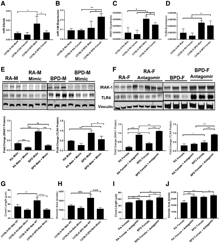
Bronchopulmonary dysplasia (BPD) is a disease prevalent in preterm babies with a need for supplemental oxygen, resulting in impaired lung development and dysregulated vascularization. Epidemiologic studies have shown that males are more prone to BPD and have a delayed recovery compared with females, for reasons unknown. Herein, we tried to recapitulate mild, moderate, and severe BPD, using two different strains of mice, in males and females: CD1 (outbred) and C57BL/6 (inbred). Aside from higher body weight in the CD1 strain, there were no other gross morphologic differences with respect to alveolar development between the two strains. With respect to lung morphology after oxygen exposure, females had less injury with better preservation of alveolar chord length and decreased alveolar protein leak and inflammatory cells in the bronchoalveolar lavage fluid. In addition, housekeeping genes, which are routinely used as loading controls, were expressed differently in males and females. In the BPD mouse model, gonadotropin-releasing hormone was increased in females compared with males. Specific miRNAs (miR-146 and miR-34a) were expressed differently in the sexes. In the severe BPD mouse model, administering miR-146 mimic to males attenuated lung damage, whereas administering miR-146 inhibitor to females increased pulmonary injury.


Figures

Figure 1
Figure 1
Gross difference between males and females in response to hyperoxia in different strains of mice. A: The average body weight of CD1 mice is significantly higher than that of C57BL/6 mice in both room air (RA) and bronchopulmonary dysplasia (BPD) groups. B and C: There is no difference in the body weight between C57BL/6 (B) or CD1 (C) males and females in both RA and BPD groups. D: There is increased bronchoalveolar lavage (BAL) protein in the BPD group of both the strains. E: Males show a significant increase of the protein over females in the C57BL/6 strain in both RA and BPD groups. F: There is no difference between the two sexes on exposure to hyperoxia in the CD1 strain. G: The chord length is increased in the BPD group of both C57BL/6 and CD1 strains compared with RA, with no difference between the strains. H: The males have significantly increased chord length over the females in both RA and BPD groups only in the C57Bl/6 strain. I: In the CD1 strain, there is no difference between the two sexes in RA; the BPD group, however, shows increased chord length over the RA group. J: The alveolar area is increased in the BPD group of both C57BL/6 and CD1 strains compared with RA, with no difference between the strains. K: The males have significantly increased alveolar area over the females in both RA and BPD groups. In BPD males, the alveolar area is significantly higher than in BPD females. In the CD1 strain, the BPD males show increased alveolar area compared with RA males. There is no difference between the males and the females of the BPD group. L: The chord length is increased in the BPD group of both C57BL/6 and CD1 strains compared with RA, with no difference between the two strains. The males have significantly increased chord length over the females in both RA and BPD groups only in the C57BL/6 strain. n = 7 to 8 (AL). P < 0.05, ∗∗P < 0.01, ∗∗∗P < 0.001, and ∗∗∗∗P < 0.0001.
Figure 2
Figure 2
Structural, functional, and hemodynamic changes in the vasculature. A and B: The mean arterial thickness increases in both bronchopulmonary dysplasia (BPD) males (M) and females (F) over room air (RA) controls in both the C57BL/6 (A) and the CD1 (B) strains. C and D: Right ventricular hypertrophy was assessed using right ventricle (RV)/left ventricle (LV) and the Fulton index [RV/LV + interventricular septum (IVS)]. The ratios are higher in the BPD group of the C57BL/6 strain compared with the CD1 strain. E: Both the RA and the BPD groups of the CD1 strain show significantly less pulmonary arterial acceleration time (PAAT) over the C57BL/6 strain. F and G: Both the BPD males and females show decreased PAAT compared with their RA controls in the C57BL/6 (F) and the CD1 (G) strains. H: There is no difference in the PAAT/pulmonary arterial ejection time (PAET) ratio in both males and females of RA and BPD groups of both the strains used in this study. I: Representative image of Western blot analysis, showing expression of angiopoietin 1 (Ang1) and Ang2 in males and females of RA and BPD groups (top panel), along with fold-change quantification (bottom panels). Ang1 is higher in females than males, whereas the reverse is true for Ang2 in the RA group. However, Ang2 is higher in BPD males compared with females, whereas there is no change in Ang1 expression between the males and females in the BPD group. n = 7 to 8 (A–H); n = 3 (I). P < 0.05, ∗∗P < 0.01, ∗∗∗P < 0.001, and ∗∗∗∗P < 0.0001.
Figure 3
Figure 3
Housekeeping genes as loading controls for bronchopulmonary dysplasia (BPD): Real-time quantitative PCR (qPCR; A–H) and Western blot analyses (I–K) with corresponding quantification bar graphs showing the expression of commonly used housekeeping genes in the C57BL/6 strain. A–C: β-Actin (A) and glyceraldehyde-3-phosphate dehydrogenase (GAPDH; B) are significantly increased (1.5-fold) in females (F) compared with room air (RA) males (M) in both the RA and the BPD groups, whereas there is no change in vinculin expression (C). D–G: qPCR also shows differences in the expression of hypoxanthine-guanine phosphoribosyl transferase (HPRT; D), phosphoglycerate kinase 1 (Pgk1; E), ribosomal protein L13a (RPL13a; F), and β-2-microglobulin (B2M; G) in the lungs of both males and females in both RA and BPD groups. H: There is no change in the expression of 18S in the lungs. I–K: Similarly, at the protein level, there are significant differences between β-actin and GAPDH in males and females of RA and BPD groups in all of the BPD experiments [mild (I), moderate (J), and severe (K)], whereas vinculin remains unchanged (I–K). Vinculin was used as a loading control for Western blot analyses in lung tissues, whereas 18S was used as a reference gene for normalization in qPCR assays for lungs. n = 8 (A–H); n = 3 (I–K). P < 0.05, ∗∗P < 0.01, and ∗∗∗P < 0.001. cT, cycle threshold.
Figure 4
Figure 4
Males (M) and females (F) differ in alveolar septation. A–H: Hematoxylin and eosin staining showing differences in secondary septa during lung development in males and females of the C57BL/6 strain. A–H: Room air (RA) males (A and E) show thinner secondary septa than RA females (C and G), whereas bronchopulmonary dysplasia (BPD) males (B and F) show short and thick septa with club-shaped ends (arrowheads), compared with BPD females (D and H) with pointed tips (arrows). E–H: Higher magnification of the septa tips, indicated with arrows and arrowheads in A–D. Alveolar septation in lung alveogenesis is controlled by genes, such as platelet-derived growth factor-A (PDGFA) and secreted phosphoprotein 1 (Spp1). Akt is considered to play a role in overall lung development. I–O: Real-time quantitative PCR showing expression of PGDF-A (I), along with Western blot analyses of PGDF-A (J), Spp1 (K), and Akt (L), with corresponding densitometric quantifications (M–O). PDGF-A, Spp1, and Akt are expressed more in females than in males in both RA and BPD groups. Vinculin is the loading control. n = 8 to 10 (A–D); n = 3 (J–L). ∗∗P < 0.01, ∗∗∗P < 0.001. Scale bars = 20 μm (A–H).
Figure 5
Figure 5
miRNAs (miR-34a and miR-146) are differentially expressed in males (M) and females (F). Real-time quantitative PCR showing expression of miR-34a, miR-146, IL-1 receptor–associated kinase 1 (IRAK1), and toll-like receptor 4 (TLR4). A and B: miR-34a is expressed more in bronchopulmonary dysplasia (BPD) males than in BPD females (A), whereas miR-146 is expressed more in the BPD females than males (B). There is no change in the expression of both miRNAs in room air (RA). IRAK1 is a downstream target of miR-146 and is involved in the inflammation pathway. C and D: IRAK1 and TLR4 are significantly up-regulated in males and females in the BPD group, but the expression of IRAK1 is significantly decreased in females when compared with males in the BPD group. Lung morphometry showing measurement of chord length and alveolar area in both males and females after treatment with miR-146 mimic and inhibitor, respectively, after exposure to hyperoxia. E and F: Both the chord length (E) and the alveolar area (F) are significantly increased in the BPD males compared with RA males, but after treatment with mimic, they are restored to normal. G and H: The females given the antagomir show increased chord lengths and alveolar areas compared with wild type (WT). I and J: Representative images of Western blot analyses, with corresponding densitometric quantification showing expression of IRAK1 and TLR4 after treating the males with miR-146 mimic (I) and the females with antagomir (J). The BPD males show increased expression of IRAK1 and TLR4 compared with RA, but after treatment with miR-146 mimic, the expression is significantly decreased. On the contrary, in females treated with miR-146 antagomir, IRAK1 and TLR4 are significantly up-regulated in both the RA and BPD groups. Vinculin was used as the loading control. n = 5 to 8 (A–F). P < 0.05, ∗∗P < 0.01, ∗∗∗P < 0.001, and ∗∗∗∗P < 0.0001.

Similar articles

Cited by

References

    1. Sigmund C.D. Viewpoint: are studies in genetically altered mice out of control? Arterioscler Thromb Vasc Biol. 2000;20:1425–1429. - PubMed
    1. Schadt E.E., Monks S.A., Drake T.A., Lusis A.J., Che N., Colinayo V., Ruff T.G., Milligan S.B., Lamb J.R., Cavet G., Linsley P.S., Mao M., Stoughton R.B., Friend S.H. Genetics of gene expression surveyed in maize, mouse and man. Nature. 2003;422:297–302. - PubMed
    1. Whitehead G.S., Burch L.H., Berman K.G., Piantadosi C.A., Schwartz D.A. Genetic basis of murine responses to hyperoxia-induced lung injury. Immunogenetics. 2006;58:793–804. - PMC - PubMed
    1. Chia R., Achilli F., Festing M.F.W., Fisher E.M.C. The origins and uses of mouse outbred stocks. Nat Genet. 2005;37:1181–1186. - PubMed
    1. Johnston C.J., Stripp B.R., Piedbeouf B., Wright T.W., Mango G.W., Reed C.K., Finkelstein J.N. Inflammatory and epithelial responses in mouse strains that differ in sensitivity to hyperoxic injury. Exp Lung Res. 1998;24:189–202. - PubMed

Publication types