Nanotube assisted microwave electroporation for single cell pathogen identification and antimicrobial susceptibility testing
- PMID: 30794964
- PMCID: PMC6520151
- DOI: 10.1016/j.nano.2019.01.015
Nanotube assisted microwave electroporation for single cell pathogen identification and antimicrobial susceptibility testing
Abstract
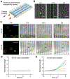
A nanotube assisted microwave electroporation (NAME) technique is demonstrated for delivering molecular biosensors into viable bacteria for multiplex single cell pathogen identification to advance rapid diagnostics in clinical microbiology. Due to the small volume of a bacterial cell (~femtoliter), the intracellular concentration of the target molecule is high, which results in a strong signal for single cell detection without amplification. The NAME procedure can be completed in as little as 30 minutes and can achieve over 90% transformation efficiency. We demonstrate the feasibility of NAME for identifying clinical isolates of bloodborne and uropathogenic pathogens and detecting bacterial pathogens directly from patient's samples. In conjunction with a microfluidic single cell trapping technique, NAME allows single cell pathogen identification and antimicrobial susceptibility testing concurrently. Using this approach, the time for microbiological analysis reduces from days to hours, which will have a significant impact on the clinical management of bacterial infections.
Keywords: Antibiotic resistance; Bacteria; Infection; Microfluidic; Urinary tract infection.
Copyright © 2019 Elsevier Inc. All rights reserved.
Figures


Similar articles
-
Interference Disturbance Analysis Enables Single-Cell Level Growth and Mobility Characterization for Rapid Antimicrobial Susceptibility Testing.Nano Lett. 2019 Feb 13;19(2):643-651. doi: 10.1021/acs.nanolett.8b02815. Epub 2019 Jan 8. Nano Lett. 2019. PMID: 30525694
-
Single cell antimicrobial susceptibility testing by confined microchannels and electrokinetic loading.Anal Chem. 2013 Apr 16;85(8):3971-6. doi: 10.1021/ac4004248. Epub 2013 Feb 27. Anal Chem. 2013. PMID: 23445209 Free PMC article.
-
Microfluidic chip systems for color-based antimicrobial susceptibility test a review.Biosens Bioelectron. 2025 Apr 1;273:117160. doi: 10.1016/j.bios.2025.117160. Epub 2025 Jan 11. Biosens Bioelectron. 2025. PMID: 39827743 Review.
-
Emerging Analytical Techniques for Rapid Pathogen Identification and Susceptibility Testing.Annu Rev Anal Chem (Palo Alto Calif). 2019 Jun 12;12(1):41-67. doi: 10.1146/annurev-anchem-061318-115529. Epub 2019 Apr 2. Annu Rev Anal Chem (Palo Alto Calif). 2019. PMID: 30939033 Free PMC article. Review.
-
Progress on the development of rapid methods for antimicrobial susceptibility testing.J Antimicrob Chemother. 2013 Dec;68(12):2710-7. doi: 10.1093/jac/dkt253. Epub 2013 Jun 30. J Antimicrob Chemother. 2013. PMID: 23818283 Review.
Cited by
-
Current state of the art in rapid diagnostics for antimicrobial resistance.Lab Chip. 2020 Aug 7;20(15):2607-2625. doi: 10.1039/d0lc00034e. Epub 2020 Jul 9. Lab Chip. 2020. PMID: 32644060 Free PMC article. Review.
-
Innovative and rapid antimicrobial susceptibility testing systems.Nat Rev Microbiol. 2020 May;18(5):299-311. doi: 10.1038/s41579-020-0327-x. Epub 2020 Feb 13. Nat Rev Microbiol. 2020. PMID: 32055026 Review.
-
A Rapid Single-Cell Antimicrobial Susceptibility Testing Workflow for Bloodstream Infections.Biosensors (Basel). 2021 Aug 22;11(8):288. doi: 10.3390/bios11080288. Biosensors (Basel). 2021. PMID: 34436090 Free PMC article.
-
Microwaves, a potential treatment for bacteria: A review.Front Microbiol. 2022 Jul 25;13:888266. doi: 10.3389/fmicb.2022.888266. eCollection 2022. Front Microbiol. 2022. PMID: 35958124 Free PMC article. Review.
-
Single-cell pathogen diagnostics for combating antibiotic resistance.Nat Rev Methods Primers. 2023;3:6. doi: 10.1038/s43586-022-00190-y. Epub 2023 Feb 2. Nat Rev Methods Primers. 2023. PMID: 39917628 Free PMC article.
References
-
- Bush K, Courvalin P, Dantas G, Davies J, Eisenstein B, Huovinen P, Jacoby GA, Kishony R, Kreiswirth BN, Kutter E, Lerner SA, Levy S, Lewis K, Lomovskaya O, Miller JH, Mobashery S, Piddock LJ, Projan S, Thomas CM, Tomasz A, Tulkens PM, Walsh TR, Watson JD, Witkowski J, Witte W, Wright G, Yeh P & Zgurskaya HI Tackling antibiotic resistance. Nat Rev Microbiol 9, 894–896 (2011). - PMC - PubMed
Publication types
MeSH terms
Substances
Grants and funding
LinkOut - more resources
Full Text Sources
Medical

