

Usutu Virus Isolated from Rodents in Senegal
- PMID: 30795524
- PMCID: PMC6409855
- DOI: 10.3390/v11020181
Usutu Virus Isolated from Rodents in Senegal
Abstract
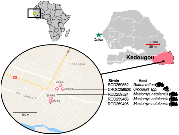
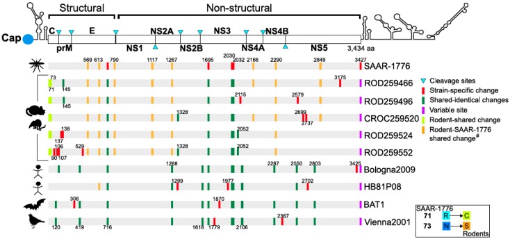
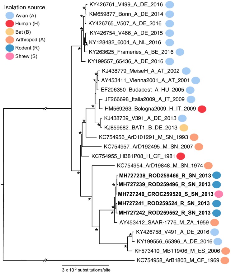
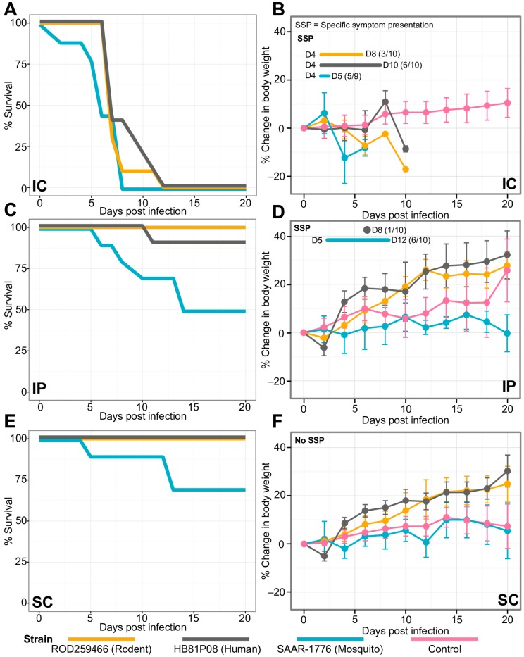
Usutu virus (USUV) is a Culex-associated mosquito-borne flavivirus of the Flaviviridae family. Since its discovery in 1959, the virus has been isolated from birds, arthropods and humans in Europe and Africa. An increasing number of Usutu virus infections in humans with neurological presentations have been reported. Recently, the virus has been detected in bats and horses, which deviates from the currently proposed enzootic cycle of USUV involving several different avian and mosquito species. Despite this increasing number of viral detections in different mammalian hosts, the existence of a non-avian reservoir remains unresolved. In Kedougou, a tropical region in the southeast corner of Senegal, Usutu virus was detected, isolated and sequenced from five asymptomatic small mammals: Two different rodent species and a single species of shrew. Additional molecular characterization and in vivo growth dynamics showed that these rodents/shrew-derived viruses are closely related to the reference strain (accession number: AF013412) and are as pathogenic as other characterized strains associated with neurological invasions in human. This is the first evidence of Usutu virus isolation from rodents or shrews. Our findings emphasize the need to consider a closer monitoring of terrestrial small mammals in future active surveillance, public health, and epidemiological efforts in response to USUV in both Africa and Europe.
Keywords: Senegal; Usutu virus; arbovirus; in vivo experiment; rodents.
Conflict of interest statement
The authors declare no conflicts of interest.
Figures
References
-
- Woodall J.P. The viruses isolated from arthropods at the East African Virus Research Institute in the 26 years ending December 1963. Proc. E Afr. Acad. 1964;2:141–146.
-
- Saiz J.C., Blazquez A.B. Usutu virus: Current knowledge and future perspectives. Virus Adapt. Treat. 2017;9:27–40. doi: 10.2147/VAAT.S123619. - DOI
-
- Cornet M., Robin Y., Chateau R., Heme G., Adam C., Valade M., Gonidec G. Isolement d’arbovirus au Sénégal Oriental a partir de moustiques (1972–1977) et notes sur l’épidémiologie des virus transmis par les Aedes en particulier du virus amaril. Cahiers ORSTOM Série Entomologie Médicale et Parasitologie. 1979;17:149–163.
Publication types
MeSH terms
Substances
Supplementary concepts
LinkOut - more resources
Full Text Sources
