Review
. 2019 Feb 21;12(4):654.
doi: 10.3390/ma12040654.

Salivary Exosomes as Nanocarriers for Cancer Biomarker Delivery

Affiliations
Review

Salivary Exosomes as Nanocarriers for Cancer Biomarker Delivery

Jordan Cheng et al. Materials (Basel). .

Abstract

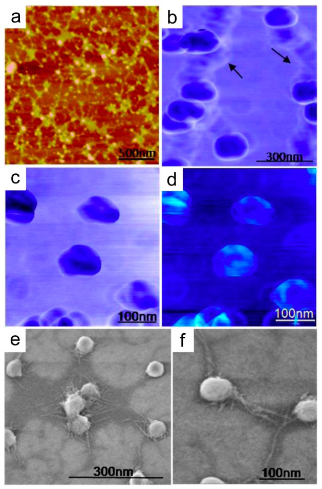
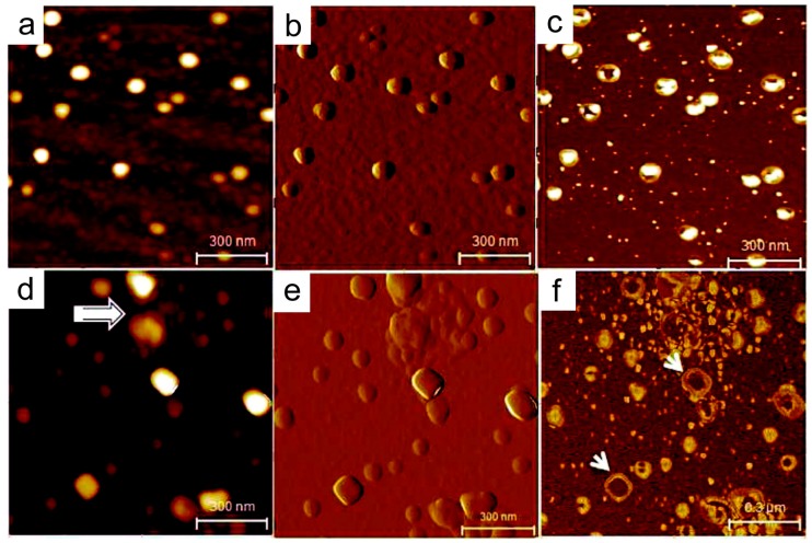
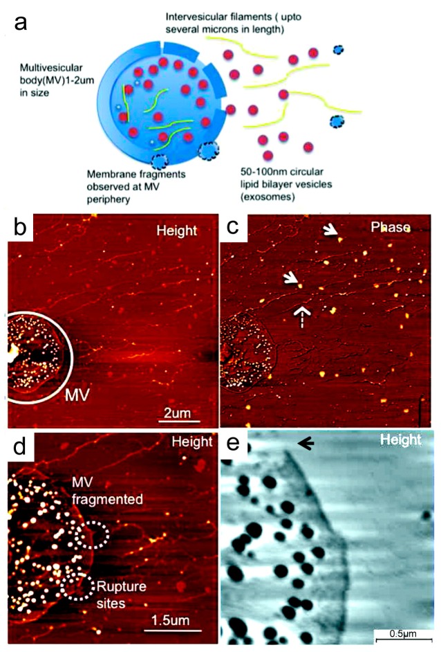
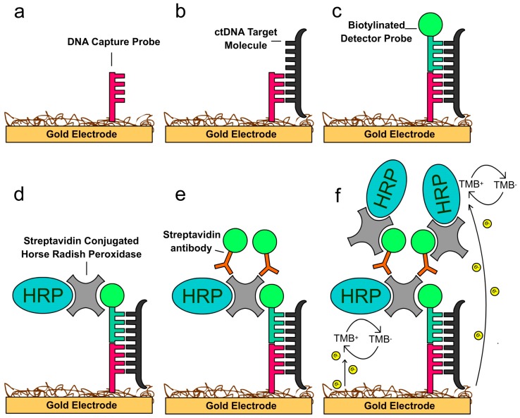
Human saliva is an ideal body fluid for developing non-invasive diagnostics. Saliva contains naturally-occurring nanoparticles with unique structural and biochemical characteristics. The salivary exosome, a nanoscale extracellular vesicle, has been identified as a highly informative nanovesicle with clinically-relevant information. Salivary exosomes have brought forth a pathway and mechanism by which cancer-derived biomarkers can be shuttled through the systemic circulation into the oral cavity. Despite such clinical potential, routine and reliable analyses of exosomes remain challenging due to their small sizes. Characterization of individual exosome nanostructures provides critical data for understanding their pathophysiological condition and diagnostic potential. In this review, we summarize a current array of discovered salivary biomarkers and nanostructural properties of salivary exosomes associated with specific cancers. In addition, we describe a novel electrochemical sensing technology, EFIRM (electric field-induced release and measurement), that advances saliva liquid biopsy, covering the current landscape of point-of-care saliva testing.

Keywords: biomarker; cancer; liquid biopsy; point-of-care; saliva-exosomics; salivaomics; salivary diagnostics.


Conflict of interest statement

D.T.W.W. is the co-founder of RNAmeTRIX Inc., a molecular diagnostic company. D.T.W.W. holds equity in RNAmeTRIX and serves as a company Director and Scientific Advisor. The University of California also holds equity in RNAmeTRIX. Intellectual property that D.T.W.W. invented and that was patented by the University of California has been licensed to RNAmeTRIX. D.T.W.W. is a consultant to GlaxoSmithKline, PeriRx, Wrigley, and Colgate-Palmolive.

Figures

Figure 1
Figure 1
Nanostructure of individual salivary exosomes observed under tapping mode AFM and FESEM. (a) Tapping mode topographic low-force AFM image showing the round morphology of isolated exosomes. (b) AFM phase image of aggregated exosomes. Interconnections (arrows) lacking the characteristic phase shift probably indicate some extravesicular protein content. (c) At higher forces under AFM (~2 nN), representative single-exosome phase images reveal trilobed substructure within the center of the vesicles. The contrast in images may be presumably attributed to variable constitutive elements (lipid, protein, RNA ratio) making up these structures. (d) Corresponding height images show a central depression of the vesicles. (e) FESEM exosome image showing clumping exosomes and (f) single isolated vesicles as round bulging structures with well-resolved intervesicular connections. Reprinted with permission from [106]. Copyright 2010, American Chemical Society.
Figure 2
Figure 2
Structural characteristics of the human salivary exosome at the single-vesicle level. (ac) AFM topographic (z = 0–10 nm), amplitude, and phase images of salivary exosomes from healthy donors. The exosomes appear as homogeneous circular structures with a distinct phase contrast between the less dense periphery and the denser core region. (df) Exosomes from an oral cancer patient show an irregular morphology with varying shapes and vesicle aggregation (arrows). (e) The amplitude image shows the clumping of vesicles. (f) In the phase image, the larger vesicles appear hollow (arrows) without the dense core region typically seen in normal exosomes. All images were obtained over mica substrates under ambient conditions. Reprinted with permission from [107]. Copyright 2011, American Chemical Society.
Figure 3
Figure 3
Release of exosomes from multivesicular bodies (MVs) seen in the saliva of an oral cancer patient. (a) Schematic of single MV membrane rupture and exosome release along with intervesicular filaments from MV lumen. (b) AFM topographic and phase image of a single MV filled with exosome vesicles. (c) Elongated intervesicular filaments (dashed arrow) and exosome-like vesicles (arrows) are observed. (d) At high resolution, the ruptures and fragmentation of the MV membrane are clearly observed (dashed circles). Additionally, the intervesicular filaments are seen in the MV lumen. (e) At higher resolution, a large rupture is seen in the MV membrane (arrow). Samples were imaged under ambient conditions. Reprinted with permission from [107]. Copyright 2011, American Chemical Society.
Figure 4
Figure 4
Schematic of the EFIRM assay procedure. (a) An electrical field is applied to polymerize pyrrole in order to anchor a single-stranded oligonucleotide capture probe specific for a ctDNA onto a gold electrode. (b) The saliva containing ctDNA target molecules is added and hybridizes with the capture probe in the presence of a cyclical square wave. (c) A complementary biotinylated single-stranded oligonucleotide detector probe hybridizes with the target under an electric field. (d) HRP (horseradish peroxidase)-streptavidin binds to biotin on the detector probe. (e,f) A subsequent layer of biotinylated anti-streptavidin antibody and HRP-streptavidin amplifies the signal. The 3,3’,5,5’-tetramethylbenzidine (TMB) substrate is added to generate a continuous, quantifiable electric current through a reduction reaction with HRP.
Figure 5
Figure 5
Schematic of the integrated magnetic-electrochemical electric field-induced release and measurement (EFIRM). (a) Magnetic beads conjugated with an exosome surface marker CD63 antibody capture salivary exosome. (b) Magnetic field capture of exosomes and electric field release of RNA.
Figure 6
Figure 6
EFIRM can disrupt exosomes to release exosomal GAPDH mRNA from human saliva. (a) Schematic illustration of an exosome disrupted with an electric field (E-field) and GADPH mRNA released. (b) Transmission electron microscopy (TEM) images before (i and iii) and after (ii and iv); E-field (top) or Triton X-100 detergent (bottom) treatment. (i and iii) show exosomes (arrows) attached to anti-CD63 antibody-conjugated magnetic beads; (ii and iv) indicate the absence of exosomes disrupted after treatment with (ii) a CSW E-field for 200 s or (iv) with Triton X-100 for 20 min. Background webbing indicates lacey support film for TEM. (c) The levels of GAPDH mRNAs were measured at different time points by EFIRM after the application of E-field or Triton X-100. The kinetics of the GAPDH mRNA signal reduction by EFIRM (top) or Triton X-100 (bottom) indicates that bare RNAs (released by E-field or Triton X-100) decay rapidly.

Similar articles

Cited by

References

    1. Gerlinger M., Rowan A.J., Horswell S., Math M., Larkin J., Endesfelder D., Gronroos E., Martinez P., Matthews N., Stewart A., et al. Intratumor heterogeneity and branched evolution revealed by multiregion sequencing. N. Engl. J. Med. 2012;366:883–892. doi: 10.1056/NEJMoa1113205. - DOI - PMC - PubMed
    1. Brindle K. New approaches for imaging tumour responses to treatment. Nat. Rev. Cancer. 2008;8:94–107. doi: 10.1038/nrc2289. - DOI - PubMed
    1. Condeelis J., Weissleder R. In vivo imaging in cancer. Cold Spring Harb. Perspect. Biol. 2010;2:a003848. doi: 10.1101/cshperspect.a003848. - DOI - PMC - PubMed
    1. Weissleder R., Pittet M.J. Imaging in the era of molecular oncology. Nature. 2008;452:580–589. doi: 10.1038/nature06917. - DOI - PMC - PubMed
    1. Nonaka T., Wong D.T.W. Liquid Biopsy in Head and Neck Cancer: Promises and Challenges. J. Dent. Res. 2018;97:701–708. doi: 10.1177/0022034518762071. - DOI - PMC - PubMed