. 2019 Feb 21;8(2):191.
doi: 10.3390/cells8020191.

The MEK-ERK-MST1 Axis Potentiates the Activation of the Extrinsic Apoptotic Pathway during GDC-0941 Treatment in Jurkat T Cells

Affiliations

The MEK-ERK-MST1 Axis Potentiates the Activation of the Extrinsic Apoptotic Pathway during GDC-0941 Treatment in Jurkat T Cells

Jana Nováková et al. Cells. .

Abstract

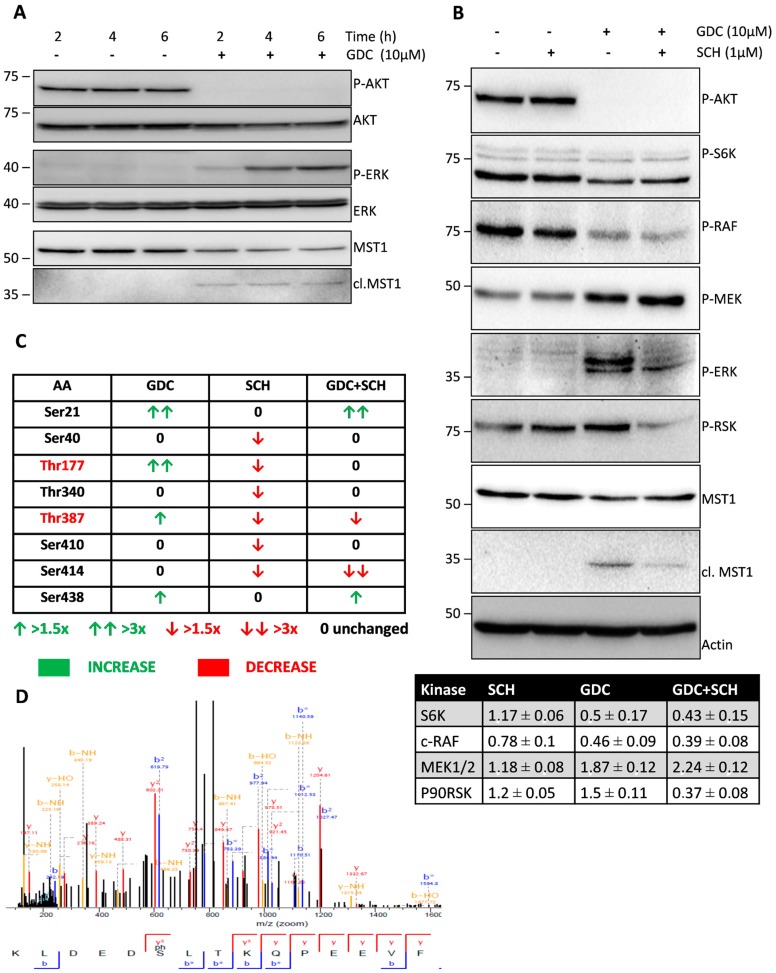
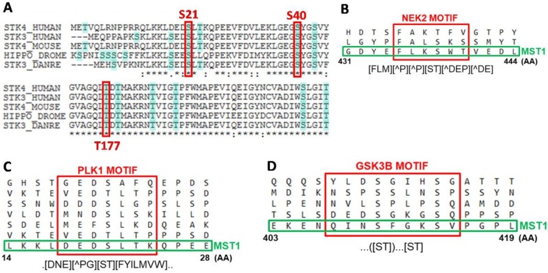
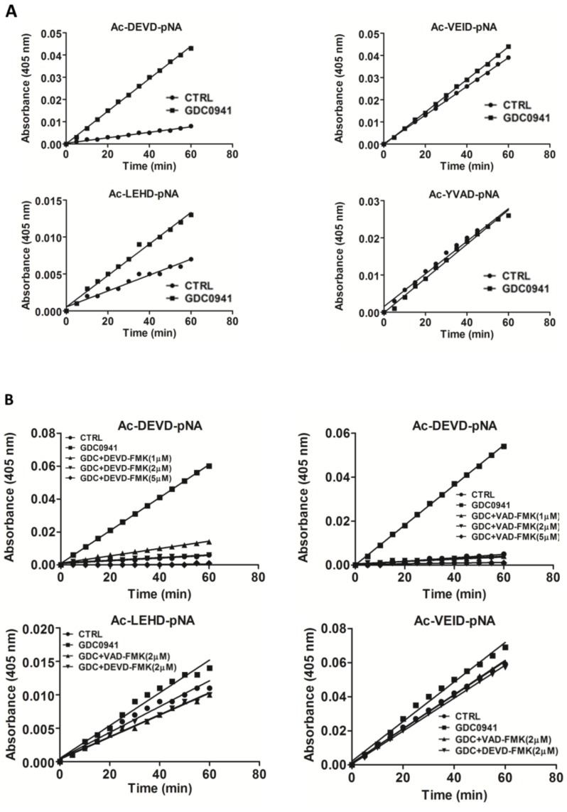
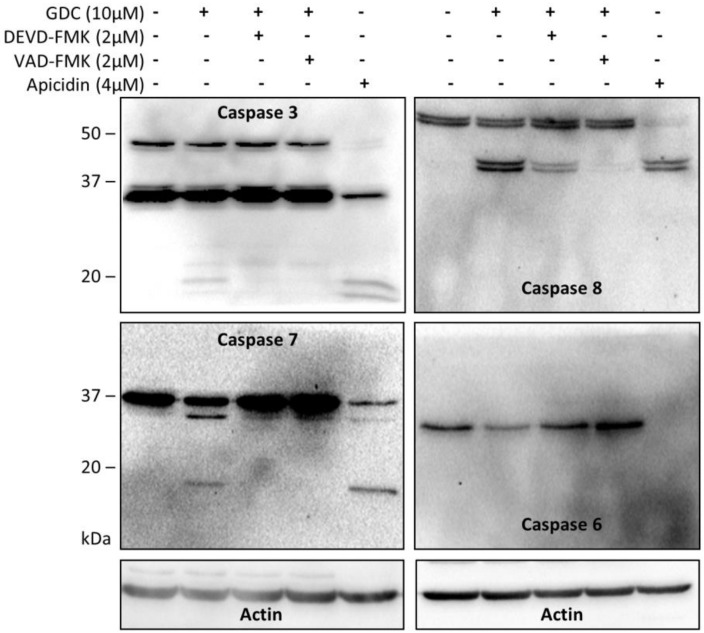
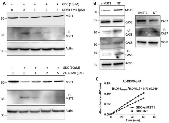
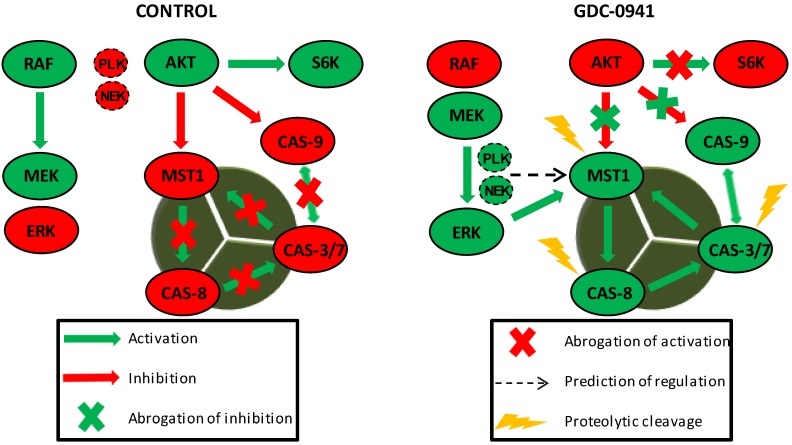
The discrete activation of individual caspases is essential during T-cell development, activation, and apoptosis. Humans carrying nonfunctional caspase-8 and caspase-8 conditional knockout mice exhibit several defects in the progression of naive CD4⁺ T cells to the effector stage. MST1, a key kinase of the Hippo signaling pathway, is often presented as a substrate of caspases, and its cleavage by caspases potentiates its activity. Several studies have focused on the involvement of MST1 in caspase activation and also reported several defects in the immune system function caused by MST1 deficiency. Here, we show the rapid activation of the MEK-ERK-MST1 axis together with the cleavage and activation of caspase-3, -6, -7, -8, and -9 after PI3K signaling blockade by the selective inhibitor GDC-0941 in Jurkat T cells. We determined the phosphorylation pattern of MST1 using a phosphoproteomic approach and identified two amino acid residues phosphorylated in an ERK-dependent manner after GDC-0941 treatment together with a novel phosphorylation site at S21 residue, which was extensively phosphorylated in an ERK-independent manner during PI3K signaling blockade. Using caspase inhibitors and the inhibition of MST1 expression using siRNA, we identified an exclusive role of the MEK-ERK-MST1 axis in the activation of initiator caspase-8, which in turn activates executive caspase-3/-7 that finally potentiate MST1 proteolytic cleavage. This mechanism forms a positive feed-back loop that amplifies the activation of MST1 together with apoptotic response in Jurkat T cells during PI3K inhibition. Altogether, we propose a novel MEK-ERK-MST1-CASP8-CASP3/7 apoptotic pathway in Jurkat T cells and believe that the regulation of this pathway can open novel possibilities in systemic and cancer therapies.

Keywords: AKT; ERK; Hippo/MST1; MEK; PI3K; apoptosis; caspase.


Conflict of interest statement

The authors declare no potential conflicts of interest.

Figures

Figure 1
Figure 1
Effect of GDC-0941, SCH-772984, and their combination on the status of selected kinases. (A) Cells were treated with GDC-0941 for the time periods shown. Levels of phosphorylated and total kinases were determined by immunoblotting. (B) Cells were treated with GDC-0941, SCH-772984, and their combination for 2 h. Effect on the phosphorylation levels of individual kinases and MST1 cleavage was detected by immunoblotting. Actin was used as a loading control. Relative changes in the phosphorylation levels of selected kinases normalized to control are shown in the table. Mean of three independent experiments ± SD. (C) Phosphorylation at individual AA residues of MST1 was quantified by LC–MS/MS analysis. Relative changes in the phosphorylation levels against control cells are demonstrated by arrows. Residues phosphorylated in an ERK1/2-dependent manner are shown in red color. (D) The MS/MS spectrum of a novel phosphorylation site of MST1 at S21 residue.
Figure 2
Figure 2
Bioinformatic analysis of MST1 protein residues identified to be phosphorylated. (A) Conservation of positions of S21, S40, and T177 residues in STK3/STK4 sequences of humans, mice, flies, and fish. (BD) Sequences of motifs recognized by individual kinases around S438, S21 and S414 residues according to the ELM resource. Common motifs are shown in the syntax of Regular Expression Parser and Visualizer (https://github.com/CJex/regulex). (AA) symbol represents AA constrained in the motif, (AA) symbol represents AA excluded from the motif and dot symbol represents random AA.
Figure 3
Figure 3
Effect of selected protease inhibitors on the activity of individual caspases during GDC-0941 treatment. (A) Proteolytic activities in cell lysates toward individual substrates were determined 2 h after GDC-0941 treatment. (B) Effect of pre-treatment of cells with selected concentrations of the Z-DEVD-FMK and Z-VAD-FMK protease inhibitors on the proteolytic activity in cell lysates. Activity was determined 2 h after GDC-0941 treatment.
Figure 4
Figure 4
Effects of selected protease inhibitors on the proteolytic activation of individual caspases during GDC-0941 treatment. The proteolytic activation of caspases in control cells, cells treated with GDC-0941 for 2 h, and cells pre-treated with the Z-DEVD-FMK and Z-VAD-FMK protease inhibitors before GDC-0941 treatment was detected by immunoblotting. Cells treated with apicidin for 24 h were used as a positive control.
Figure 5
Figure 5
Effect of protease inhibitors on the proteolytic activation of MST1 and regulation of selected caspases by MST1 levels after GDC-0941 treatment. (A) The proteolytic activation of MST1 in non-treated cells and cells pre-treated with selected concentrations of Z-DEVD-FMK and Z-VAD-FMK was detected by immunoblotting after 2 h of GDC-0941 treatment. (B) The activation of caspases in cells transfected with siRNAs against MST1 (siMST1) and non-target (NT) siRNAs. The activation of individual caspases was detected by immunoblotting after 2 h of GDC-0941 treatment. (C) Proteolytic activity against the Ac-DEVD-pNA substrate in cells transfected with siMST1 and NT siRNAs. Activity was determined in cell lysates after 2 h of GDC-0941 treatment. Slope ratio was determined in three independent experiments.
Figure 6
Figure 6
MST1 acts as a regulator of the extrinsic apoptotic pathway in Jurkat T cells. PI3K signaling blockade by GDC-0941 resulted in the abrogation of AKT kinase phosphorylation at S473 and significantly attenuated the phosphorylation of P70S6K and c-RAF kinase. This led to a paradoxical increase in the phosphorylation of MEK1/2 at S217/S221 and ERK1/2 at T202/Y204 followed by the activation of MEK-ERK signaling. Active ERK1/2 signaling stimulated the proteolytic activation of MST1 which was insensitive to the experimentally determined concentration of the Z-DEVD-FMK inhibitor. At selected concentrations, Z-DEVD-FMK effectively blocked the activity of caspase-3, -7 and -9 but was insufficient to block the proteolytic activation of caspase-8, suggesting the potential activation of MST1 by caspase-8. On the other hand, the inhibition of MST1 levels by siRNA significantly reduced the proteolytic activation of caspase-3, -7 and -8, rendering MST1 a potential regulator of caspase-8 activity in Jurkat T cells. Moreover, activity of caspase-3/-7 potentiates the proteolytic activation of MST1, hence we presume the existence of a positive feed-back loop orchestrated by MST1 to amplify the apoptotic signal in Jurkat T cells. PLK1 and NEK2 were predicted as potential regulators of MST1 activity during PI3K signaling blockade. Cleavage of caspase-9 was not determined in this study.

Similar articles

Cited by

References

    1. Nagata S., Tanaka M. Programmed cell death and the immune system. Nat. Rev. Immunol. 2017;17:333–340. doi: 10.1038/nri.2016.153. - DOI - PubMed
    1. Sharon A., Finkelstein A., Shlezinger N., Hatam I. Fungal apoptosis: Function, genes and gene function. FEMS Microbiol. Rev. 2009;33:833–854. doi: 10.1111/j.1574-6976.2009.00180.x. - DOI - PubMed
    1. Li J., Yuan J. Caspases in apoptosis and beyond. Oncogene. 2008;27:6194–6206. doi: 10.1038/onc.2008.297. - DOI - PubMed
    1. Pfeffer C.M., Singh A.T.K. Apoptosis: A Target for Anticancer Therapy. Int. J. Mol. Sci. 2018;19:448. doi: 10.3390/ijms19020448. - DOI - PMC - PubMed
    1. Salvesen G.S., Walsh C.M. Functions of caspase 8: The identified and the mysterious. Semin. Immunol. 2014;26:246–252. doi: 10.1016/j.smim.2014.03.005. - DOI - PMC - PubMed

Publication types

MeSH terms

Substances

LinkOut - more resources