

Next Generation Sequencing in AML-On the Way to Becoming a New Standard for Treatment Initiation and/or Modulation?
- PMID: 30795628
- PMCID: PMC6406956
- DOI: 10.3390/cancers11020252
Next Generation Sequencing in AML-On the Way to Becoming a New Standard for Treatment Initiation and/or Modulation?
Abstract
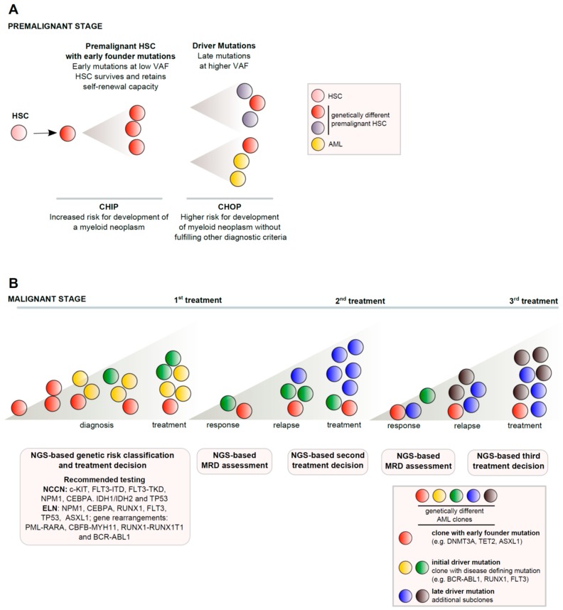
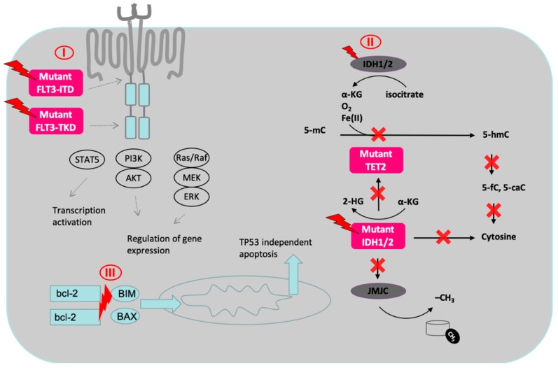
Acute myeloid leukemia (AML) is a clonal disease caused by genetic abberations occurring predominantly in the elderly. Next generation sequencing (NGS) analysis has led to a deeper genetic understanding of the pathogenesis and the role of recently discovered genetic precursor lesions (clonal hematopoiesis of indeterminate/oncogenic potential (CHIP/CHOP)) in the evolution of AML. These advances are reflected by the inclusion of certain mutations in the updated World Health Organization (WHO) 2016 classification and current treatment guidelines by the European Leukemia Net (ELN) and National Comprehensive Cancer Network (NCCN) and results of mutational testing are already influencing the choice and timing of (targeted) treatment. Genetic profiling and stratification of patients into molecularly defined subgroups are expected to gain ever more weight in daily clinical practice. Our aim is to provide a concise summary of current evidence regarding the relevance of NGS for the diagnosis, risk stratification, treatment planning and response assessment in AML, including minimal residual disease (MRD) guided approaches. We also summarize recently approved drugs targeting genetically defined patient populations with risk adapted- and individualized treatment strategies.
Keywords: AML; NGS; acute myeloid leukemia; minimal residual disease; next generation sequencing; targeted therapy.
Conflict of interest statement
M.L.: reports receiving travel support from Celgene and Novartis and reports receiving honoraria from Bristol-Myers-Squibb and Novartis. L.P.: has been a consultant for Agios, Celgene, Bristol-Myers Squibb, and Novartis, and reports receiving honoraria and travel support from Agios, Celgene, Novartis. R.G.: reports receiving honoraria from Bristol-Myers-Squibb, Cephalon, Amgen, Eisai, Mundipharma, Merck, Janssen-Cilag, Genentech, Novartis, AstraZeneca, Boehringer Ingelheim, Pfizer, Roche, and Sanofi Aventis, and research funding from Cephalon, Celgene, Amgen, Mundipharma, Genentech, Pfizer, GSK, and Ratiopharm, and has been a consultant for Bristol-Myers-Squibb, Cephalon, and Celgene. B.J.: none. N.Z.: none.
Figures
References
-
- Patel J.P., Gönen M., Figueroa M.E., Fernandez H., Sun Z., Racevskis J., Van Vlierberghe P., Dolgalev I., Thomas S., Aminova O., et al. Prognostic relevance of integrated genetic profiling in acute myeloid leukemia. N. Engl. J. Med. 2012;366:1079–1089. doi: 10.1056/NEJMoa1112304. - DOI - PMC - PubMed
-
- Arber D.A., Orazi A., Hasserjian R., Thiele J., Borowitz M.J., Le Beau M.M., Bloomfield C.D., Cazzola M., Vardiman J.W. The 2016 revision to the World Health Organization classification of myeloid neoplasms and acute leukemia. Blood. 2016;127:2391–2405. doi: 10.1182/blood-2016-03-643544. - DOI - PubMed
-
- Gaidzik V.I., Teleanu V., Papaemmanuil E., Weber D., Paschka P., Hahn J., Wallrabenstein T., Kolbinger B., Köhne C.H., Horst H.A., et al. RUNX1 mutations in acute myeloid leukemia are associated with distinct clinico-pathologic and genetic features. Leukemia. 2016;30:2160–2168. doi: 10.1038/leu.2016.126. - DOI - PubMed
Publication types
LinkOut - more resources
Full Text Sources
Research Materials
