. 2019 Feb 22;23(1):63.
doi: 10.1186/s13054-019-2356-2.

Acute neuropathological consequences of short-term mechanical ventilation in wild-type and Alzheimer's disease mice

Affiliations

Acute neuropathological consequences of short-term mechanical ventilation in wild-type and Alzheimer's disease mice

Shouri Lahiri et al. Crit Care. .

Abstract

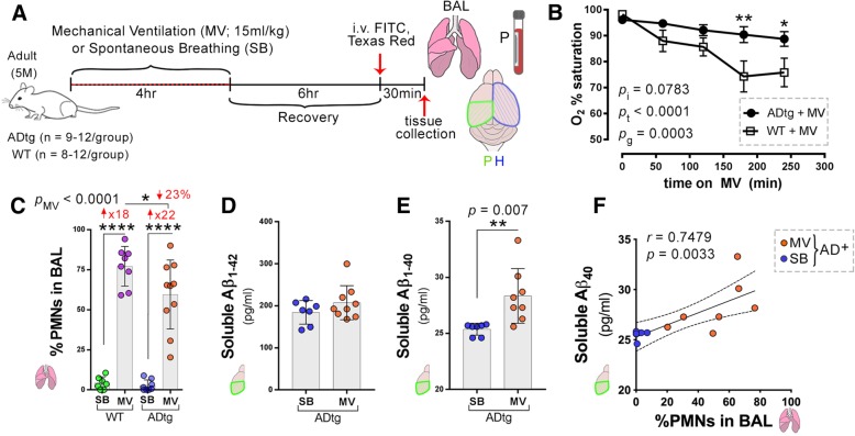
Background: Mechanical ventilation is strongly associated with cognitive decline after critical illness. This finding is particularly evident among older individuals who have pre-existing cognitive impairment, most commonly characterized by varying degrees of cerebral amyloid-β accumulation, neuroinflammation, and blood-brain barrier dysfunction. We sought to test the hypothesis that short-term mechanical ventilation contributes to the neuropathology of cognitive impairment by (i) increasing cerebral amyloid-β accumulation in mice with pre-existing Alzheimer's disease pathology, (ii) increasing neurologic and systemic inflammation in wild-type mice and mice with pre-existing Alzheimer's disease pathology, and (iii) increasing hippocampal blood-brain barrier permeability in wild-type mice and mice with pre-existing Alzheimer's disease pathology.

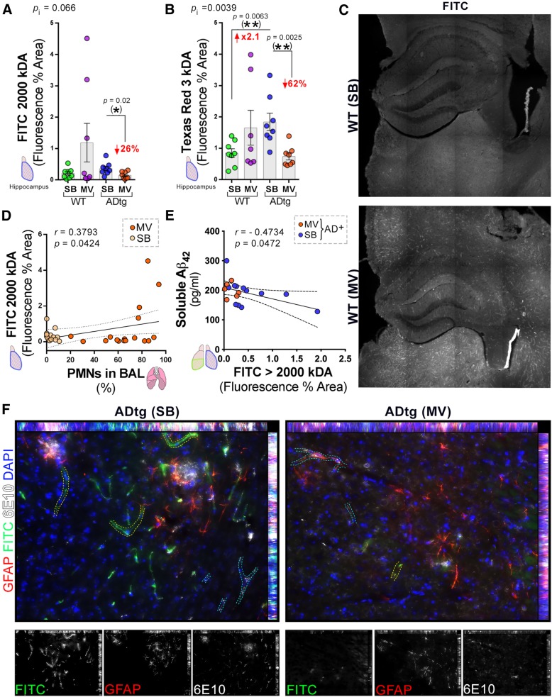
Methods: We subjected double transgenic Alzheimer's disease (APP/PSEN1) and wild-type mice to mechanical ventilation for 4 h and compared to non-mechanically ventilated Alzheimer's disease model and wild-type mice. Cerebral soluble/insoluble amyloid-β1-40/amyloid-β1-42 and neurological and systemic markers of inflammation were quantified. Hippocampal blood-brain barrier permeability was quantified using a novel methodology that enabled assessment of small and large molecule permeability across the blood-brain barrier.

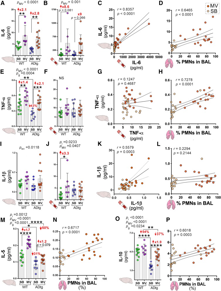
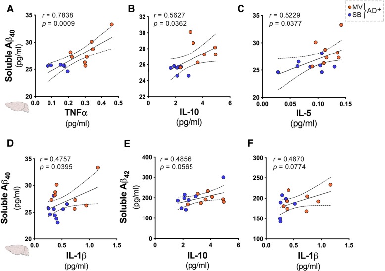
Results: Mechanical ventilation resulted in (i) a significant increase in cerebral soluble amyloid-β1-40 (p = 0.007) and (ii) significant increases in neuroinflammatory cytokines in both wild-type and Alzheimer's disease mice which, in most cases, were not reflected in the plasma. There were (i) direct correlations between polymorphonuclear cells in the bronchoalveolar fluid and cerebral soluble amyloid-β1-40 (p = 0.0033), and several Alzheimer's disease-relevant neuroinflammatory biomarkers including cerebral TNF-α and IL-6; (iii) significant decreases in blood-brain barrier permeability in mechanically ventilated Alzheimer's disease mice and a trend towards increased blood-brain barrier permeability in mechanically ventilated wild-type mice.

Conclusions: These results provide the first evidence that short-term mechanical ventilation independently promotes the neuropathology of Alzheimer's disease in subjects with and without pre-existing cerebral Alzheimer's disease pathology. Future studies are needed to further clarify the specific mechanisms by which this occurs and to develop neuroprotective mechanical ventilation strategies that mitigate the risk of cognitive decline after critical illness.

Keywords: Alzheimer’s disease; Cognitive impairment; Critical illness; Mechanical ventilation.


Conflict of interest statement

Ethics approval and consent to participate

All experiments were conducted in accordance with Cedars-Sinai Medical Center Institutional Animal Care and Use Committee (IACUC) guidelines under an approved protocol and complied with current United States law.

Consent for publication

not applicable

Competing interests

The authors declare that they have no competing interests.

Publisher’s Note

Springer Nature remains neutral with regard to jurisdictional claims in published maps and institutional affiliations.

Figures

Fig. 1
Fig. 1
Mechanical ventilation induces pulmonary inflammation and increases cerebral soluble Aβ1–40 in ADtg mice. a Schematic illustration of experimental design and timeline: 5-month-old ADtg and wild-type (WT) mice underwent mechanical ventilation (MV) with a tidal volume of 15 ml/kg for 4 h while control groups of ADtg and WT mice were spontaneously breathing (SB) (n = 8–12 mice per group). Mice recovered for 6 h and 30 min prior to perfusion, they received intravenous injections of Texas Red-dextran (3 kD) and FITC-dextran (2000 kD) tracers (0.25% each). Bronchoalveolar lavage (BAL) fluid specimens were analyzed for cell count, plasma and left hindbrains were collected for protein analysis (P), and the right brain hemispheres were isolated for histology (H). b Mean arterial oxygen saturation for ADtg and WT mice are presented for each hour of MV, analyzed by two-way ANOVA. c Percent of polymorphonuclear cells (PMNs), or neutrophils, in the BAL was measured for each group, ADtg and WT in both MV and SB conditions (n = 8–12 mice/group). d Sandwich ELISA analysis of human soluble Aβ1–42 levels in the brains of age-matched ADtg mice (n = 8–9 mice/group). e ELISA analysis of cerebral soluble Aβ1–40 levels in age-matched ADtg mice (n = 8–9 mice/group). f Pearson’s r correlation analysis between cerebral soluble Aβ1–40 and % PMNs in BAL in age-matched ADtg mice in both conditions, MV (orange dots) and SB (yellow dots) with 95% confidence interval (CI) in dashed lines. Data from individual mice and group means with standard error of measurements are shown, as well as p values (pi = p value for interaction; pMV = p value for MV intervention effect; pg = p value for genotype effect). Fold increases in MV compared to SB-control groups are shown in red. *p < 0.05, **p < 0.01, ****p < 0.0001, using two-way ANOVA with Holm-Sidak’s post hoc multiple comparisons correction, unpaired two-tailed Student t tests for two-group comparison, and Pearson’s correlation analysis
Fig. 2
Fig. 2
Mechanical ventilation affects key cognition-relevant cytokine responses in the brain and plasma of WT and ADtg mice. The Meso Scale Discovery (MSD) multiplex inflammatory assay was performed on plasma and brain specimens from all experimental groups (n = 8–12 mice/group). a, b IL-6 expression. c Pearson’s r correlation analysis between cerebral and plasma IL-6 levels and d cerebral IL-6 and % PMNs in BAL. e, f TNF-α expression. g Correlations between cerebral and plasma TNF-α levels and h cerebral TNF-α and % PMNs in BAL. i, j IL-1β expression. k Correlations between cerebral and plasma IL-1β levels and l cerebral IL-1β and % PMNs in BAL. m IL-5 expression in the brain. n Correlation between brain IL-5 and % PMNs in BAL. o IL-10 expression in the brain. p Correlation between brain IL-10 and % PMNs in BAL. Data from individual mice and group means with standard error of measurements are shown as well as p values (pi = p value for interaction; pMV = p value for MV intervention effect; pg = p value for genotype effect); mice in the MV group are indicated by orange dots and mice in the SB group by yellow dots. Fold increase and percentage decrease between the groups are shown in red. *p < 0.05, **p < 0.01, ***p < 0.005, ****p < 0.0001, using two-way ANOVA with Holm-Sidak’s post hoc multiple comparisons correction or Pearson’s r correlation analysis with 95% CI
Fig. 3
Fig. 3
Associations between cerebral soluble Aβ and key cytokines in ADtg mice following mechanical ventilation. Pearson’s r correlations between human soluble Aβ1–40 and Aβ1–42 levels and cerebral cytokine levels measured by the Meso Scale Discovery (MSD) multiplex inflammatory assay in age-matched ADtg mice (n = 8–9 mice/group). ad Correlations between cerebral soluble Aβ1–40 levels in ADtg groups (MV and SB) and the following cerebral cytokines: a TNF-α, b IL-10, c IL-5, and d IL-1β. e, f Correlations between cerebral soluble Aβ1–42 and the following cerebral cytokines: e IL-10 and f IL-1β. Data from individual mice in MV (orange dots) and SB (yellow dots) groups are shown. Both r and p values, as well as 95% CI, dashed lines are shown as measured by Pearson’s r correlations
Fig. 4
Fig. 4
Blood-brain barrier permeability in the hippocampus following mechanical ventilation in WT and ADtg mice. Blood-brain barrier damage in the hippocampus was assessed by quantitative analysis of percent area of extravasation of either high molecular weight FITC-dextran (2000 kD) or low molecular weight Texas Red-dextran (3 kD) tracers. a FITC-dextran and b Texas Red-dextran. c Representative confocal images of FITC-dextran tracer in WT mice in the SB (top) and MV (bottom) conditions. d, e Pearson’s r correlation analysis between FITC-dextran hippocampal tracer leakage and d % PMNs in BAL of all animals (n = 8–12 mice/group) and e soluble cerebral Aβ1–42 in both groups of ADtg mice (n = 8–9 mice/group). f Representative fluorescent micrographs of hippocampal ADtg-SB (left) and ADtg-MV (right) immunolabeled for GFAP+ astrocytes (red), 6E10+ human Aβ (white), FITC-dextran extravasation (green) and nuclei (blue). Increased extravasation of FITC-dextran is seen in vessels without 6E10+ Aβ aggregates (traced in yellow dotted lines) while less extravasation of FITC-dextran is seen in vessels with 6E10+ Aβ aggregates (traced in blue dotted lines). Individual channel micrographs are shown below. Data from individual mice in MV (orange dots) and SB (yellow dots) groups are shown, as well as p values (pi = p value for interaction; pMV = p value for MV intervention effect; pg = p value for genotype effect). Fold increase and percentage decreases compared to control groups are shown in red. *p < 0.05, **p < 0.01, using two-way ANOVA with Holm-Sidak’s post hoc multiple comparisons correction, while the asterisk in parenthesis signifies an unpaired two-tailed Student t test. For Pearson’s r correlations, both r and p values, as well as 95% CI dashed lines are presented

References

    1. Wunsch H, Linde-Zwirble WT, Angus DC, Hartman ME, Milbrandt EB, Kahn JM. The epidemiology of mechanical ventilation use in the United States. Crit Care Med. 2010;38(10):1947–1953. doi: 10.1097/CCM.0b013e3181ef4460. - DOI - PubMed
    1. Pandharipande PP, Girard TD, Jackson JC, Morandi A, Thompson JL, Pun BT, Brummel NE, Hughes CG, Vasilevskis EE, Shintani AK, et al. Long-term cognitive impairment after critical illness. N Engl J Med. 2013;369(14):1306–1316. doi: 10.1056/NEJMoa1301372. - DOI - PMC - PubMed
    1. Tarasoff-Conway JM, Carare RO, Osorio RS, Glodzik L, Butler T, Fieremans E, Axel L, Rusinek H, Nicholson C, Zlokovic BV, et al. Clearance systems in the brain--implications for Alzheimer diseaser. Nat Rev Neurol. 2016;12(4):248. doi: 10.1038/nrneurol.2016.36. - DOI - PubMed
    1. Hopkins RO, Wade D, Jackson JC. What's new in cognitive function in ICU survivors. Intensive Care Med. 2017;43(2):223–225. doi: 10.1007/s00134-016-4550-x. - DOI - PubMed
    1. Prince M, Bryce R, Albanese E, Wimo A, Ribeiro W, Ferri CP. The global prevalence of dementia: a systematic review and metaanalysis. Alzheimers Dement. 2013;9(1):63–75. doi: 10.1016/j.jalz.2012.11.007. - DOI - PubMed

Publication types