. 2019 Mar 5;116(5):760-771.
doi: 10.1016/j.bpj.2019.01.027. Epub 2019 Feb 1.

Nanomechanics of Diaminopurine-Substituted DNA

Affiliations

Nanomechanics of Diaminopurine-Substituted DNA

Matteo Cristofalo et al. Biophys J. .

Abstract

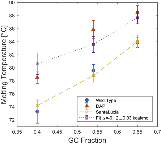
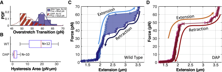
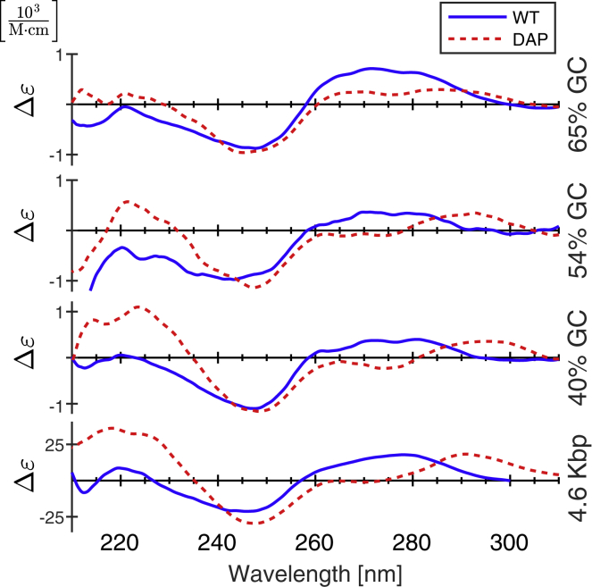
2,6-diaminopurine (DAP) is a nucleobase analog of adenine. When incorporated into double-stranded DNA (dsDNA), it forms three hydrogen bonds with thymine. Rare in nature, DAP substitution alters the physical characteristics of a DNA molecule without sacrificing sequence specificity. Here, we show that in addition to stabilizing double-strand hybridization, DAP substitution also changes the mechanical and conformational properties of dsDNA. Thermal melting experiments reveal that DAP substitution raises melting temperatures without diminishing sequence-dependent effects. Using a combination of atomic force microscopy (AFM), magnetic tweezer (MT) nanomechanical assays, and circular dichroism spectroscopy, we demonstrate that DAP substitution increases the flexural rigidity of dsDNA yet also facilitates conformational shifts, which manifest as changes in molecule length. DAP substitution increases both the static and dynamic persistence length of DNA (measured by AFM and MT, respectively). In the static case (AFM), in which tension is not applied to the molecule, the contour length of DAP-DNA appears shorter than wild-type (WT)-DNA; under tension (MT), they have similar dynamic contour lengths. At tensions above 60 pN, WT-DNA undergoes characteristic overstretching because of strand separation (tension-induced melting) and spontaneous adoption of a conformation termed S-DNA. Cyclic overstretching and relaxation of WT-DNA at near-zero loading rates typically yields hysteresis, indicative of tension-induced melting; conversely, cyclic stretching of DAP-DNA showed little or no hysteresis, consistent with the adoption of the S-form, similar to what has been reported for GC-rich sequences. However, DAP-DNA overstretching is distinct from GC-rich overstretching in that it happens at a significantly lower tension. In physiological salt conditions, evenly mixed AT/GC DNA typically overstretches around 60 pN. GC-rich sequences overstretch at similar if not slightly higher tensions. Here, we show that DAP-DNA overstretches at 52 pN. In summary, DAP substitution decreases the overall stability of the B-form double helix, biasing toward non-B-form DNA helix conformations at zero tension and facilitating the B-to-S transition at high tension.


Figures

Figure 1
Figure 1
2,6-diaminopurine (bottom) is an analog of adenine with an additional amino group at position 2 of the purine molecule. When paired with thymine, it forms an additional hydrogen bond along the minor-groove side of the molecule. To see this figure in color, go online.
Figure 2
Figure 2
Melting curves of three sequences of WT (blue, solid circle) and DAP-DNA (red, solid triangles) with GC fractions of 0.4, 0.54, and 0.65 were measured using the fluorescence of Syto-84 (1 μM); error bars indicate SD. WT results closely match Tm predicted by the SantaLucia nearest-neighbor model (yellow); error bars indicate model accuracy (61). The DAP melting temperatures were fit with a modified SantaLucia model (purple). A least-squares fit yields α = −0.12 ± 0.03 kcal/mol. To see this figure in color, go online.
Figure 3
Figure 3
Worm-like chain fits of force versus length MT data. (A) Representative force versus length data for wild-type (blue) and DAP (red) DNA molecules with corresponding WLC fits (solid line for WT and dashed line for DAP) are shown. Fit errors correspond to 95% confidence limits. (B) Histogram of fit persistence length values is shown; DAP substitution increases Lp to 56 ± 5 from 45 ± 4 nm for WT (mean ± SD). Distributions are statistically distinct, p = 5.22 × 10−16. (C) Histogram of fit contour length values is shown. DAP substitution does not significantly decrease dynamic contour length, p = 0.103. N = 35 for WT and N = 48 for DAP in both histogram plots. To see this figure in color, go online.
Figure 4
Figure 4
Representative images of (A) wild-type and (B) DAP-DNA molecules captured via AFM. To see this figure in color, go online.
Figure 5
Figure 5
Contour and persistence length measured via analysis of AFM images. (A) Contour length was calculated by tracing individual molecules in each AFM image. DAP substitution reduced the length (mean ± SD). The distributions are statistically different in a two-tailed t-test (p = 3.07 × 10−4). (B) Mean-squared distance versus separation along each molecular contour is shown. Data points (blue circles and red squares) correspond to the average square distance, aggregated for all molecules of a given type (WT and DAP, respectively). Data was fit to Eq. 4, yielding estimates of the average persistence length; ± error indicates 95% confidence interval. To see this figure in color, go online.
Figure 6
Figure 6
Overstretching under high tension. (A) Histogram of overstretching transition force is shown. Distributions are statistically distinct, p = 2.88 × 10−8. (B) Box and whisker plot of area between extension and relaxation curves are shown. Box edges indicate the 25th and 75th percentiles. Whiskers indicate the extent of data. Red line indicates the median. Nearly all WT molecules exhibited significant hysteresis H = 19 ± 9 pN μm; DAP molecules showed little hysteresis H = 2 ± 1 pN μm. (C) Example force-extension cycle for WT-DNA is shown. As tension is increased (light blue trace), the molecule undergoes force-induced melting. Upon relaxation (dark blue), molecule length decreases with a different characteristic curve owing to the presence of single-stranded DNA. (D) Example force-extension cycle for DAP-DNA is shown. Little hysteresis is observed between extension and relaxation, indicating basepair persistence and the adoption of the S-form. To see this figure in color, go online.
Figure 7
Figure 7
CD spectra of WT (blue, solid lines) and DAP-substituted molecules (red, dashed lines). To see this figure in color, go online.

Similar articles

Cited by

References

    1. Bailly C., Waring M.J. The use of diaminopurine to investigate structural properties of nucleic acids and molecular recognition between ligands and DNA. Nucleic Acids Res. 1998;26:4309–4314. - PMC - PubMed
    1. Schrum J.P., Ricardo A., Szostak J.W. Efficient and rapid template-directed nucleic acid copying using 2′-amino-2′,3′-dideoxyribonucleoside-5′-phosphorimidazolide monomers. J. Am. Chem. Soc. 2009;131:14560–14570. - PMC - PubMed
    1. Kirnos M.D., Khudyakov I.Y., Vanyushin B.F. 2-aminoadenine is an adenine substituting for a base in S-2L cyanophage DNA. Nature. 1977;270:369–370. - PubMed
    1. Khudyakov I.Y., Kirnos M.D., Vanyushin B.F. Cyanophage S-2L contains DNA with 2,6-diaminopurine substituted for adenine. Virology. 1978;88:8–18. - PubMed
    1. Cerami A., Reich E., Goldberg I.H. The interaction of actinomycin with DNA: requirement for the 2-amino group of purines. Proc. Natl. Acad. Sci. USA. 1967;57:1036–1042. - PMC - PubMed

Publication types