. 2019 May 23;133(21):2291-2304.
doi: 10.1182/blood-2018-10-882944. Epub 2019 Feb 22.

Fratricide-resistant CD1a-specific CAR T cells for the treatment of cortical T-cell acute lymphoblastic leukemia

Affiliations

Fratricide-resistant CD1a-specific CAR T cells for the treatment of cortical T-cell acute lymphoblastic leukemia

Diego Sánchez-Martínez et al. Blood. .

Abstract

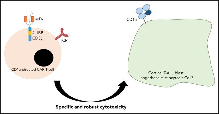
Relapsed/refractory T-cell acute lymphoblastic leukemia (T-ALL) has a dismal outcome, and no effective targeted immunotherapies for T-ALL exist. The extension of chimeric antigen receptor (CAR) T cells (CARTs) to T-ALL remains challenging because the shared expression of target antigens between CARTs and T-ALL blasts leads to CART fratricide. CD1a is exclusively expressed in cortical T-ALL (coT-ALL), a major subset of T-ALL, and retained at relapse. This article reports that the expression of CD1a is mainly restricted to developing cortical thymocytes, and neither CD34+ progenitors nor T cells express CD1a during ontogeny, confining the risk of on-target/off-tumor toxicity. We thus developed and preclinically validated a CD1a-specific CAR with robust and specific cytotoxicity in vitro and antileukemic activity in vivo in xenograft models of coT-ALL, using both cell lines and coT-ALL patient-derived primary blasts. CD1a-CARTs are fratricide resistant, persist long term in vivo (retaining antileukemic activity in re-challenge experiments), and respond to viral antigens. Our data support the therapeutic and safe use of fratricide-resistant CD1a-CARTs for relapsed/refractory coT-ALL.


Conflict of interest statement

Conflict-of-interest disclosure: The authors declare no competing financial interests.

Figures

None
Graphical abstract
Figure 1.
Figure 1.
CD1a expression in T-ALL and normal hematopoiesis and thymopoiesis. (A) Immunophenotype of de novo T-ALL samples (n = 38) for the indicated markers. Red and blue circles identify CD1a+/++ and CD1alow/+ coT-ALL patients, respectively. Green circles depict non-coTALL patients. (B) Representative FACS dot plot of a patient with coT-ALL. Red cells are CD7+CD1a+ coT-ALL blasts, and blue cells are normal mature T cells (CD3+CD7+CD1a, either CD4+ or CD8+) present in the diagnostic (DX) sample. (C) CD1a is retained at relapse (RX) (n = 5 DX-RX coT-ALL pairs). Data shown as CD1a expression in RX samples relative to the DX-matched samples (DX shown as 100% expression). (D) T cells and CD34+ HSPCs do not express CD1a across ontogeny. (E) Scheme depicting the phenotype of developing thymic T-cell populations. (F) Representative FACS for precortical (CD34highCD7++CD1a) and cortical (CD34+CD7++CD1a+) thymocytes.
Figure 2.
Figure 2.
CD1a CARTs specifically target and eliminate CD1a+ T-ALL cell lines in vitro. (A) Scheme of the CD1aCAR construct used. (B) CAR detection in 293T cells using an anti-scFv mAb and GFP. (C) Representative CAR transduction and detection in CD4+ and CD8+ T cells (n = 6). (D) Proper T-cell activation (n = 3). (E) Robust expansion of activated T cells transduced with either MOCK or CD1a CAR reveals no signs of fratricide (n = 4). (F) Surface expression of CD1a (blue line) in Jurkat, MOLT4, and NALM6 cell lines. (G) CD1a antigen density in cell lines, primary coT-ALL samples, and primografts. (H) Cytotoxicity of CD1a CARTs and MOCK T cells against coT-ALL and B-ALL cell lines at the indicated E:T ratios in 16-hour assays (n = 4). (I) Absolute counts of alive eFluor-positive target cells measured according to FACS in 72-hour cytotoxicity assays at a 1:1 E:T ratio. (J) Representative FACS analysis of cytotoxicity with target cells labeled with eFluor 670 (shown in blue). (K) Enzyme-linked immunosorbent assay showing high-level production of the inflammatory cytokines IL-2, IFN-γ, and TNFα by CD1a CARTs exposed to Jurkat and NALM6 (negative control) cells in 16-hour assays at a 1:1 E:T ratio (n = 4). n.s., not significant. *P < .05, **P < .01, ***P < .001.
Figure 3.
Figure 3.
CD1a CARTs specifically target and eliminate in vitro CD1a+ T-ALL blasts from primary samples or PDX models. (A) Expression of CD1a vs CD7 in coT-ALL blasts from primary patients/primografts. The percentage of CD1a+ blasts is indicated. (B) Cytotoxicity (in absolute counts of eFluor-positive cells) measured by using FACS in 48-hour cytotoxicity assays at a 4:1 E:T ratio (n = 3). (C) Representative FACS analysis of CD1a (shown in blue) within the eFluor-labeled target cells at the end of the cytotoxicity assay, revealing specificity of CD1a CARTs (n = 3). (D) High-level production of pro-inflammatory cytokines by CD1a CARTs analyzed according to enzyme-linked immunosorbent assay (n = 3 independent supernatants) in 16-hour assays at a 4:1 E:T ratio. *P < .05, **P < .01, ***P < .001, ****P < .0001.
Figure 4.
Figure 4.
CD1a CARTs fully control the progression of coT-ALL cells in a mouse xenograft setting. (A) Scheme of the xenograft model. NSG mice (n = 6/group) were IV injected with 3 × 10⁶ Luc/GFP–expressing Jurkat cells followed 3 days after by a single IV injection of 5 × 10⁶ MOCK or CD1a CARTs. Tumor burden was monitored every 4 to 6 days according to BLI using IVIS imaging. When MOCK-treated animals were fully leukemic, one-half of the CD1a CART-treated animals were euthanized and analyzed by using FACS (BM, PB, and spleen) for leukemic burden and CART persistence. The remaining animals were rechallenged 6 weeks later with 1.5 × 10⁶ Luc-Jurkat cells and were followed up as before. (B) IVIS imaging of tumor burden monitored by BLI at the indicated time points. (C) Total radiance quantification at the indicated time points. †Euthanization. (D) Circulating Jurkat cells in PB 17 days after CARTs infusion. (E) T-cell persistence in PB at day 17, and spleen and BM at euthanization. Data are shown as mean ± SD (n = 6 mice/group). *P < .05, **P < .01, ***P < .001.
Figure 5.
Figure 5.
CD1a CARTs fully abolish the progression of primary CD1a+ coT-ALL blasts in a PDX setting. (A) Scheme of the PDX model. NSG mice (n = 5–6/group) were IV injected with 1 × 10⁶ primary coT-ALL cells followed 3 days after by a single IV injection of 1 × 10⁶ MOCK or CD1a CARTs. Tumor burden was monitored according to FACS every other week by bleeding and BM aspirate after 6 and 9 weeks. Frequency of leukemic mice and levels of leukemia in BM (B) and PB (C) 6 and 9 weeks after infusion of CARTs. The left panels show representative FACS plots. Primary CD1a+ T-ALL blasts are shown in blue, effector T cells are shown in red, and mouse cells are shown in black. (D) Nine-week OS of coT-ALL primografts receiving either CD1a CARTs or MOCK T cells. (E) Effector T-cell persistence over time in PB (week 2 toward week 9) and BM (weeks 6 and 9). Each dot represents an independent mouse. *P < .05, **P < .01, ***P < .001, Malcolm-Cox test.
Figure 6.
Figure 6.
CD1a CARTs retain the ability to control progression of CD1a+ cell lines and coT-ALL primary samples in a rechallenge PDX setting. (A) IVIS imaging of Jurkat cells burden in the rechallenged mice. (B) Total radiance quantification over time in the mice rechallenged with Jurkat cells. (C) Circulating Jurkat cells in PB 16 days after rechallenge. (D) Robust effector T-cell persistence in PB, BM, and spleen at euthanization of the rechallenged animals. (E) Scheme of the rechallenge PDX experiments using coT-ALL primary samples. CART-bearing PDX mice were rechallenged with 1 × 10⁶ primary CD1a+ T-ALL 7 weeks after initial CART infusion. (F) Secondary coT-ALL burden in engrafted PB (left panel) and BM (right panel) 6 weeks after leukemia rechallenge. (G) Effector T-cell persistence over time in PB (weeks 2, 4, and 6) from PDXs rechallenged with coT-ALL primary samples. Each dot represents an independent mouse. *P < .05, **P < .01, ****P < .0001.
Figure 7.
Figure 7.
CD1a CARTs derived from patients with coT-ALL at presentation (specifically lyse autologous CD1a+ T-ALL blasts). (A) Scheme depicting the experimental design for the autologous cytotoxic assay. Mature (normal) CD3+CD1a T cells were FACS-purified from the PB of a patient with coT-ALL, infected with CD1a CAR, expanded, and exposed to autologous total PBMCs. (B) FACS analysis of autologous cytotoxic 48-hour assay at 1:1 and 4:1 E:T ratios. eFluor 670–labeled total PBMC target population contains CD1a+ T-ALL blasts (red) and mature CD3+CD1a T cells (blue). (C) Quantification of CD1a CART-mediated specific lysis for coT-ALL blasts (upper panel) and CD3+CD1a mature T cells (bottom panel). (D) ELISpot showing the number of IFN-γ SFC from mock vs CD1a CARTs stimulated with a pool of peptides from CMV, EBV, and flu (CEF). Staphylococcal enterotoxin B was used as positive control. SFC, spot-forming cell.

Comment in

Similar articles

Cited by

References

    1. Karrman K, Johansson B. Pediatric T-cell acute lymphoblastic leukemia. Genes Chromosomes Cancer. 2017;56(2):89-116. - PubMed
    1. Weng AP, Ferrando AA, Lee W, et al. . Activating mutations of NOTCH1 in human T cell acute lymphoblastic leukemia. Science. 2004;306(5694):269-271. - PubMed
    1. Hunger SP, Mullighan CG. Acute lymphoblastic leukemia in children. N Engl J Med. 2015;373(16):1541-1552. - PubMed
    1. Litzow MR, Ferrando AA. How I treat T-cell acute lymphoblastic leukemia in adults. Blood. 2015;126(7):833-841. - PubMed
    1. Schneider NR, Carroll AJ, Shuster JJ, et al. . New recurring cytogenetic abnormalities and association of blast cell karyotypes with prognosis in childhood T-cell acute lymphoblastic leukemia: a pediatric oncology group report of 343 cases. Blood. 2000;96(7):2543-2549. - PubMed

Publication types

MeSH terms