. 2019 May;68(5):1054-1061.
doi: 10.2337/db19-0025. Epub 2019 Feb 22.

The Hypothalamic Arcuate Nucleus-Median Eminence Is a Target for Sustained Diabetes Remission Induced by Fibroblast Growth Factor 1

Affiliations

The Hypothalamic Arcuate Nucleus-Median Eminence Is a Target for Sustained Diabetes Remission Induced by Fibroblast Growth Factor 1

Jenny M Brown et al. Diabetes. 2019 May.

Erratum in

Abstract

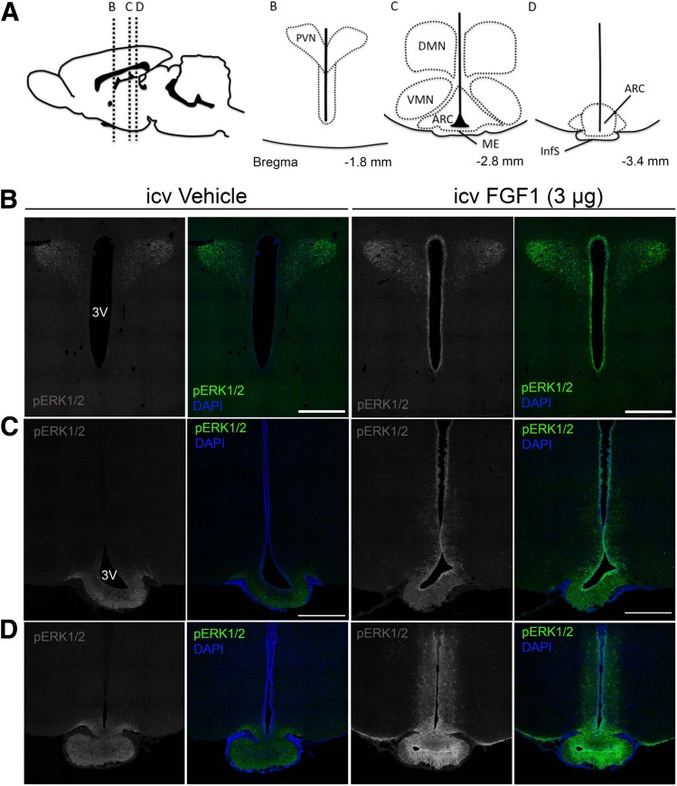
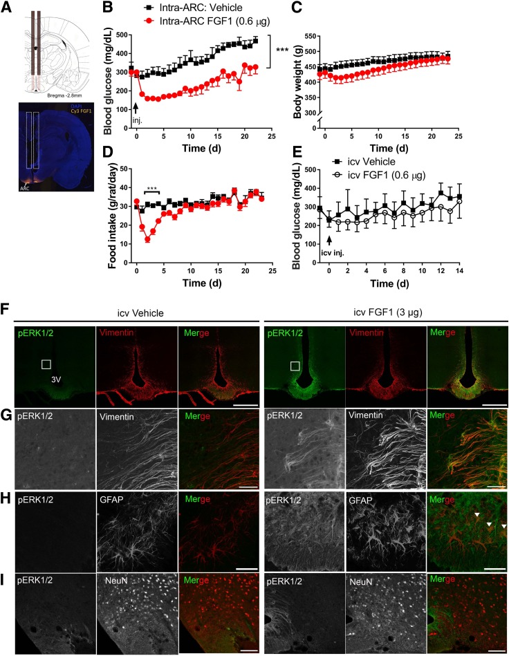
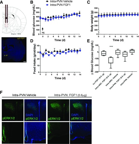
In rodent models of type 2 diabetes (T2D), sustained remission of diabetic hyperglycemia can be induced by a single intracerebroventricular (icv) injection of fibroblast growth factor 1 (FGF1). To identify the brain areas responsible for this effect, we first used immunohistochemistry to map the hypothalamic distribution of phosphorylated extracellular signal-related kinase 1/2 (pERK1/2), a marker of mitogen-activated protein kinase-ERK signal transduction downstream of FGF receptor activation. Twenty minutes after icv FGF1 injection in adult male Wistar rats, pERK1/2 staining was detected primarily in two hypothalamic areas: the arcuate nucleus-median eminence (ARC-ME) and the paraventricular nucleus (PVN). To determine whether an action of FGF1 localized to either the ARC-ME or the PVN is capable of mimicking the sustained antidiabetic effect elicited by icv FGF1, we microinjected either saline vehicle or a low dose of FGF1 (0.3 µg/side) bilaterally into either the ARC-ME area or PVN of Zucker Diabetic Fatty rats, a model of T2D, and monitored daily food intake, body weight, and blood glucose levels over a 3-week period. Whereas bilateral intra-arcuate microinjection of saline vehicle was without effect, remission of hyperglycemia lasting >3 weeks was observed following bilateral microinjection of FGF1 into the ARC-ME. This antidiabetic effect cannot be attributed to leakage of FGF1 into cerebrospinal fluid and subsequent action on other brain areas, since icv injection of the same total dose was without effect. Combined with our finding that bilateral microinjection of the same dose of FGF1 into the PVN was without effect on glycemia or other parameters, we conclude that the ARC-ME area (but not the PVN) is a target for sustained remission of diabetic hyperglycemia induced by FGF1.


Figures

Figure 1
Figure 1
Regional activation of MAPK/ERK1/2 signaling in the hypothalamus after icv FGF1. A: Diagram at top shows sagittal view of the rat brain (left) and inserts of the mediobasal hypothalamus (right) showing orientation of panel images of the PVN, ARC, ME, ventromedial nucleus (VMN), dorsomedial nucleus (DMN), and infundibular stalk (InfS). BD: Confocal images of coronal sections showing pERK1/2 (green) and DAPI (blue) obtained 20 min after icv injection of either vehicle or FGF1 (3 μg). Images are taken from the PVN (B), the ARC-ME (C), and InfS (D). Scale bars: 500 μm (B and C). 3V, third ventricle.
Figure 2
Figure 2
Effect of intra-ARC microinjection of FGF1 in ZDF rats. A: Scale diagram of bilateral intra-ARC guide cannula (top panel) and representative image of Cy3-labled FGF1 injectate spread following microinjection into the ARC area (bottom panel). Daily blood glucose (B), body weight (C), and food intake (D) from ZDF rats after a single bilateral intra-ARC microinjection of either vehicle (n = 7) (black squares) or FGF1 (0.3 μg/side, for a total of 0.6 μg; n = 8) (red circles). E: Daily blood glucose levels in ZDF rats after a single icv injection of either vehicle (n = 4) or FGF1 at a dose equal to that given by microinjection into the ARC (0.6 μg; n = 4). Significance determined by linear mixed-model analysis. Confocal images of coronal sections of the ARC-ME costained with antibodies to pERK1/2 (green) and markers of tanycytes (vimentin), astrocytes (GFAP), or neurons (NeuN) 20 min after icv injection of either vehicle (left panels, F–I) or FGF1 (3 μg) (right panels, F–I) in Wistar rats (n = 5/group). G: Higher magnification view of images shown in inset in F. Scale bars: 500 μm (F), 50 μm (G and H), 100 μm (I). Colocalization of GFAP and pERK1/2 denoted by white arrowheads in (H). Data are mean ± SEM. ***P < 0.001 vs. intra-ARC vehicle. 3V, third ventricle; d, days; inj., injection.
Figure 3
Figure 3
Effect of intra-PVN FGF1 microinjection on blood glucose levels in ZDF rats. A: Scale diagram of bilateral intra-PVN guide cannula placement (top panel) and representative image of Cy3 FGF1 injectate spread following intra-PVN microinjection (bottom panel). Scale bar: 1,000 μm. Daily levels of blood glucose (B), body weight (C), and food intake (D) of ZDF rats after a single bilateral microinjection into the PVN of either vehicle (n = 5) or FGF1 (0.3 μg/side, for a total of 0.6 μg; n = 6). No significant differences in group mean values of any of these parameters were observed. Significance determined by linear mixed-model analysis. E: Change of blood glucose levels measured 48 h after icv, intra-ARC, or intra-PVN injection of either vehicle or FGF1 in the same animals. ***P < 0.05 vs. the other four groups by one-way ANOVA with Tukey post hoc test. F: Confocal images of pERK1/2 and DAPI immunostaining in coronal sections collected 20 min after an intra-PVN microinjection of either vehicle or FGF1 (0.3 μg/side; n = 3/group) at the level of either the PVN (top panel) or ARC-ME (bottom panel) in Wistar rats. Data are mean ± SEM. d, days.

References

    1. Morton GJ, Matsen ME, Bracy DP, et al. . FGF19 action in the brain induces insulin-independent glucose lowering. J Clin Invest 2013;123:4799–4808 - PMC - PubMed
    1. Sarruf DA, Thaler JP, Morton GJ, et al. . Fibroblast growth factor 21 action in the brain increases energy expenditure and insulin sensitivity in obese rats. Diabetes 2010;59:1817–1824 - PMC - PubMed
    1. Scarlett JM, Rojas JM, Matsen ME, et al. . Central injection of fibroblast growth factor 1 induces sustained remission of diabetic hyperglycemia in rodents. Nat Med 2016;22:800–806 - PMC - PubMed
    1. Lan T, Morgan DA, Rahmouni K, et al. . FGF19, FGF21, and an FGFR1/β-klotho-activating antibody act on the nervous system to regulate body weight and glycemia. Cell Metab 2017;26:709–718.e3 - PMC - PubMed
    1. Scarlett JM, Muta K, Brown JM, et al. . Peripheral mechanisms mediating the sustained antidiabetic action of FGF1 in the brain. Diabetes 2019;68:654–664 - PMC - PubMed

Publication types

MeSH terms

Substances