. 2019 Apr 15;202(8):2276-2286.
doi: 10.4049/jimmunol.1800844. Epub 2019 Feb 22.

The Protein Phosphatase Shp1 Regulates Invariant NKT Cell Effector Differentiation Independently of TCR and Slam Signaling

Affiliations

The Protein Phosphatase Shp1 Regulates Invariant NKT Cell Effector Differentiation Independently of TCR and Slam Signaling

Mayra Cruz Tleugabulova et al. J Immunol. .

Abstract

Invariant NKT (iNKT) cells are innate lipid-reactive T cells that develop and differentiate in the thymus into iNKT1/2/17 subsets, akin to TH1/2/17 conventional CD4 T cell subsets. The factors driving the central priming of iNKT cells remain obscure, although strong/prolonged TCR signals appear to favor iNKT2 cell development. The Src homology 2 domain-containing phosphatase 1 (Shp1) is a protein tyrosine phosphatase that has been identified as a negative regulator of TCR signaling. In this study, we found that mice with a T cell-specific deletion of Shp1 had normal iNKT cell numbers and peripheral distribution. However, iNKT cell differentiation was biased toward the iNKT2/17 subsets in the thymus but not in peripheral tissues. Shp1-deficient iNKT cells were also functionally biased toward the production of TH2 cytokines, such as IL-4 and IL-13. Surprisingly, we found no evidence that Shp1 regulates the TCR and Slamf6 signaling cascades, which have been suggested to promote iNKT2 differentiation. Rather, Shp1 dampened iNKT cell proliferation in response to IL-2, IL-7, and IL-15 but not following TCR engagement. Our findings suggest that Shp1 controls iNKT cell effector differentiation independently of positive selection through the modulation of cytokine responsiveness.


Figures

Figure 1.
Figure 1.
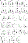
Shp1-deficient iNKT cells are biased towards iNKT2 and iNKT17 subsets. (A) Frequency (top row) and absolute numbers (bottom row) of iNKT cells in the indicated tissues of Shp1fl/fl and Shp1fl/fl CD4-cre mice. (B, C) iNKT cell subsets in the thymus (B) and spleen (C) of Shp1fl/fl (fl/fl) and Shp1fl/fl CD4-cre (cre) mice were assessed by PLZF and RORγt staining. (D) Frequency of iNKT cells in the thymus of single or mixed (mix) bone marrow chimeras. Rag1−/− mice were reconstituted with wild type CD45.1 and/or Shp1fl/fl CD4-cre CD45.2 bone marrow cells. (E, F) Frequency of iNKT2 and iNKT17 subsets in the thymus (E) and spleen (F) of 16-week-old Shp1fl/fl and Shp1fl/fl CD4-cre mice. Data shows representative dot plots, individual mice and the mean values +/− s.e.m. *p < 0.05, **p < 0.01, ***p < 0.001, two-tailed unpaired Student t test.
Figure 2.
Figure 2.
Shp1-deficient iNKT cells produce more TH2 cytokines. (A) Shp1fl/fl and Shp1fl/fl CD4-cre mice were injected i.v. with αGC (0.5 μg). Intracellular FACS analysis of IFN-γ and IL-4 in spleen iNKT cells 90 min post-injection. Data shows representative flow cytometry plots (left), as well as the corresponding frequency (individual mice and mean values +/− s.e.m.) of total, single and double producers of IFN-γ and/or IL-4 (right). (B, C) Thymocytes (B) and splenocytes (C) from Shp1fl/fl and Shp1fl/fl CD4-cre mice were stimulated with plate-bound CD1d-αGC, or PMA/ionomycin (P/I) for 6 hours. Intracellular FACS analysis of IFN-γ and IL-4 in iNKT cells. Data shows representative flow cytometry plots (left), as well as individual and mean values +/− s.e.m. of IL-4/IFN-γ ratios. (D) Shp1fl/fl and Shp1fl/fl CD4-creERT2 mice were treated with tamoxifen and subsequently injected i.v. with αGC (0.2 μg). Data shows individual and mean values +/− s.e.m. of IFN-γ+ or IL-4+ iNKT cells, as well as IL-4/IFN-γ ratios. (E) iNKT cells from Shp1fl/fl or Shp1fl/fl CD4-cre mice were sorted and cultured with BMDCs alone, BMDCs pre-loaded with αGC, or BMDCs in the presence of the indicated cytokine for 48 h. in the presence of indicated cytokines. IFN-γ, IL-4, IL-13, IL-17A and IL-22 production was assessed using a multiplex assay. (F) iNKT cells from Shp1fl/fl or Shp1fl/fl CD4-cre mice were sorted and cultured with BMDCs with isotype control, anti-CD25 or anti-CD127 antibodies (40 μg/ml) for 48 h. Data shows the mean +/− s.e.m. of triplicate values and is representative of 2 individual experiments. *p < 0.05, **p < 0.01, ***p < 0.001, two-tailed unpaired Student t test (A-D) or two-way ANOVA (E).
Figure 3.
Figure 3.
Shp1-deficient iNKT cells drive the expansion of innate memory CD8 T cells in the thymus. (A) Frequency of Eomes+ CD8 T cells in the thymus (left) and spleen (right) of Shp1fl/fl and Shp1fl/fl CD4-cre mice. (B) BrdU incorporation by CD44high CD8 T cells from the thymus (left) and spleen (right) of Shp1fl/fl and Shp1fl/fl CD4-cre mice. (C) Frequency of Eomes+ CD8 T cells in the thymus (top) and spleen (bottom) of Shp1fl/fl CD1d+/−, Shp1fl/fl CD1d−/−, Shp1fl/fl CD4-cre CD1d+/− and Shp1fl/fl CD4-cre CD1d−/− mice. Data represents individual experiments and mean values +/− s.e.m of 2 or 3 independent experiments. *p < 0.05, **p < 0.01, ***p < 0.001, two-tailed unpaired Student t test.
Figure 4.
Figure 4.
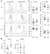
Shp1 deficiency does not affect iNKT TCR signaling. (A) Expression of Egr2 in thymic iNKT cells from Shp1fl/fl (fl/fl) and Shp1fl/fl CD4-cre (cre) mice was determined by FACS. (B) Expression of Egr2 and PLZF in thymic stage 0 (CD24hi CD44 NK1.1), stage 1 (CD24 CD44 NK1.1), stage 2 (CD24 CD44+ NK1.1) and stage 3 (CD24 CD44+ NK1.1+) iNKT cells from Shp1fl/fl (fl/fl) and Shp1fl/fl CD4-cre (cre) mice. (C) Frequency of Nur77+ and Egr2+ thymic (top) or splenic (bottom) iNKT cells upon stimulation with different concentrations of anti-CD3 antibodies. (D) Splenocytes from Shp1fl/fl (fl/fl) and Shp1fl/fl CD4-cre (cre) mice were labelled with CellTrace™ CFSE and stimulated for 72 h with the indicated concentrations of anti-CD3 (top) or anti-CD3/CD28 (bottom) antibodies. Proliferation of iNKT cells (TCRβ+ PBS57-CD1d Tetramer+) was assessed by FACS. (E) Frequency of Vβ8+ and Vβ7+ iNKT cells in the thymus (left) and spleen (right) of Shp1fl/fl and Shp1fl/fl CD4-cre mice. Data represents individual mice and mean +/− s.e.m of 2-3 independent experiments (A, E) or the mean +/− s.e.m. of 2 to 3 independent experiments (n ≥ 4). *p < 0.05, **p < 0.01, ***p < 0.001, two-tailed unpaired Student t test.
Figure 5.
Figure 5.
Shp1 deletion does not affect Slamf6 mediated upregulation of Egr2 and PLZF. (A) Frequency of live PLZF+ PSDPs from Shp1fl/fl and Shp1fl/fl CD4-cre mice after 48 hours of stimulation with the indicated immobilized antibodies. (B) PSDPs were stimulated stimulation with anti-CD3 and anti-Slamf6 antibodies for 30 min., washed and analyzed for Egr2 expression at the indicated times following stimulus withdrawal. (C) Frequency of iNKT cells in the thymus of SAP−/− Shp1fl/fl, SAP−/− Shp1fl/fl CD4-cre and SAP+/− Shp1fl/fl CD4-cre mice Representative dot plots gated on live cells are shown. Data represents individual mice and mean +/− s.e.m. of 2-3 independent experiments.
Figure 6.
Figure 6.
Shp1 regulates cytokine-mediated iNKT cell proliferation. (A) Representative histogram plots (3 mice per group) of CFSE dilution profile of iNKT cells sorted from the thymus (left) or the spleen (right) of Shp1fl/fl and Shp1fl/fl CD4-cre mice cultured for 4 days in the presence of 10ng/ml of IL-2, IL-7 or IL-15. One experiment out of two is shown. (B) BrdU incorporation by iNKT cells from the thymus (left) and spleen (right) of Shp1fl/fl and Shp1fl/fl CD4-cre mice. (C) Expression of CD25, CD127 and CD122 on thymic (left) and splenic (right) iNKT cells from Shp1fl/fl and Shp1fl/fl CD4-cre mice. Data shows representative histogram plots as well as individual mice and the mean values +/− s.e.m of 2-3 independent experiments.

Similar articles

Cited by

References

    1. Brennan PJ, Brigl M, and Brenner MB. 2013. Invariant natural killer T cells: an innate activation scheme linked to diverse effector functions. Nat. Rev. Immunol 13: 101–117. - PubMed
    1. Bendelac A 1995. Positive selection of mouse NK1+ T cells by CD1-expressing cortical thymocytes. J. Exp. Med 182: 2091–2096. - PMC - PubMed
    1. Pasquier B, Yin L, Fondanèche M-C, Relouzat F, Bloch-Queyrat C, Lambert N, Fischer A, de Saint-Basile G, and Latour S. 2005. Defective NKT cell development in mice and humans lacking the adapter SAP, the X-linked lymphoproliferative syndrome gene product. J. Exp. Med 201: 695–701. - PMC - PubMed
    1. Nichols KE, Hom J, Gong S-Y, Ganguly A, Ma CS, Cannons JL, Tangye SG, Schwartzberg PL, Koretzky GA, and Stein PL. 2005. Regulation of NKT cell development by SAP, the protein defective in XLP. Nat. Med 11: 340–345. - PMC - PubMed
    1. Griewank K, Borowski C, Rietdijk S, Wang N, Julien A, Wei DG, Mamchak AA, Terhorst C, and Bendelac A. 2007. Homotypic Interactions Mediated by Slamf1 and Slamf6 Receptors Control NKT Cell Lineage Development. Immunity 27: 751–762. - PMC - PubMed

Publication types

MeSH terms

Substances