. 2019 Feb 22;10(3):185.
doi: 10.1038/s41419-019-1426-3.

Confounding off-target effects of BH3 mimetics at commonly used concentrations: MIM1, UMI-77, and A-1210477

Affiliations

Confounding off-target effects of BH3 mimetics at commonly used concentrations: MIM1, UMI-77, and A-1210477

David J Mallick et al. Cell Death Dis. .

Abstract

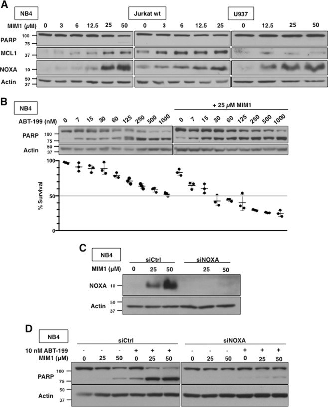
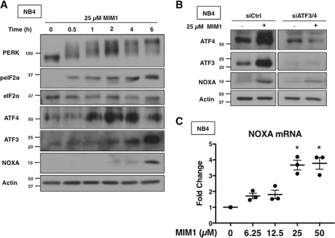
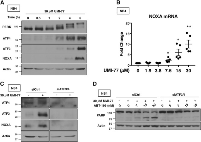
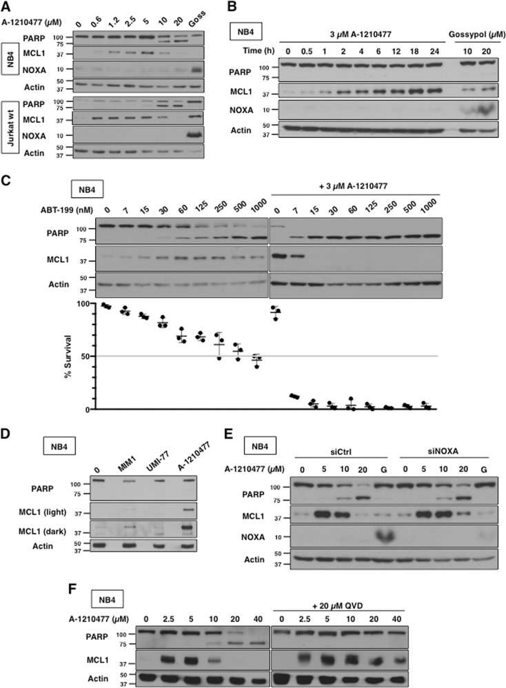
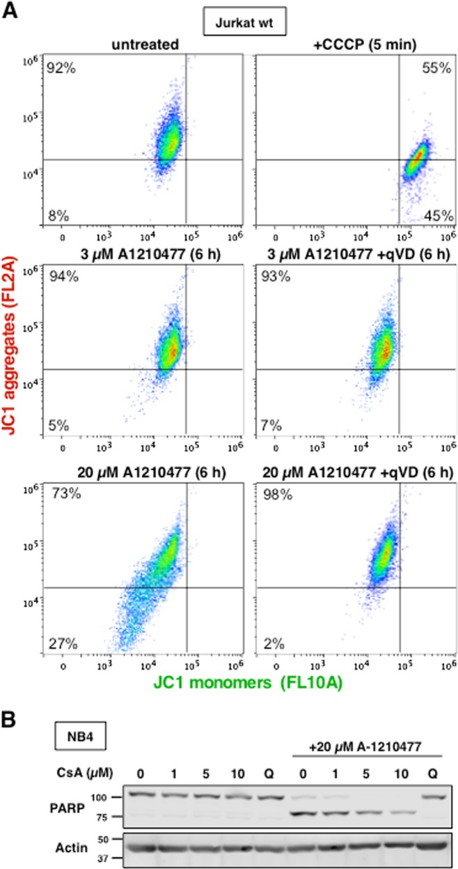
Targeting anti-apoptotic BCL2 family proteins has become an attractive therapeutic strategy for many cancers, and the BCL2-selective inhibitor ABT-199 (venetoclax) has obtained clinical success. However, MCL1 can promote drug resistance and overall cancer cell survival. Thus, there is a critical need to develop an effective drug that antagonizes MCL1. However, most putative MCL1 inhibitors have been misclassified as they fail to directly inhibit MCL1 in cells, but rather induce the pro-apoptotic protein NOXA. We have investigated three putative MCL1 inhibitors: MIM1, UMI-77, and A-1210477. All three compounds were developed in cell-free assays and then found to be cytotoxic, and hence assumed to directly target MCL1 in cells. Here, we investigated whether these compounds directly inhibit MCL1 or inhibit MCL1 indirectly through the induction of NOXA. Both MIM1- and UMI-77-induced NOXA through the unfolded protein response pathway, and sensitized leukemia cells to ABT-199; this cytotoxicity was dependent on NOXA suggesting that these compounds do not directly target MCL1. A-1210477 was the only compound that did not induce NOXA, but it still sensitized cells to ABT-199. A-1210477 induced accumulation of MCL1 protein consistent with it binding and preventing MCL1 degradation. However, at concentrations used in several prior studies, A-1210477 also induced cytochrome c release, caspase activation, and apoptosis in a BAX/BAK-independent manner. Furthermore, the release of cytochrome c occurred without loss of mitochondrial membrane potential. This apoptosis was extremely rapid, sometimes occurring within 0.5-1 h. Hence, we have identified a novel mechanism of apoptosis that circumvents the known mechanisms of cytochrome c release. It remains to be determined whether these unexpected mechanisms of action of putative BH3 mimetics will have therapeutic potential.


Conflict of interest statement

The authors declare that they have no conflict of interest.

Figures

Fig. 1
Fig. 1. MIM1-induced apoptosis requires NOXA and sensitizes leukemia cells to ABT-199.
a NB4, Jurkat and U937 cells were incubated with MIM1 for 6 h and assessed for PARP cleavage as a marker of apoptosis, MCL1 and NOXA expression via western blotting. b NB4 cells were incubated with MIM1, ABT-199 or both for 6 h. Apoptosis was assessed by PARP cleavage and chromatin condensation, and the percentage of surviving cells is shown (n = 3, errors bars represent standard deviation). c, d NB4 cells were transfected with non-targeting siRNA (siCtrl) or siRNA against NOXA (siNOXA). Cells were then incubated with 25 or 50 µM MIM1 for 6 h alone (c) or in combination with 10 nM ABT-199 (d)
Fig. 2
Fig. 2. MIM1 induces NOXA via activation of ATF3 and ATF4.
a NB4 cells were incubated with 25 µM MIM1 for 0–6 h and analyzed for markers of UPR via western blotting. b NB4 cells were co-transfected with siRNA against ATF3 and ATF4 then incubated with 25 µM MIM1 for 6 h. c NB4 cells were incubated with 0–50 µM MIM1 for 6 h and NOXA mRNA was quantified via qPCR. Error bars represent 1 standard deviation of the mean (n = 3, *p < 0.05)
Fig. 3
Fig. 3. UMI-77 requires NOXA for apoptosis and sensitizes leukemia cells to ABT-199.
a NB4, Jurkat and U937 cells were incubated with UMI-77 for 6 h. b NB4 cells were incubated with 30 µM UMI-77 for 0–24 h. c NB4 cells were incubated with UMI-77, ABT-199 or both for 6 h. Apoptosis was assessed by PARP cleavage and chromatin condensation, and the percentage of surviving cells is shown (n = 3, error bars represent standard deviation). d, e NB4 cells were transfected with nontargeting siRNA (siCtrl) or siRNA against NOXA (siNOXA) then incubated with 30 µM UMI-77 alone (d) or in combination with ABT-199 for 6 h (e)
Fig. 4
Fig. 4. UMI-77 transcriptionally upregulates NOXA via ATF3 and ATF4.
a NB4 cells were incubated with 30 µM UMI-77 for 0–6 h and analyzed for markers of UPR. b NB4 cells were incubated with 0–30 µM UMI-77 for 6 h and NOXA mRNA was quantified via qPCR. Error bars represent standard deviation of the mean (n = 5, *p < 0.05, **p < 0.01). c NB4 cells were co-transfected with nontargeting siRNA (siCtrl) or siRNA against ATF3 and ATF4 (siATF3/4) and incubated with 30 µM UMI-77 for 6 h. d NB4 cells co-transfected in (b) were incubated with 30 µM UMI-77 alone or in combination with ABT-199 for 6 h
Fig. 5
Fig. 5. A-1210477 induces MCL1 protein accumulation and but not induce NOXA.
a NB4 and Jurkat cells were incubated with 0–20 µM A-1210477 for 6 h. Incubation with 20 µM gossypol was used as a positive control. b NB4 cells were incubated with 3 µM A-1210477 for 0–24 h or 10–20 µM Gossypol for 6 h. c NB4 cells were incubated with 0–1000 nM ABT-199 alone or in combination with 3 µM A-1210477 for 6 h. Apoptosis was assessed by PARP cleavage and chromatin condensation, and the percentage of surviving cells is shown (n = 3, errors bars represent standard deviation). d NB4 cells were incubated with MIM1, UMI-77, and A-1210477 and compared for the level of MCL1 induction after 6 h. e NB4 cells were transfected with non-targeting siRNA (siCtrl) or siRNA against NOXA (siNOXA) then incubated with 0–20 µM A-1210477. Gossypol (G) at a concentration of 20 µM was included as a positive control. f NB4 cells were incubated with 0–40 µM A-1201477 for 6 h, either alone or with 10 µM of the pan-caspase inhibitor QVD
Fig. 6
Fig. 6. A-1210477 induces cytochrome c release and apoptosis in the absence of BAX and BAK.
a Jurkat wt, Jurkat c3, Jurkat c4, and ML-1 cells were analyzed for BAX/BAK expression. b Jurkat wt, Jurkat c3, and Jurkat c4 cells were incubated with either ABT-737 or ABT-199 for 0–24 h as a positive control showing the need for BAX/BAK during apoptosis in these cells. c Jurkat c3 and c4 cells were incubated with 0–20 µM A-1210477 for 6 h. d Jurkat wt, c3, and c4 cells were incubated with 0–20 µM A-1210477 for 6 h. Digitonin permeabilization was performed to assess cytochrome c release. All incubations included 10 µM QVD to prevent loss of cytochrome c from apoptotic cells. PARP, MEK1/2, and Tom20 were used as fraction controls. e HCT116 wt and HCT116 BAX/BAK DKO were incubated with 0–20 µM A-1210477 for 6 h. A 20 µM gossypol was used as a positive control for NOXA induction. f Six cell lines were incubated with 20 µM A-1210477 for 0–6 h and analyzed for PARP cleavage
Fig. 7
Fig. 7. A-1210477-induced apoptosis does not involve mitochondrial dysfunction.
a Jurkat wt cells were incubated with 0, 3, or 20 µM A-1210477 for 6 h or 50 µM CCCP for 5 min as indicated then stained with 2.5 µM JC-1 for 15–30 min. Additionally, cells were incubated concurrently with 20 µM QVD. Each sample was analyzed by flow cytometry to detect JC1 monomers and aggregates. b NB4 cells were incubated with 0, 1, 5, or 10 µM cyclosporine A alone or with 20 µM A-1210477 and analyzed for PARP cleavage. The pan-caspase inhibitor, QVD, was used as a positive control

References

    1. Birkinshaw RW, Czabotar PE. The BCL2 family of proteins and mitochondrial outer membrane permeabilisation. Semin. Cell Dev. Biol. 2017;72:152–162. doi: 10.1016/j.semcdb.2017.04.001. - DOI - PubMed
    1. Hanahan D, Weinberg RA. Hallmarks of cancer: the next generation. Cell. 2011;144:646–674. doi: 10.1016/j.cell.2011.02.013. - DOI - PubMed
    1. Kvansakul M, Hinds MG. The BCL2 family: structures, interactions and targets for drug discovery. Apoptosis. 2015;20:136–150. doi: 10.1007/s10495-014-1051-7. - DOI - PubMed
    1. Deeks ED. Venetoclax: first global approval. Drugs. 2016;76:979–987. doi: 10.1007/s40265-016-0596-x. - DOI - PubMed
    1. Yecies D, Carlson NE, Deng J, Letai A. Acquired resistance to ABT-737 in lymphoma cells that up-regulate MCL1 and BFL-1. Blood. 2010;115:3304–3313. doi: 10.1182/blood-2009-07-233304. - DOI - PMC - PubMed

Publication types

MeSH terms