. 2019 Feb 22;10(1):907.
doi: 10.1038/s41467-019-08796-9.

Rationally designed azobenzene photoswitches for efficient two-photon neuronal excitation

Affiliations

Rationally designed azobenzene photoswitches for efficient two-photon neuronal excitation

Gisela Cabré et al. Nat Commun. .

Abstract

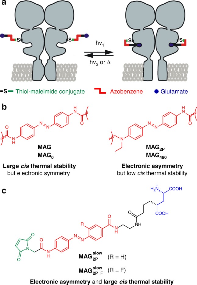
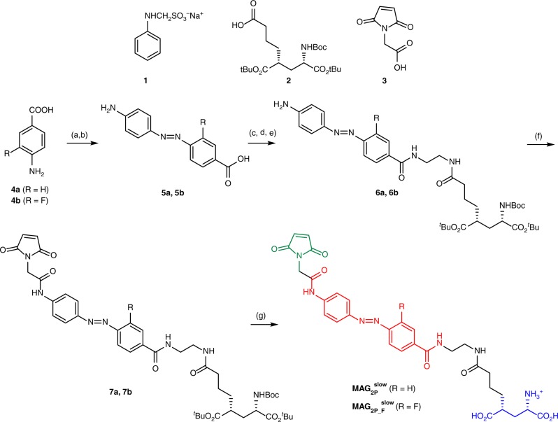
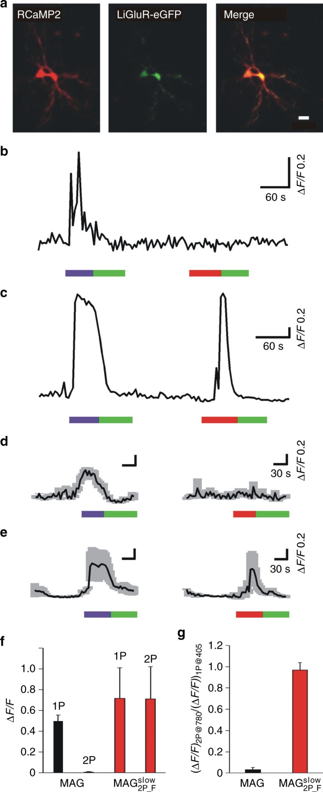
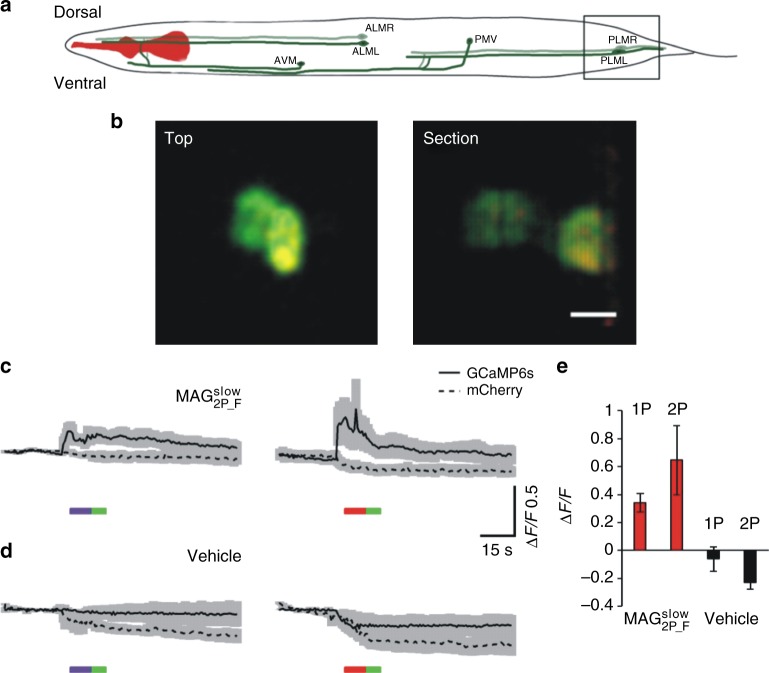
Manipulation of neuronal activity using two-photon excitation of azobenzene photoswitches with near-infrared light has been recently demonstrated, but their practical use in neuronal tissue to photostimulate individual neurons with three-dimensional precision has been hampered by firstly, the low efficacy and reliability of NIR-induced azobenzene photoisomerization compared to one-photon excitation, and secondly, the short cis state lifetime of the two-photon responsive azo switches. Here we report the rational design based on theoretical calculations and the synthesis of azobenzene photoswitches endowed with both high two-photon absorption cross section and slow thermal back-isomerization. These compounds provide optimized and sustained two-photon neuronal stimulation both in light-scattering brain tissue and in Caenorhabditis elegans nematodes, displaying photoresponse intensities that are comparable to those achieved under one-photon excitation. This finding opens the way to use both genetically targeted and pharmacologically selective azobenzene photoswitches to dissect intact neuronal circuits in three dimensions.


Conflict of interest statement

The authors declare no competing interest.

Figures

Fig. 1
Fig. 1
Strategy toward optimized azobenzene photoswitches for the two-photon (2P) excitation of light-gated glutamate receptors (LiGluR). a Operating mode of MAG-type photoswitchable tethered ligands (PTLs) on LiGluR, which are composed of three covalently tethered units: a glutamate ligand, an azobenzene core, and a maleimide group that binds to a cysteine residue genetically engineered in the receptor. Ultraviolet-visible (one-photon; 1P) or near-infrared (2P) light excitation induces glutamate recognition and channel opening via trans → cis isomerization, which results in ion flow across the membrane. This process is reverted by illumination with visible light excitation (1P) or thermal back-isomerization of the cis state of the switch. b Structures of the azobenzene cores of MAG, MAG0, MAG2P, and MAG460 PTLs proposed for the photoswitching of LiGluR under 1P and 2P excitation conditions. c Structures of PTLs MAG2Pslow and MAG2P-Fslow
Fig. 2
Fig. 2
Synthesis of MAG2Pslow and MAG2P-Fslow. Reagents and conditions: (a) 5.5 M HCl, NaNO2; (b) (i) 1, 0.86 M NaOAc; (ii) 1 M NaOH; (iii) 5.5 M HCl (38%, over the two steps, for 5a, 47% for 5b); (c) tert-butyl (2-aminoethyl)carbamate, N-ethyl-N′-(3-dimethyldiaminopropyl)-carbodiimide HCl (EDCI), 1-hydroxybenzotriazole hydrate (HOBt), diisopropylethylamine (DIPEA), THF; (d) 37% HCl, MeOH; (e) 2, EDCI, HOBt, DIPEA, THF (47%, over the three steps, for 6a, 49% for 6b); (f) (i) 3, ClCOCOCl, CH2Cl2, DMF; (ii) DIPEA, THF (86% for 7a, 84% for 7b); (g) trifluoroacetic acid (TFA), CH2Cl2 (80% for MAG2P-Fslow, quantitative yield for MAG2P-Fslow)
Fig. 3
Fig. 3
Photochemical and physiological characterization of MAG2Pslow and MAG2P-Fslow under one-photon (1P) stimulation. a Normalized absorption spectra of trans-MAG, trans-MAG2Pslow, trans-MAG2P-Fslow, and trans-MAG2P in 99% phosphate-buffered solution (PBS):1% dimethylsulfoxide (DMSO). b Thermal lifetimes of cis-MAG, cis-MAG2Pslow, cis-MAG2P-Fslow, and cis-MAG2P at room temperature in 99% PBS:1% DMSO. Errors from the monoexponentials fits to obtain τcis are shown. c Normalized 1P action spectra recorded using whole-cell patch-clamp in human embryonic kidney 293 (HEK293) cells expressing GluK2-L439C after conjugation to MAG, MAG2Pslow, MAG2P-Fslow, and MAG2P (n = 5, 7, 3, and 8 biologically independent cells, respectively). Errors are standard error of the mean (SEM). d Normalized 1P action spectra recorded using calcium imaging in HEK293 cells co-expressing GluK2-L439C and GCaMP6s after conjugation to MAG, MAG2Pslow, and MAG2P-Fslow (n = 33, 20, and 25 biologically independent cells, respectively). Errors are SEM. In c and d wavelength-dependent photoresponses were normalized to the maximum signal along the spectral range measured for each cell before averaging over different cells. Source data for c and d are provided as a source Data file
Fig. 4
Fig. 4
One-photon (1P) and two-photon (2P) stimulation of MAG, MAG2Pslow, and MAG2P-Fslow in cultured cells. a Individual (thin lines) and average (thick lines) calcium imaging fluorescence traces recorded for human embryonic kidney 293 (HEK293) cells co-expressing GluK2-L439C and R-GECO1 after conjugation to MAG (n = 16 biologically independent cells), MAG2Pslow (n= 14 biologically independent cells), and MAG2P-Fslow (n= 34 biologically independent cells). The bands around average traces plot the corresponding SEM. Both 1P (violet, 405 nm, power density = 0.37 mW μm−2) and 2P excitation scans (red, 780 nm, power density = 2.8 mW μm−2) were applied to open LiGluR channels and trigger calcium-induced R-GECO1 fluorescence enhancement, while 1P excitation scans (green, 514 nm, power density = 0.35 mW μm−2) were applied to revert back the process. b Repetitive 2P-induced calcium imaging fluorescence responses recorded in five different HEK293 cells co-expressing GluK2-L439C and R-GECO1 after conjugation to MAG2P-Fslow. Source data for a are provided as a source Data file
Fig. 5
Fig. 5
Average two-photon (2P) activity of MAG, MAG2Pslow, and MAG2P-Fslow in cultured cells. a 2P action spectra of MAG, MAG2Pslow, and MAG2P-Fslow after conjugation to GluK2-L439C-expressing human embryonic kidney 293 (HEK293) cells. Fluorescence calcium responses were measured using R-GECO1. Before averaging over different cells, the 2P responses of each cell were normalized with respect to the 1P response at 405 nm (MAG: 740, 760, 780, 800, and 820 nm; n = 28, 33, 40, 7, and 12 biologically independent cells, respectively; MAG2Pslow: 740, 760, 780, 800, and 820 nm; n = 20, 9, 12, 16, and 17 biologically independent cells, respectively; and MAG2P-Fslow: 720, 740, 760, 780, 800, 820, and 840 nm; n = 14, 17, 18, 86, 15, 23, and 17 biologically independent cells, respectively). Errors are SEM. b Ratio between the 2P and 1P responses of MAG, MAG2Pslow, and MAG2P-Fslow for the same cells excited at 780 and 405 nm, respectively. Errors are SEM. c Reliability of the 2P calcium imaging response elicited in GluK2-L439C-expressing HEK293 cells after conjugation to MAG, MAG2Pslow, and MAG2P-Fslow (n= 72, 25, and 86 biologically independent cells, respectively). Reliability is expressed as the percentage of transfected cells showing measurable 2P stimulation signals. Source data for a and b are provided as a source Data file
Fig. 6
Fig. 6
Two-photon (2P) Ca2+ photoresponses in rat hippocampal organotypic slices expressing GluK2-L439C-eGFP and RCaMP2. a Microphotograph of a neuron expressing both RCaMP2 (red) and GluK2-L439C-eGFP (green) (scale bar = 20 μm). b, c Real time traces of a single-cell neuronal activity of slices incubated with b MAG or c MAG2P-Fslow. d, e Average one-photon (1P) and 2P responses of neurons incubated with d MAG (n= 3 biologically independent cells) or e MAG2P-Fslow (n = 6 biologically independent cells). In be 1P stimulation was performed at 405  nm (purple bar, 0.81 mW μm−2) and 514 nm (green bar, 0.35 mW μm−2), and 2P stimulation at 780 nm (red bar, 2.8 mW μm−2). f, g Quantification of photoresponses in slices incubated with MAG (black bars, n = 5 biologically independent cells) and MAG2P-Fslow (red bars, n = 6 biologically independent cells): f fluorescence enhancement; g ratio between the 2P and 1P responses of MAG and MAG2P-Fslow for the same cells. Error bars are SEM. Source data for dg are provided as a source Data file
Fig. 7
Fig. 7
In vivo calcium induced photoresponses by two-photon (2P) stimulation of MAG2P-Fslow in C. elegans. a Schematics of C. elegans in which touch receptor neurons (TRNs) are depicted. Squared region is magnified in b. b Microphotograph of an animal expressing LiGluR-mCherry (red) and GCaMP6s (green) (scale bar = 5 μm). Top and lateral section view of TRN from the tail. c, d Average traces of one-photon (1P)- and 2P-induced photoactivation of TRNs in animals treated with c MAG2P-Fslow (n = 5 and 6 cells from four different animals experiments for 1P and 2P traces, respectively) or d with vehicle (n = 5 and 6 cells from four different animals experiments for 1P and 2P traces, respectively). Continuous line trace indicates GCaMP6s fluorescence signal and dashed trace mCherry fluorescence. In c and d 2P stimulation was performed at 780 nm (red bar, 2.8 mW mm−2) and 1P stimulation at 405 nm (purple bar, 15 µW mm−2) and 514 nm (green bar, 1.21 µW mm−2). e Quantification of photoresponses (fluorescence enhancement) in animals injected with MAG2P-Fslow (red bars, n = 5 cells from four different animals experiments) and vehicle (black bars, n = 5 cells from four different animals experiments). Error bars are SEM. Source data for ce are provided as a source Data file

References

    1. Bandara HMD, Burdette SC. Photoisomerization in different classes of azobenzene. Chem. Soc. Rev. 2012;41:1809–1825. doi: 10.1039/C1CS15179G. - DOI - PubMed
    1. Ankenbruck N, Courtney T, Naro Y, Deiters A. Optochemical control of biological processes in cells and animals. Angew. Chem. Int. Ed. 2018;57:2768–2798. doi: 10.1002/anie.201700171. - DOI - PMC - PubMed
    1. Broichhagen J, Frank JA, Trauner D. A roadmap to success in photopharmacology. Acc. Chem. Res. 2015;48:1947–1960. doi: 10.1021/acs.accounts.5b00129. - DOI - PubMed
    1. Lerch MM, Hansen MJ, van Dam GM, Szymanski W, Feringa BL. Emerging targets in photopharmacology. Angew. Chem. Int. Ed. 2016;55:10978–10999. doi: 10.1002/anie.201601931. - DOI - PubMed
    1. Gorostiza P, Isacoff EY. Optical switches for remote and noninvasive control of cell signaling. Science. 2008;322:395–399. doi: 10.1126/science.1166022. - DOI - PMC - PubMed

Publication types