

Moraxella catarrhalis NucM is an entry nuclease involved in extracellular DNA and RNA degradation, cell competence and biofilm scaffolding
- PMID: 30796312
- PMCID: PMC6384898
- DOI: 10.1038/s41598-019-39374-0
Moraxella catarrhalis NucM is an entry nuclease involved in extracellular DNA and RNA degradation, cell competence and biofilm scaffolding
Abstract
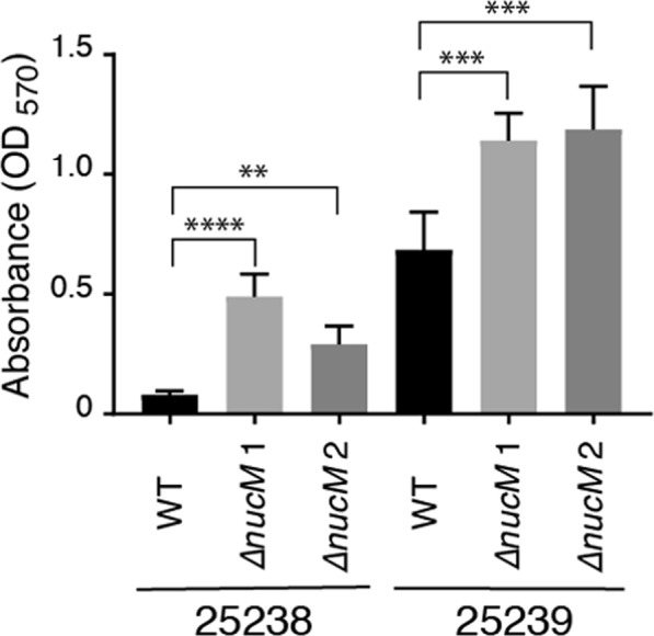
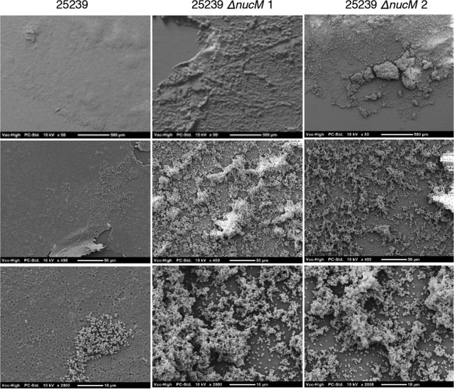
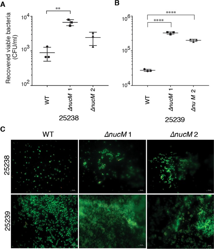
Moraxella catarrhalis is a host-adapted bacterial pathogen that causes otitis media and exacerbations of chronic obstructive pulmonary disease. This study characterises the conserved M. catarrhalis extracellular nuclease, a member of the ββα metal finger family of nucleases, that we have named NucM. NucM shares conserved sequence motifs from the ββα nuclease family, including the DRGH catalytic core and Mg2+ co-ordination site, but otherwise shares little primary sequence identity with other family members, such as the Serratia Nuc and pneumococcal EndA nucleases. NucM is secreted from the cell and digests linear and circular nucleic acid. However, it appears that a proportion of NucM is also associated with the cell membrane and acts as an entry nuclease, facilitating transformation of M. catarrhalis cells. This is the first example of a ββα nuclease in a Gram negative bacteria that acts as an entry nuclease. In addition to its role in competence, NucM affects cell aggregation and biofilm formation by M. catarrhalis, with ΔnucM mutants having increased biofilm biomass. NucM is likely to increase the ability of cells to survive and persist in vivo, increasing the virulence of M. catarrhalis and potentially affecting the behaviour of other pathogens that co-colonise the otorhinolaryngological niche.
Conflict of interest statement
The authors declare no competing interests.
Figures
Similar articles
-
The Moraxella catarrhalis phase-variable DNA methyltransferase ModM3 is an epigenetic regulator that affects bacterial survival in an in vivo model of otitis media.BMC Microbiol. 2019 Dec 9;19(1):276. doi: 10.1186/s12866-019-1660-y. BMC Microbiol. 2019. PMID: 31818247 Free PMC article.
-
The Respiratory Pathogen Moraxella catarrhalis Targets Collagen for Maximal Adherence to Host Tissues.mBio. 2016 Mar 22;7(2):e00066. doi: 10.1128/mBio.00066-16. mBio. 2016. PMID: 27006460 Free PMC article.
-
Contribution of Moraxella catarrhalis type IV pili to nasopharyngeal colonization and biofilm formation.Infect Immun. 2007 Dec;75(12):5559-64. doi: 10.1128/IAI.00946-07. Epub 2007 Oct 1. Infect Immun. 2007. PMID: 17908808 Free PMC article.
-
Otitis media pathogens - A life entrapped in biofilm communities.Crit Rev Microbiol. 2019 Sep-Nov;45(5-6):595-612. doi: 10.1080/1040841X.2019.1660616. Epub 2019 Sep 10. Crit Rev Microbiol. 2019. PMID: 31502909 Review.
-
Molecular aspects of Moraxella catarrhalis pathogenesis.Microbiol Mol Biol Rev. 2009 Sep;73(3):389-406, Table of Contents. doi: 10.1128/MMBR.00007-09. Microbiol Mol Biol Rev. 2009. PMID: 19721084 Free PMC article. Review.
Cited by
-
Panel 7 - Pathogenesis of otitis media - a review of the literature between 2015 and 2019.Int J Pediatr Otorhinolaryngol. 2020 Mar;130 Suppl 1:109838. doi: 10.1016/j.ijporl.2019.109838. Epub 2019 Dec 19. Int J Pediatr Otorhinolaryngol. 2020. PMID: 31879085 Free PMC article. Review.
-
The majority of microorganisms in gas hydrate-bearing subseafloor sediments ferment macromolecules.Microbiome. 2023 Mar 2;11(1):37. doi: 10.1186/s40168-023-01482-5. Microbiome. 2023. PMID: 36864529 Free PMC article.
-
Biological Characteristics of Listeria monocytogenes Following Deletion of TatD-like Protein Gene.Curr Microbiol. 2023 Feb 28;80(4):118. doi: 10.1007/s00284-023-03229-9. Curr Microbiol. 2023. PMID: 36853439
-
The Moraxella catarrhalis phase-variable DNA methyltransferase ModM3 is an epigenetic regulator that affects bacterial survival in an in vivo model of otitis media.BMC Microbiol. 2019 Dec 9;19(1):276. doi: 10.1186/s12866-019-1660-y. BMC Microbiol. 2019. PMID: 31818247 Free PMC article.
-
Studies on Bd0934 and Bd3507, Two Secreted Nucleases from Bdellovibrio bacteriovorus, Reveal Sequential Release of Nucleases during the Predatory Cycle.J Bacteriol. 2020 Aug 25;202(18):e00150-20. doi: 10.1128/JB.00150-20. Print 2020 Aug 25. J Bacteriol. 2020. PMID: 32601070 Free PMC article.
References
-
- Baldwin RL. Effects of otitis media on child development. Am. J. Otol. 1993;14:601–604. - PubMed
Publication types
MeSH terms
Substances
LinkOut - more resources
Full Text Sources
Miscellaneous
