. 2019 Feb 22;9(1):2600.
doi: 10.1038/s41598-019-39580-w.

Absence of Global Stress Regulation in Escherichia coli Promotes Pathoadaptation and Novel c-di-GMP-dependent Metabolic Capability

Affiliations

Absence of Global Stress Regulation in Escherichia coli Promotes Pathoadaptation and Novel c-di-GMP-dependent Metabolic Capability

Nikola Zlatkov et al. Sci Rep. .

Abstract

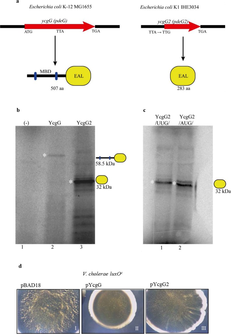
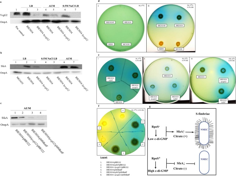
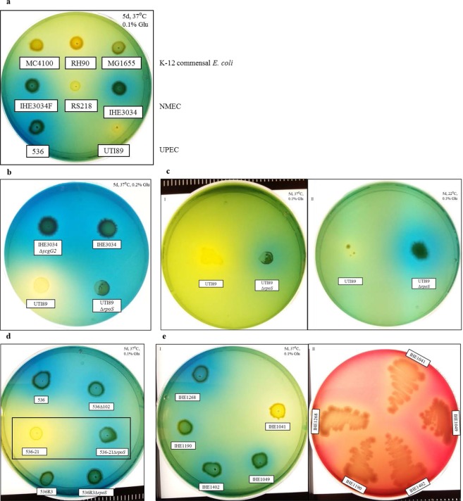
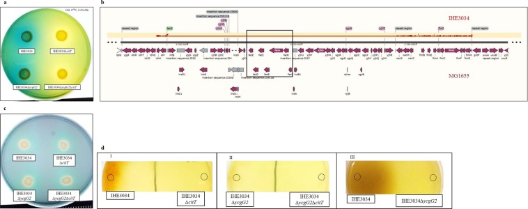
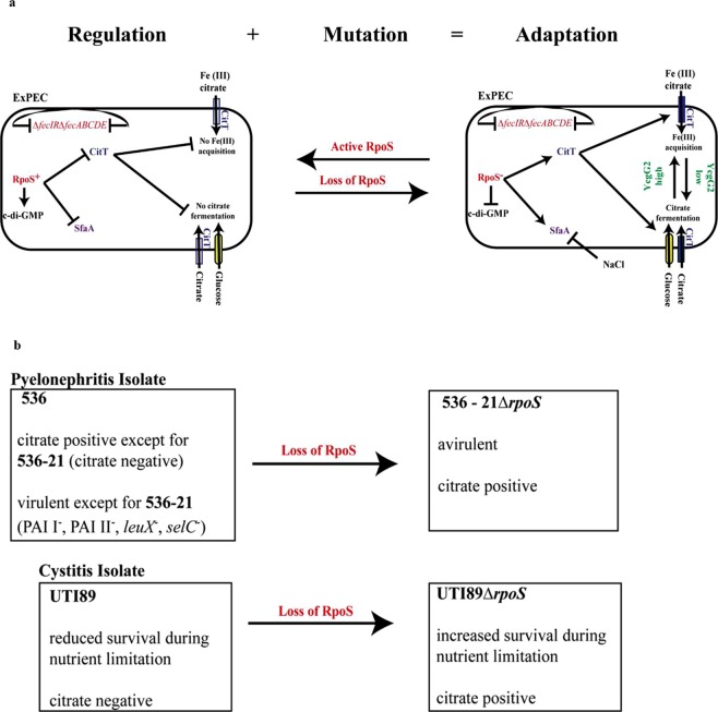
Pathoadaptive mutations linked to c-di-GMP signalling were investigated in neonatal meningitis-causing Escherichia coli (NMEC). The results indicated that NMEC strains deficient in RpoS (the global stress regulator) maintained remarkably low levels of c-di-GMP, a major bacterial sessility-motility switch. Deletion of ycgG2, shown here to encode a YcgG allozyme with c-di-GMP phosphodiesterase activity, and the restoration of RpoS led to a decrease in S-fimbriae, robustly produced in artificial urine, hinting that the urinary tract could serve as a habitat for NMEC. We showed that NMEC were skilled in aerobic citrate utilization in the presence of glucose, a property that normally does not exist in E. coli. Our data suggest that this metabolic novelty is a property of extraintestinal pathogenic E. coli since we reconstituted this ability in E. coli UTI89 (a cystitis isolate) via deactivation rpoS; additionally, a set of pyelonephritis E. coli isolates were shown here to aerobically use citrate in the presence of glucose. We found that the main reason for this metabolic capability is RpoS inactivation leading to the production of the citrate transporter CitT, exploited by NMEC for ferric citrate uptake dependent on YcgG2 (an allozyme with c-di-GMP phosphodiesterase activity).


Conflict of interest statement

The authors declare no competing interests.

Figures

Figure 1
Figure 1
RpoS deficiency in NMEC. (a) Low c-di-GMP levels of NMEC strains (RS218, IHE3034F and IHE3034) compared to those of UPEC strains (536 and UTI89) due to RpoS deactivation. When RpoS activity was restored, the c-di-GMP levels of IHE3034 reached the levels of the K-12 MG1655 strain. (Data are represented as the mean +/− SD). (b) Schematic comparison between NMEC and UPEC. NMEC is schematically present in blue, while a UPEC cell that has the same K1 serotype (such as UTI89) of NMEC is shown in red. The common features are present in the intersection. (c) Immunoblotting against RpoS of different E. coli groups. Strain RH90 (MC4100∆rpoS) was used as a negative control. H-NS levels were used as a loading control. (d) Rugose colony formation by IHE3034 derivative strains with restored RpoS. Only the strains having pMMkatF showed rugose colony formation at 22 °C.
Figure 2
Figure 2
Properties of ycgG2. (a) Schematic comparison between ycgG and ycgG2 and their products. Abbreviations: MBD – membrane-binding domain; EAL – a c-di-GMP PDE domain. (b) In vitro transcription/translation of ycgG (lane 2) and ycgG2 (lane 3). Protein products indicated with a white star. The identity of YcgG2 was confirmed via ESI-MS analysis as described in the Methods. (c) In vitro transcription/translation of ycgG2 from its native TTG start codon (lane 2) and from the ATG start codon (lane 3). Protein products indicated with a white star. (d) Ectopic expression of YcgG and Ycg2 in the V. cholerae luxOc background.
Figure 3
Figure 3
Effects of the absence of RpoS and YcgG2 on IHE3034 bacteria. For the citrate utilization assays, all conditions, i.e., days (d), temperature (°C) and percentage of glucose added (%), are stated in the upper corner of the subfigures. (a) YcgG2 levels of IHE3034 bacteria cultured in LB (sample 2), AUM (sample 4) and LB supplemented with 0.5 M NaCl (sample 6). Positive control – in vitro-produced YcgG2. ∆ycgG2 bacterial extract, used as a negative control, showed very faint bands due to cross-reactivity of the antibody (samples 3 and 5). (b) SfaA levels of IHE3034 and ∆ycgG2 bacteria cultured in LB, AUM and LB supplemented with 0.5 M NaCl. (c) SfaA levels of IHE3034 and ∆ycgG2 bacteria with restored RpoS (strains 3 and 4, respectively) activity cultured in AUM compared to the levels of the empty-vector strains (strains 1 and 2). (d) Citrate utilization by IHE3034 cells. Box I: Lack of bacterial growth when citrate was provided as a single carbon and energy source. Box II: Citrate utilization by IHE3034 bacteria when glucose was provided as a co-substrate. Representatives of the rest of the strains utilizing only glucose, i.e., MC4100 and RH90 (MC4100∆rpoS), were used as negative controls. (e) Citrate utilization by IHE3034, ∆ycgG2 and ∆sfaY bacteria on Simmon’s plates supplemented with 0.1% glucose and incubated at 37 °C (Box I) and at 22 °C (Box II), and with 0.2% glucose at 22 °C (Box III). (f) Citrate utilization by IHE3034, ∆ycgG2 and ∆sfaY bacteria with restored RpoS activity compared to that of their empty-vector derivatives. (g) Schematic illustration of the effects of RpoS activity on S-fimbrial expression.
Figure 4
Figure 4
Aerobic citrate utilization by different ExPEC bacteria on Simmon’s plates when a co-substrate (glucose) is provided. All conditions, i.e., days (d), temperature (°C) and percentage of glucose added (%), are stated in the upper corner of the subfigures. (a) Citrate utilization by different E. coli groups, i.e., NMEC (IHE3034, IHE3034F and RS218), UPEC (UTI89 and 536), compared to that of the commensal MG1655. (b) Reconstitution of citrate uptake signalling events in the uropathogenic E. coli K1 strain UTI89. The wild-type UTI89 strain did not utilize citrate, while its RpoS-deficient variant (UTI89∆rpoS) showed positive results compared to those of IHE3034 and IHE3034 ∆ycgG2. (c) UTI89 and UTI89∆rpoS bacteria tested for citrate utilization in the presence of 0.1% glucose at 37 °C (Box I) and at 22 °C (Box II). (d) The 536 pyelonephritis isolate, its less virulent derivatives, 536102 and 536R3, and its avirulent derivative, 536-21, were tested for citrate utilization. The 536-21 derivative tested negative, but its ∆rpoS mutant was positive for citrate utilization (marked with black box), while the 536R3∆rpoS strain still tested positive for citrate, like its parental strain. (e) Different pyelonephritis clinical isolates tested for citrate utilization (Box I) and for haemolysis (Box II).
Figure 5
Figure 5
Analysis of IHE3034 bacteria for Fe(III) uptake. (a) Citrate utilization by IHE3034 and IHE3034∆ycgG2 bacteria compared to that of their ∆citT mutant derivatives. (b) Graphic representation of the local alignment of the sequence (shown in red) of the IHE3034 genome (coordinates 4927723–4959109) carrying the fecIRfecABCDE deletion against the genome of MG1655. Deletion of the operons in the IHE3034 genome is indicated with a black frame. (c) The general ability of IHE3034 and its ∆citT, ∆ycgG2 and ∆ycgG2citT mutants to undergo Fe(III) acquisition, as tested on a CAS plate. The yellow halo around the bacteria indicated a positive reaction for siderophore production. (d) Fe(III) citrate utilization assay (sites of inoculation marked with rings). Box I: Fe(III) citrate utilization by IHE3034 compared to that of its ∆citT mutant. Box II: Fe(III) citrate utilization by IHE3034 ∆ycgG2 compared to that of its ∆citT mutant. Box III: Reduced Fe(III) citrate utilization by wild-type IHE3034∆ycgG2 compared to that of wild-type IHE3034.
Figure 6
Figure 6
A model representing the pathoadaptive events in RpoS-deficient ExPEC strains. (a) Regulatory and mutational events define bacterial adaptation, exemplified by ExPEC and especially NMEC, which has naturally lost the RpoS regulator. Loss of RpoS leads to citrate utilization and ferric citrate uptake in nutrient-limited environments. RpoS deactivation (in red) leads to low c-di-GMP levels and triggers the production of CitT (in blue) and SfaA (in purple). Once CitT (in blue) is expressed, the bacteria can take in citrate and further ferment it, stimulated by low YcgG2 levels (in green). Low YcgG2 levels also lead to a decrease in SfaA (in purple) production. Due to the lack of fecIRfecABCDE (shown in red), the bacteria utilize CitT (in blue) to import ferrous iron citrate, whose utilization is further increased in the absence of YcgG2 (shown in green). These results are abolished when RpoS is restored. (b) The signalling events leading to metabolic novelty can be restored in UPEC. In the case of pyelonephritis isolates, citrate utilization is naturally occurring in the virulent 536 derivatives, while in the avirulent 536-21 strain, which is citrate-negative, utilization can be restored upon loss of RpoS. The UTI89 cystitis isolate does not utilize citrate, but utilization can be restored in the absence of RpoS, which also increases the survival of UTI89 in restrictive environments.

Similar articles

Cited by

References

    1. Boone, D. R., Castenholz, R. W. & Garrity, G. M. Bergey’s manual of systematic bacteriology. 2nd edn, (Springer, 2001).
    1. Koser SA. Correlation of Citrate Utilization by Members of the Colon-Aerogenes Group with Other Differential Characteristics and with Habitat. J Bacteriol. 1924;9:59–77. - PMC - PubMed
    1. Lutgens M, Gottschalk G. Why a co-substrate is required for anaerobic growth of Escherichia coli on citrate. J Gen Microbiol. 1980;119:63–70. doi: 10.1099/00221287-119-1-63. - DOI - PubMed
    1. Pos KM, Dimroth P, Bott M. The Escherichia coli citrate carrier CitT: a member of a novel eubacterial transporter family related to the 2-oxoglutarate/malate translocator from spinach chloroplasts. J Bacteriol. 1998;180:4160–4165. - PMC - PubMed
    1. Ishiguro N, Oka C, Sato G. Isolation of citrate-positive variants of Escherichia coli from domestic pigeons, pigs, cattle, and horses. Appl Environ Microbiol. 1978;36:217–222. - PMC - PubMed

Publication types

MeSH terms