. 2019 Sep;10(9):649-667.
doi: 10.1007/s13238-019-0610-7. Epub 2019 Feb 22.

Telomere-dependent and telomere-independent roles of RAP1 in regulating human stem cell homeostasis

Affiliations

Telomere-dependent and telomere-independent roles of RAP1 in regulating human stem cell homeostasis

Xing Zhang et al. Protein Cell. 2019 Sep.

Abstract

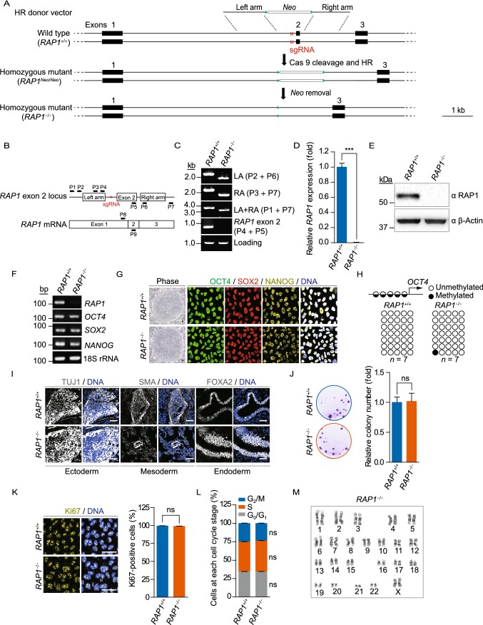
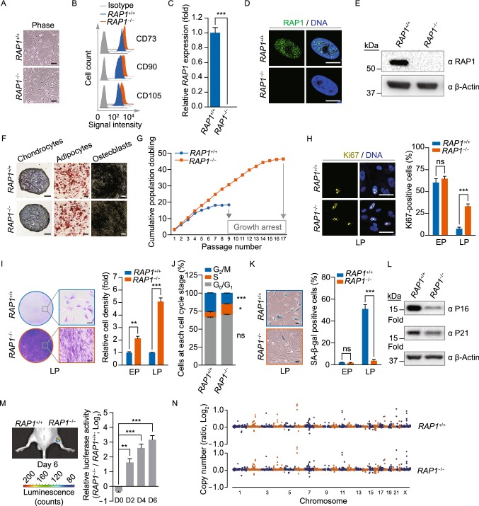
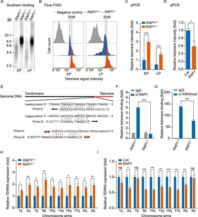
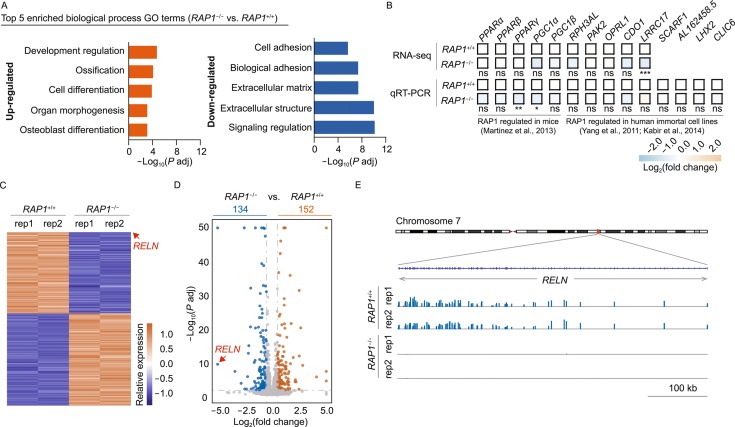
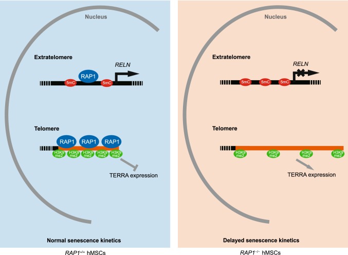
RAP1 is a well-known telomere-binding protein, but its functions in human stem cells have remained unclear. Here we generated RAP1-deficient human embryonic stem cells (hESCs) by using CRISPR/Cas9 technique and obtained RAP1-deficient human mesenchymal stem cells (hMSCs) and neural stem cells (hNSCs) via directed differentiation. In both hMSCs and hNSCs, RAP1 not only negatively regulated telomere length but also acted as a transcriptional regulator of RELN by tuning the methylation status of its gene promoter. RAP1 deficiency enhanced self-renewal and delayed senescence in hMSCs, but not in hNSCs, suggesting complicated lineage-specific effects of RAP1 in adult stem cells. Altogether, these results demonstrate for the first time that RAP1 plays both telomeric and nontelomeric roles in regulating human stem cell homeostasis.

Keywords: RAP1; RELN; methylation; stem cell; telomere.


Conflict of interest statement

Xing Zhang, Zunpeng Liu, Xiaoqian Liu, Si Wang, Yiyuan Zhang, Xiaojuan He, Shuhui Sun, Shuai Ma, Ng Shyh-Chang, Feng Liu, Qiang Wang, Xiaoqun Wang, Lin Liu, Weiqi Zhang, Moshi Song, Guang-Hui Liu and Jing Qu declare that they have no conflict of interest. All institutional and national guidelines for the care and use of laboratory animals were followed.

Figures

Figure 1
Figure 1
Generation and characterization of RAP1−/− hESCs. (A) Schematic representation of the deletion of the exon 2 of RAP1 in hESCs via CRISPR/Cas9-facilitated HR. The green triangles represented FRT sites and the red cross demonstrated the region of sgRNA. (B) Schematic representation of the primers used for genomic PCR and qRT-PCR to confirm RAP1 knockout. (C) Genomic PCR analysis demonstrated that the exon 2 of RAP1 was deleted from the genome. LA and RA represented left and right homology arm, respectively. (D) qRT-PCR analysis demonstrated the deletion of RAP1 at the transcriptional level in RAP1−/− hESCs by primers P8 and P9. Data were presented as the mean ± SEM, n = 3. ***P < 0.001. (E) Western blotting analysis verified the absence of RAP1 in RAP1−/− hESCs. β-Actin was used as a loading control. (F) RT-PCR for pluripotency markers demonstrated comparable expression in WT and RAP1−/− hESCs. 18S rRNA was used as a loading control. (G) Representative brightfield and immunofluorescence micrographs of WT and RAP1−/− hESCs showed normal morphology of hESCs and comparable expression of pluripotency markers, respectively. Scale bar, 50 µm. (H) DNA methylation status of the OCT4 promoter in WT and RAP1−/− hESCs showed normal hypomethylation. (I) Immunostaining of representative markers of the three germ layers in teratomas formed by WT and RAP1−/− hESCs. Scale bar, 50 µm. (J) Clonal expansion analysis of WT and RAP1−/− hESCs. Data were presented as the mean ± SEM, n = 3. NS, not significant. (K) Immunostaining of the proliferation marker Ki67 in WT and RAP1−/− hESCs. Scale bar, 50 µm. Data were presented as the mean ± SEM, n = 6. NS, not significant. (L) Cell cycle analysis of WT and RAP1−/− hESCs. Data were presented as the mean ± SEM, n = 3. NS, not significant. (M) Karyotype analysis of RAP1−/− hESCs showed normal karyotype. n = 10
Figure 2
Figure 2
RAP1−/− hMSCs exhibited retarded cellular senescence. (A) Brightfield micrographs of hMSCs showed normal morphology. Scale bar, 50 µm. (B) Flow cytometry demonstrated that sorted hMSCs uniformly expressed the MSC-specific surface markers CD73, CD90 and CD105. (C) qRT-PCR analysis demonstrated the deletion of RAP1 at the transcriptional level in RAP1−/− hMSCs by primers P8 and P9. Data were presented as the mean ± SEM, n = 3. ***P < 0.001. (D) Immunofluorescence micrographs of RAP1 in WT and RAP1−/− hMSCs. Scale bar, 10 µm. (E) Western blotting analysis demonstrated the absence of RAP1 in RAP1−/− hMSCs. β-Actin was used as a loading control. (F) Characterization of the differentiation potential of hMSCs. Toluidine blue O, Oil Red O and Von Kossa staining were used to detect chondrocytes, adipocytes and osteoblasts, respectively. Scale bar, 200 µm. (G) Cell growth curves showed the enhanced proliferation ability of RAP1−/− hMSCs. (H) Immunostaining of the proliferation marker Ki67 in WT and RAP1−/− hMSCs. EP (early passage) and LP (late passage) represented P2 and P9, respectively. Scale bar, 50 µm. Data were presented as the mean ± SEM, n = 6. NS, not significant, ***P < 0.001. (I) Clonal expansion analysis of WT and RAP1−/− hMSCs. EP and LP represented P2 and P9, respectively. Scale bar, 50 µm. Data were presented as the mean ± SEM, n = 3. **P < 0.01, ***P < 0.001. (J) Cell cycle analysis of WT and RAP1−/− hMSCs (LP). Data were presented as the mean ± SEM, n = 3. NS, not significant, *P < 0.05, ***P < 0.001. (K) SA-β-gal staining of WT and RAP1−/− hMSCs. EP and LP represented P2 and P9, respectively. Scale bar, 50 μm. Data were presented as the mean ± SEM, n = 6. NS, not significant, ***P < 0.001. (L) Western blotting analysis showed decreased expression of P16 and P21 in RAP1−/− hMSCs (LP). β-Actin was used as a loading control. (M) Photon flux from TA muscle implanted with WT (left) and RAP1−/− (right) hMSCs (LP) expressing luciferase. Data were presented as the mean ± SEM of RAP1−/− to WT ratios (Log2(fold change)), n = 5. **P < 0.01, ***P < 0.001. (N) Identification of CNVs by whole-genome sequencing analysis in WT and RAP1−/− hMSCs (EP) showed genomic integrity in RAP1−/− hMSCs
Figure 3
Figure 3
RAP1−/− hMSCs contained longer telomeres. (A) Terminal restriction fragment analysis of WT and RAP1−/− hMSCs by Southern blotting demonstrated longer telomeres in RAP1−/− hMSCs. EP and LP represented P2 and P9, respectively. (B) Telomere length analysis of WT and RAP1−/− hMSCs by flow FISH showed a right shift of the telomere signal in RAP1−/− hMSCs. EP and LP represented P2 and P9, respectively. (C) Telomere length analysis of WT and RAP1−/− hMSCs by qPCR. EP and LP represented P2 and P9, respectively. Data were presented as the mean ± SEM, n = 3. ***P < 0.001. (D) Telomere length analysis of RAP1−/− hMSCs expressing luciferase (Luc) or RAP1 by qPCR. Data were presented as the mean ± SEM, n = 3. *P < 0.05. (E) Schematic representation of the primers designed for telomere detection explaining how primer A and B specifically amplified telomeres rather than forming primer dimers. (F) ChIP-PCR analysis of RAP1 enrichment at the telomeres in WT and RAP1−/− hMSCs (EP). Data were presented as mean ± SEM, n = 3. ***P < 0.001. (G) ChIP-PCR analysis of H3K9me2 enrichment at the telomeres in WT and RAP1−/− hMSCs (EP) indicated a looser telomere structure in RAP1−/− hMSCs. Data were presented as the mean ± SEM, n = 3. ***P < 0.001. (H) qRT-PCR analysis showed that RAP1−/− hMSCs (EP) expressed more TERRA. Data were presented as the mean ± SEM, n = 3. NS, not significant, *P < 0.05, **P < 0.01. (I) TERRA analysis of RAP1−/− hMSCs expressing luciferase or RAP1 by qRT-PCR. Data were presented as the mean ± SEM, n = 3. NS, not significant, *P < 0.05, **P < 0.01, ***P < 0.001
Figure 4
Figure 4
RAP1 regulated the expression of RELN in hMSCs. (A) Biological process GO enrichment analysis of differentially expressed genes in RAP1−/− vs. WT hMSCs (EP). The top 5 enriched biological process GO terms were shown. (B) Heatmap of reported genes regulated by RAP1 in mice or human immortalized cell lines. Relative expression of indicated genes tested by RNA-seq and qRT-PCR demonstrated comparable levels in WT and RAP1−/− hMSCs (EP). n = 3 (qRT-PCR) or 2 (RNA-seq). NS, not significant, *P(qRT-PCR) or P adj (RNA-seq) < 0.05, **P or P adj < 0.01, ***P or P adj < 0.001. (C) Heatmap of differentially expressed genes in RAP1−/− vs. WT hMSCs (EP). RELN was labeled by an arrow. (D) Volcano plot of differentially expressed genes in RAP1−/− vs. WT hMSCs (EP). RELN was labeled by an arrow. (E) Transcriptional signals of RELN in RAP1−/− vs. WT hMSCs (EP). Transcriptional signals were normalized by RPKM at a bin size of 10 bp. (F) qRT-PCR analysis verified that RELN was downregulated in RAP1−/− hMSCs (EP). Data were presented as the mean ± SEM, n = 3. ***P < 0.001. (G) qRT-PCR analysis demonstrated that expressing RAP1 partially rescued the expression of RELN in RAP1−/− hMSCs. Data were presented as the mean ± SEM, n = 3. *P < 0.05. (H) Diagram showing that RAP1 bound immediately upstream of the TSS of RELN in WT hMSCs (EP). ChIP-PCR analysis showed that RAP1 was enriched at the RELN promoter region. Data were presented as the mean ± SEM, n = 3. **P < 0.01. (I) Methylation-specific PCR analysis of the RELN promoter in WT and RAP1−/− hMSCs (EP) demonstrated hypermethylation of the RELN promoter in RAP1−/− hMSCs. Data were presented as the mean ± SEM, n = 3. **P < 0.01. (J) qRT-PCR analysis demonstrated that RELN shRNA (sh RELN) effectively decreased the mRNA of RELN than control shRNA (sh CTRL). Data were presented as the mean ± SEM, n = 3. **P < 0.01. (K) Cell growth curves showed that reducing the expression of RELN enhanced the proliferation ability of WT hMSCs. (L) Immunostaining of the proliferation marker Ki67 in WT hMSCs transfected with control or RELN shRNA. Scale bar, 50 μm. Data were presented as the mean ± SEM, n = 6. ***P < 0.001. (M) Clonal expansion analysis of WT hMSCs transfected with control or RELN shRNA. Scale bar, 50 μm. Data were presented as the mean ± SEM, n = 3. **P < 0.01. (N) SA-β-gal staining of WT hMSCs transfected with control or RELN shRNA. Scale bar, 50 μm. Data were presented as the mean ± SEM, n = 6. ***P < 0.001
Figure 4
Figure 4
RAP1 regulated the expression of RELN in hMSCs. (A) Biological process GO enrichment analysis of differentially expressed genes in RAP1−/− vs. WT hMSCs (EP). The top 5 enriched biological process GO terms were shown. (B) Heatmap of reported genes regulated by RAP1 in mice or human immortalized cell lines. Relative expression of indicated genes tested by RNA-seq and qRT-PCR demonstrated comparable levels in WT and RAP1−/− hMSCs (EP). n = 3 (qRT-PCR) or 2 (RNA-seq). NS, not significant, *P(qRT-PCR) or P adj (RNA-seq) < 0.05, **P or P adj < 0.01, ***P or P adj < 0.001. (C) Heatmap of differentially expressed genes in RAP1−/− vs. WT hMSCs (EP). RELN was labeled by an arrow. (D) Volcano plot of differentially expressed genes in RAP1−/− vs. WT hMSCs (EP). RELN was labeled by an arrow. (E) Transcriptional signals of RELN in RAP1−/− vs. WT hMSCs (EP). Transcriptional signals were normalized by RPKM at a bin size of 10 bp. (F) qRT-PCR analysis verified that RELN was downregulated in RAP1−/− hMSCs (EP). Data were presented as the mean ± SEM, n = 3. ***P < 0.001. (G) qRT-PCR analysis demonstrated that expressing RAP1 partially rescued the expression of RELN in RAP1−/− hMSCs. Data were presented as the mean ± SEM, n = 3. *P < 0.05. (H) Diagram showing that RAP1 bound immediately upstream of the TSS of RELN in WT hMSCs (EP). ChIP-PCR analysis showed that RAP1 was enriched at the RELN promoter region. Data were presented as the mean ± SEM, n = 3. **P < 0.01. (I) Methylation-specific PCR analysis of the RELN promoter in WT and RAP1−/− hMSCs (EP) demonstrated hypermethylation of the RELN promoter in RAP1−/− hMSCs. Data were presented as the mean ± SEM, n = 3. **P < 0.01. (J) qRT-PCR analysis demonstrated that RELN shRNA (sh RELN) effectively decreased the mRNA of RELN than control shRNA (sh CTRL). Data were presented as the mean ± SEM, n = 3. **P < 0.01. (K) Cell growth curves showed that reducing the expression of RELN enhanced the proliferation ability of WT hMSCs. (L) Immunostaining of the proliferation marker Ki67 in WT hMSCs transfected with control or RELN shRNA. Scale bar, 50 μm. Data were presented as the mean ± SEM, n = 6. ***P < 0.001. (M) Clonal expansion analysis of WT hMSCs transfected with control or RELN shRNA. Scale bar, 50 μm. Data were presented as the mean ± SEM, n = 3. **P < 0.01. (N) SA-β-gal staining of WT hMSCs transfected with control or RELN shRNA. Scale bar, 50 μm. Data were presented as the mean ± SEM, n = 6. ***P < 0.001
Figure 5
Figure 5
RAP1 deficiency did not influence the proliferation or senescence of hNSCs. (A) Brightfield and immunofluorescence micrographs of WT and RAP1−/− hNSCs showed normal morphology and expression of the NSC markers PAX6, SOX2 and Nestin. Scale bar, 50 μm. (B) Immunofluorescence micrographs of RAP1 in WT and RAP1−/− hNSCs. Scale bar, 10 μm. (C) Western blotting analysis demonstrated the absence of RAP1 in RAP1−/− hNSCs. β-Tubulin was used as a loading control. (D) Venn diagram showing differentially expressed genes in RAP1−/− vs. WT hMSCs and hNSCs. RELN was labeled by an arrow. (E) Biological process GO enrichment analysis of differentially expressed genes in RAP1−/− vs. WT hNSCs. The top 5 enriched biological process GO terms were shown. (F) Heatmap of differentially expressed genes in RAP1−/− vs. WT hNSCs. RELN was labeled by an arrow. (G) Volcano plot of differentially expressed genes in RAP1−/− vs. WT hNSCs. RELN was labeled by an arrow. (H) qRT-PCR analysis verified that RELN was downregulated in RAP1−/− hNSCs. Data were presented as the mean ± SEM, n = 3. ***P < 0.001. (I) ChIP-PCR of RAP1 enrichment at the RELN promoter region in WT and RAP1−/− hNSCs. Data were presented as the mean ± SEM, n = 3. **P < 0.01. (J) Methylation-specific PCR analysis of the RELN promoter in WT and RAP1−/− hNSCs demonstrated hypermethylation of the RELN promoter in RAP1−/− hNSCs. Data were presented as the mean ± SEM, n = 3. **P < 0.01. (K) ChIP-PCR analysis of RAP1 enrichment at the telomeres in WT and RAP1−/− hNSCs. Data were presented as the mean ± SEM, n = 3. ***P < 0.001. (L) Terminal restriction fragment analysis of WT and RAP1−/− hNSCs by Southern blotting demonstrated the elongated telomere length in RAP1−/− hNSCs. (M) Telomere length analysis of WT and RAP1−/− hNSCs by qPCR. Data were presented as the mean ± SEM, n = 3. ***P < 0.001. (N) Cell growth curves of WT and RAP1−/− hNSCs showed comparable proliferation ability of WT and RAP1−/− hNSCs. (O) Immunostaining of the proliferation marker Ki67 in WT and RAP1−/− hNSCs. Scale bar, 50 μm. Data were presented as the mean ± SEM, n = 6. NS, not significant. (P) Clonal expansion analysis of WT and RAP1−/− hNSCs. Data were presented as the mean ± SEM, n = 3. NS, not significant. (Q) SA-β-gal staining of WT and RAP1−/− hNSCs. Scale bar, 50 μm. Data were presented as the mean ± SEM, n = 6. NS, not significant
Figure 6
Figure 6
A proposed model showing how RAP1 regulates hMSC homeostasis

Similar articles

Cited by

References

    1. Anders S, Pyl PT, Huber W. HTSeq–a Python framework to work with high-throughput sequencing data. Nat Methods. 2015;31:166–169. - PMC - PubMed
    1. Arnoult N, Van Beneden A, Decottignies A. Telomere length regulates TERRA levels through increased trimethylation of telomeric H3K9 and HP1alpha. Nat Struct Mol Biol. 2012;19:948–956. - PubMed
    1. Bandaria JN, Qin P, Berk V, Chu S, Yildiz A. Shelterin Protects Chromosome Ends by Compacting Telomeric Chromatin. Cell. 2016;164:735–746. - PMC - PubMed
    1. Blackburn EH. Switching and signaling at the telomere. Cell. 2001;106:661–673. - PubMed
    1. Blackburn EH, Epel ES, Lin J. Human telomere biology: A contributory and interactive factor in aging, disease risks, and protection. Science. 2015;350:1193–1198. - PubMed

Publication types