Review
. 2019 Feb 22;10(1):26.
doi: 10.1186/s13244-019-0715-9.

CT imaging findings of epiploic appendagitis: an unusual cause of abdominal pain

Affiliations
Review

CT imaging findings of epiploic appendagitis: an unusual cause of abdominal pain

Dario Giambelluca et al. Insights Imaging. .

Abstract

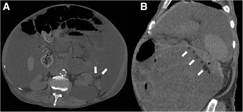
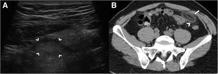
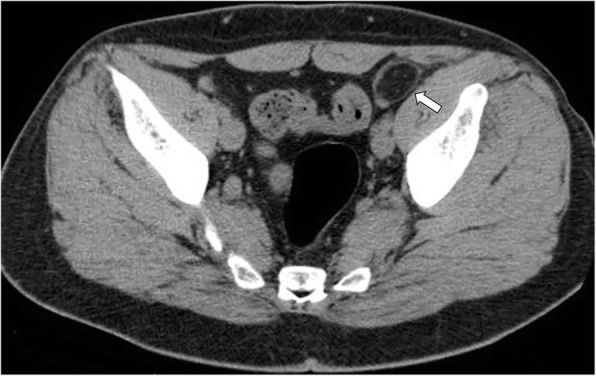
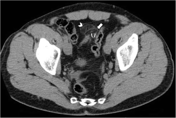
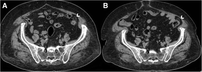
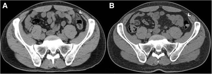
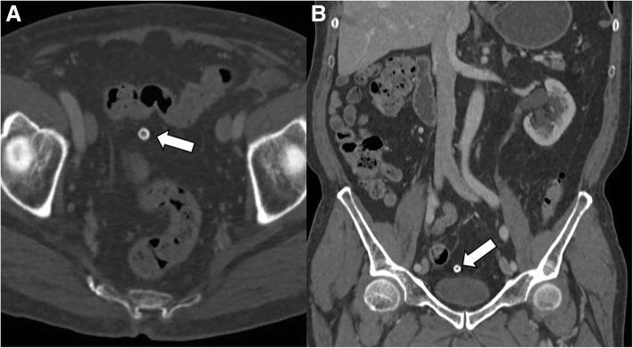
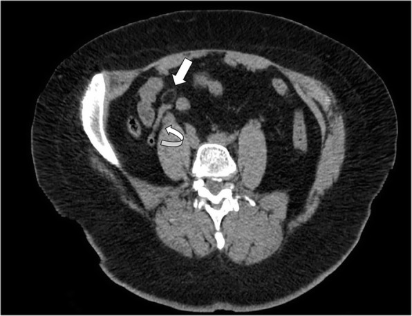
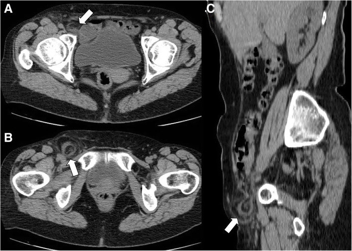
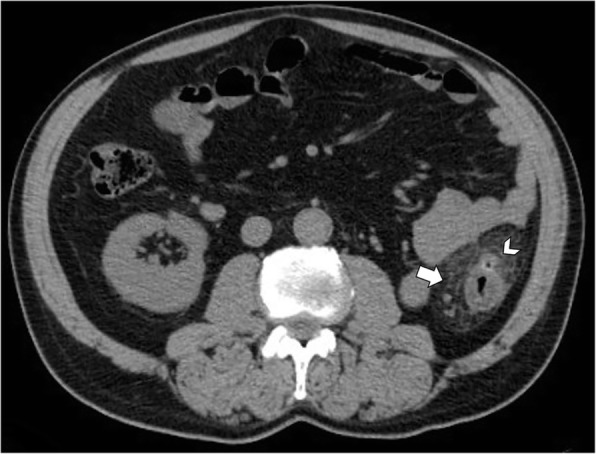
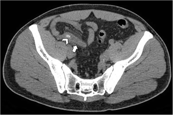
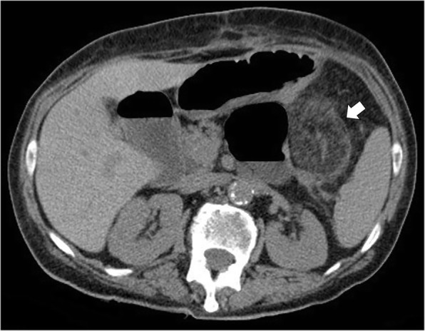
Epiploic appendagitis is a rare cause of acute abdominal pain, determined by a benign self-limiting inflammation of the epiploic appendages. It may manifest with heterogeneous clinical presentations, mimicking other more severe entities responsible of acute abdominal pain, such as acute diverticulitis or appendicitis. Given its importance as clinical mimicker, imaging plays a crucial role to avoid inaccurate diagnosis that may lead to unnecessary hospitalization, antibiotic therapy, and surgery. CT represents the gold standard technique for the evaluation of patients with indeterminate acute abdominal pain. Imaging findings include the presence of an oval lesion with fat-attenuation surrounded by a thin hyperdense rim on CT ("hyperattenuating ring sign") abutting anteriorly the large bowel, usually associated with inflammation of the adjacent mesentery. A central high-attenuation focus within the fatty lesion ("central dot sign") can sometimes be observed and is indicative of a central thrombosed vein within the inflamed epiploic appendage. Rarely, epiploic appendagitis may be located within a hernia sac or attached to the vermiform appendix. Chronically infarcted epiploic appendage may detach, appearing as an intraperitoneal loose calcified body in the abdominal cavity. In this review, we aim to provide an overview of the clinical presentation and key imaging features that may help the radiologist to make an accurate diagnosis and guide the clinical management of those patients.

Keywords: Abdominal pain; Acute abdomen; Adipose tissue; Differential diagnosis; Epiploic appendices; Large intestine.


Conflict of interest statement

The authors declare that they have no competing interests.

Figures

Fig. 1
Fig. 1
Axial (a) and sagittal reformatted (b) non-contrast CT images demonstrates massive ascites revealing the normal epiploic appendages (arrows), abutting the splenic flexure of the colon
Fig. 2
Fig. 2
Acute epiploic appendagitis in a 41-year-old man. a Ultrasound transverse scan, at the point of maximum tenderness, shows a well-defined hyperechoic ovoid fat lesion, surrounded by hypoechoic rim (arrowheads), adjacent to the anterior abdominal wall. b Axial non-contrast CT image shows an inflamed fatty ovoid lesion near the sigmoid colon with hyperattenuating ring (arrow) and nearby fat stranding (arrowhead)
Fig. 3
Fig. 3
Acute epiploic appendagitis in a 43-year-old man with presumptive clinical diagnosis of colonic diverticulitis. Axial non-contrast CT image shows a pericolonic fat-density lesion (arrow) that abuts anteriorly the colon-sigmoid junction in the left iliac fossa
Fig. 4
Fig. 4
Acute epiploic appendagitis in a 53-year-old man. Axial non-contrast CT image demonstrates a fat-density lesion (arrow) that abuts the hepatic flexure of the transverse colon. Mild fat stranding is surrounding the lesion (arrowhead)
Fig. 5
Fig. 5
Acute epiploic appendagitis in a 40-year-old man. Axial non-contrast CT image demonstrates a fatty ovoid lesion with high density rim (arrow) that abuts the sigmoid colon and contain central focal area of hyperattenuation (curved arrow), consistent with the “central dot sign” suggestive of thrombosed vascular pedicle. Note also surrounding mild fat inflammation (arrowhead)
Fig. 6
Fig. 6
Evolution of acute epiploic appendagitis in a 72-year-old woman. a Axial non-contrast CT image shows a pericolonic fat-density lesion (arrowhead) that abuts anteriorly the colon-sigmoid junction in the left iliac fossa. b At 3-month follow-up, CT image demonstrates reduction in size of the lesion (arrowhead)
Fig. 7
Fig. 7
Evolution of acute epiploic appendagitis in a 37-year-old man. a Axial non-contrast CT image shows an inflamed fatty ovoid lesion near the sigmoid colon (arrowhead), with hyperattenuating ring sign and nearby fat stranding. b CT scan, performed 1 year later for other reasons, demonstrates complete resolution of imaging findings of epiploic appendagitis (arrowhead)
Fig. 8
Fig. 8
Chronic calcified, amputated epiploic appendage in an 81-year-old man. a Axial contrast-enhanced CT image (bone window) shows a calcified, ring-like lesion (arrow) medial to the sigmoid colon which could be mistaken for a mesenteric lymph node or drop gallstone. b Coronal reformatted CT image of the same patient depicts the peritoneal loose body in pelvic cavity, likely due to chronic infarcted epiploic appendage (arrow)
Fig. 9
Fig. 9
Acute epiploic appendagitis adjacent to the vermiform appendix in a 39-year-old woman. Curved reformatted non-contrast CT image shows an oval lesion with fat attenuation and peripheral hyperdense rim (arrow), that is depending directly from the vermiform appendix tip (curved arrow)
Fig. 10
Fig. 10
Acute epiploic appendagitis in a 59-year-old woman. Axial non-contrast CT images at two different levels (a, b) and sagittal reformatted image (c) show a right inguinal hernia containing an epiploic appendage surrounded by a thin hyperdense rim (arrows). Fat stranding is seen within the hernia sac. Surgical hernia repair and pathological examination confirmed the presence of an epiploic appendagitis incarcerated in inguinal hernia
Fig. 11
Fig. 11
Acute diverticulitis in a 55-year-old man. Axial non-contrast CT image shows segmental thickening of descending colon, with pericolonic fat stranding (arrow). An inflamed diverticulum is identified abutting the anterior colonic wall (arrowhead)
Fig. 12
Fig. 12
Acute appendicitis in a 43-year-old man. Axial non-contrast CT image shows an inflamed vermiform appendix (arrow), with thickened wall and nearby fat stranding. Appendicoliths are identified within the appendix (arrowhead)
Fig. 13
Fig. 13
Acute omental infarction in a 67-year-old woman with previous history of total colectomy. Axial non-contrast CT image shows a 6.0 cm heterogeneous fatty ovoid lesion in the left upper quadrant (arrow), associated with inflammatory changes in the nearby fatty tissue

Similar articles

Cited by

References

    1. Singh AK, Gervais DA, Hahn PF, Sagar P, Mueller PR, Novelline RA. Acute epiploic Appendagitis and its mimics. Radiographics. 2005;25:1521–1534. - PubMed
    1. Almeida AT, Melão L, Viamonte B, Cunha R, Pereira JM. Epiploic appendagitis: an entity frequently unknown to clinicians—diagnostic imaging, pitfalls, and look-alikes. AJR Am J Roentgenol. 2009;193:1243–1251. - PubMed
    1. Schnedl WJ, Krause R, Tafeit E, Tillich M, Lipp RW, Wallner-Liebmann SJ. Insights into epiploic appendagitis. Nat Rev Gastroenterol Hepatol. 2011;8:45–49. - PubMed
    1. Pereira JM, Sirlin CB, Pinto PS, Jeffrey RB, Stella DL, Casola G. Disproportionate fat stranding: a helpful CT sign in patients with acute abdominal pain. Radiographics. 2004;24:703–715. - PubMed
    1. Ghahremani GG, White EM, Hoff FL, Gore RM, Miller JW, Christ ML. Appendices epiploicae of the colon: radiologic and pathologic features. Radiographics. 1992;12:59–77. - PubMed

LinkOut - more resources