. 2019 Feb;34(1):66-77.
doi: 10.1007/s12250-019-00089-1. Epub 2019 Feb 22.

Polypyrimidine Tract-Binding Protein Regulates Enterovirus 71 Translation Through Interaction with the Internal Ribosomal Entry Site

Affiliations

Polypyrimidine Tract-Binding Protein Regulates Enterovirus 71 Translation Through Interaction with the Internal Ribosomal Entry Site

Juemin Xi et al. Virol Sin. 2019 Feb.

Abstract

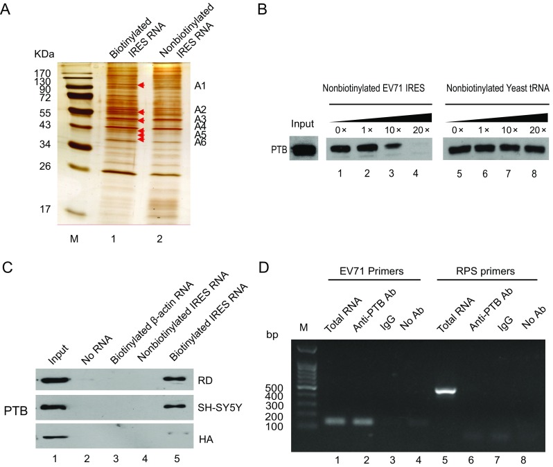
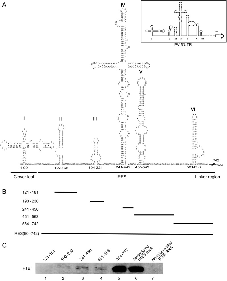
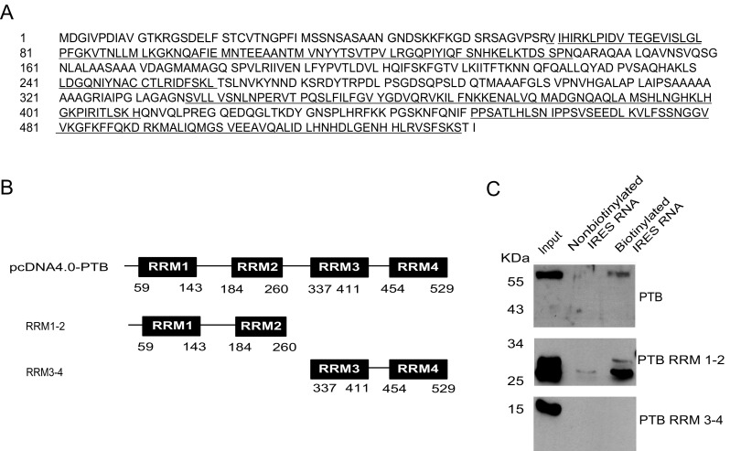
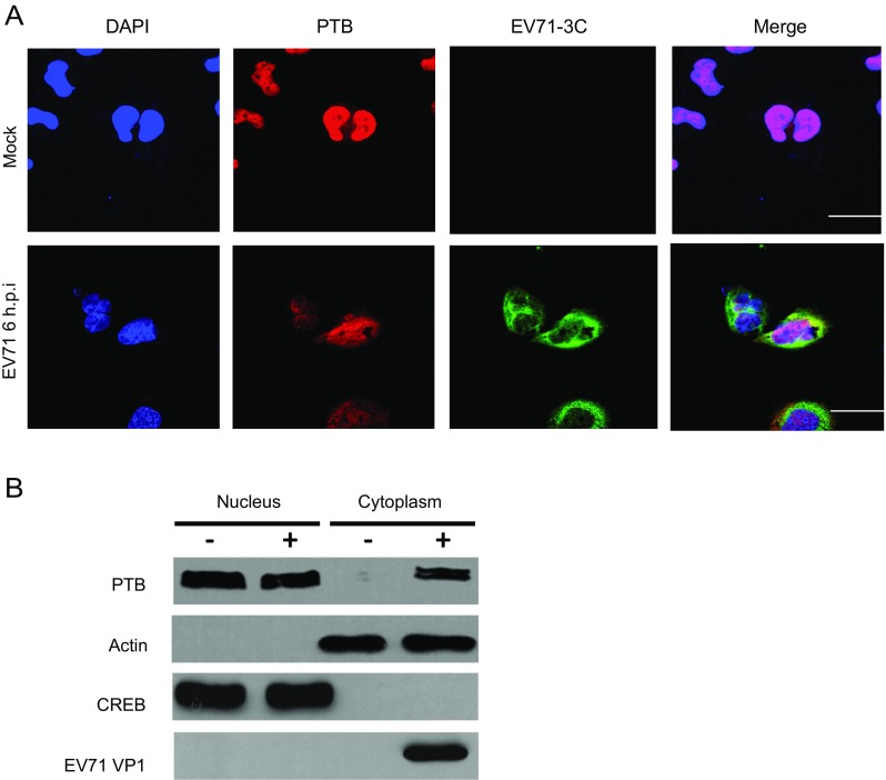
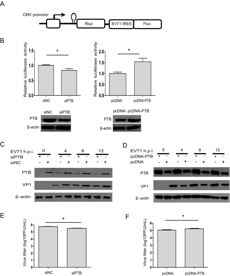
Enterovirus 71 (EV71), a major causative agent of hand, foot, and mouth disease, has caused periodic infection outbreaks in children in the Asia-Pacific region. In order to describe the largely unknown life cycle of EV71, the molecular basis of its virus-host interactions must first be determined. The 5' untranslated region of EV71 contains a cloverleaf-like structure and internal ribosomal entry site (IRES), which play an important role in transcription and translation of viral protein. We found that polypyrimidine tract-binding protein 1 (PTB) bound to the IRES of EV71. RNA recognition motifs 1 and 2 of PTB were responsible for its binding to the EV71 IRES. Moreover, PTB protein was shuttled from nucleus to cytoplasm after EV71 infection. Additionally, IRES activity and viral protein production were inhibited by PTB knockdown. These results suggest that PTB interacts with the EV71 IRES, and positively regulates viral protein translation.

Keywords: EV71; IRES; PTB; Translation.


Conflict of interest statement

Conflict of interest

The authors declare that they have no conflict of interest.

Animal and Human Rights Statement

The authors declare that they have no conflict of interest. This article does not contain any studies with human or animal subjects performed by any of the authors.

Figures

Fig. 1
Fig. 1
Interaction of PTB with the EV71 IRES region. A Pull down of proteins with the EV71 IRES in T98G cell lysate. The specific bands interacting with EV71 IRES are shown by silver staining. Lane 1: biotinylated IRES RNA, Lane 2: nonbiotinylated EV71 IRES RNA. B A specific association between PTB and the EV71 IRES region was confirmed by Western blotting and a competition assay. Increasing amounts of unlabelled RNA were added to compete with the biotin-labeled EV71 IRES RNA interacting with PTB. The eluted proteins were separated by 12% SDS-PAGE. Lanes are as follows: lanes 1–4, unlabeled EV71 IRES RNA; lanes 5–8, unlabeled yeast tRNA. C Extracts of RD cells, SH-SY5Y cells, and HA cells were prepared and then incubated without RNA (lane 2), with biotinylated actin RNA (lane 3), non-biotinylated EV71 IRES RNA (lane 4), or biotinylated EV71 IRES RNA (lane 5). After pull-down assay, the bound proteins were eluted, boiled, and subjected to 12% SDS-PAGE. PTB protein was detected by western blot with a rabbit anti-PTB antibody. The inputs were cell extracts of RD, SH-SY5Y and HA (lane 1). D EV71 IRES RNA was pulled down with PTB from EV71-infected T98G cell lysate. T98G cells were infected with EV71 at an MOI of 20 for 6 h and then cell extracts were incubated with rabbit anti-PTB antibody (lanes 2 and 6), normal rabbit IgG (lanes 3 and 7), or without antibody (− Ab) (lanes 4 and 8). Following washing and dissociation, the RNA extract was prepared and subjected to RT-PCR analysis with primers specific for the ribosomal protein S16 (PRS16) RNA (lanes1–4) or for EV71 IRES region RNA (lanes 5–8). Lane 1, cell lysate without immunoprecipitation as a positive RT-PCR control; Lane 2, anti-PTB antibody incubated with 200 mg infected-cell lysate; Lane 3, negative control with rabbit IgG; Lane 4, negative control with no antibody.
Fig. 2
Fig. 2
Determination of the EV71 IRES sequences required for the binding of PTB. A Prediction of the RNA secondary structure of the EV71 5′UTR by the mfold web serve. The modified schematic representation of the secondary structure of poliovirus (PV) 5′UTR (modified from Hellen et al. 1994) is shown in the top right corner. B Plasmids carrying different deletions in the five stem-loops of EV71 IRES: pGEM-3zf-(SL II, 121–181), (SL III, 190–230), (SL IV, 241–450), (SL V, 451–463), (SL VI, 564–742). C Analysis of the regions responsible for the interaction in the EV71 IRES region using various truncated RNA forms, transcribed in vitro and biotinylated. T98G cell lysate were incubated with these biotin-labeled RNAs and the non-biotinylated IRES RNA probes were used as controls. After being pulled down by streptavidin, the protein complex was separated by SDS-PAGE and Western blot was carried out to detect PTB in the pulled-down complex (lanes 1–7).
Fig. 3
Fig. 3
Analysis of the expression of PTB in neuro-related cells and RRMs required for its binding to EV71 IRES. A The RRM domains of full-length PTB protein as described. The amino acids of the four RRM domains, RRM1, RRM2, RRM3, and RRM4, are underlined. B Three constructed plasmids expressing the full-length PTB, or truncated PTB proteins in which RRM3–4 was deleted (RRM1–2), or RRM1–2 was deleted (RRM3–4), respectively. C RNA–protein binding assay showing PTB protein as determined by Western blot analysis using anti-His antibody, from 293ET cells transfected with plasmids coding the His-Tagged proteins, pcDNA4.0-PTB, RRM1–2, RRM3–4, respectively.
Fig. 4
Fig. 4
Subcellular distribution of PTB protein during EV71 infection. T98G cells mock-infected (upper panels) or infected with EV71 at an MOI of 20 (lower panels). At 6 h post infection, cells were fixed with formaldehyde, washed, and detected with antibody against PTB or EV71 3C protein. FITC-conjugated goat anti-rabbit IgG or TRITC-conjugated goat anti-mouse IgG was used as secondary antibody and stained with DAPI. Images were captured by confocal laser scanning microscopy. PTB (red), EV71 3C (green), DAPI (blue). Bar = 10 µm. B T98G cells infected with or without EV71 at an MOI of 20 for 6 h. Whole cell lysate, cytoplasm, and nucleus extractions were prepared from mock infected and EV71-infected cells, respectively. Endogenous proteins as detected by Western blot analysis using antibodies to CREB, EV71 VP1, PTB, or β-actin. The blot is representative of three independent experiments with similar results.
Fig. 5
Fig. 5
Analysis of PTB in the regulation of EV71 viral translation and virus production. A Schematic diagram of dicistronic reporter plasmids pRF-EV71 IRES. B T98G cells were transfected with either PTB siRNA or pcDNA4.0-PTB. 48 h later, dicistronic construct pRF or pRF-EV71 IRES and siRNA duplexes were co-transfected into T98G cells. After 48 h, the relative ISRE activity were analyzed by monitoring the luciferase activity. Western blot were used to analyze the expression level of PTB and β-actin. CF T98G cells were transfected with siRNA targeting PTB (siPTB) and control siRNA (siNC), respectively. 48 h later, T98G cells were transfected with plasmids containing either full-length PTB cloned into pcDNA4.0 vector (pcDNA-PTB), or vector without PTB (pcDNA). After 24 h, cells were infected with EV71 at an MOI of 20 for 4, 8 and 12 h. Cell culture supernatants from EV71-infected cells were prepared and subjected to plaque assays. Western blotting was used to determine the expression level of EV71 VP1 protein, PTB or β-actin protein. E, F The effects of PTB on EV71 growth. Cell culture supernatants from C and D were determined by plaque assay. Each bar represents the average of triplicate data points with the standard deviation represented as the error bar. *P < 0.05 and **P < 0.01 versus negative control.

Similar articles

Cited by

References

    1. Agis-Juarez RA, Galvan I, Medina F, Daikoku T, Padmanabhan R, Ludert JE, del Angel RM. Polypyrimidine tract-binding protein is relocated to the cytoplasm and is required during dengue virus infection in Vero cells. J Gen Virol. 2009;90:2893–2901. doi: 10.1099/vir.0.013433-0. - DOI - PubMed
    1. Auweter SD, Allain FH. Structure-function relationships of the polypyrimidine tract binding protein. Cell Mol Life Sci. 2008;65:516–527. doi: 10.1007/s00018-007-7378-2. - DOI - PMC - PubMed
    1. Bellaousov S, Reuter JS, Seetin MG, Mathews DH. RNAstructure: Web servers for RNA secondary structure prediction and analysis. Nucleic Acids Res. 2013;41:W471–W474. doi: 10.1093/nar/gkt290. - DOI - PMC - PubMed
    1. Black DL. Mechanisms of alternative pre-messenger RNA splicing. Annu Rev Biochem. 2003;72:291–336. doi: 10.1146/annurev.biochem.72.121801.161720. - DOI - PubMed
    1. Clery A, Blatter M, Allain FH. RNA recognition motifs: boring? Not quite. Curr Opin Struct Biol. 2008;18:290–298. doi: 10.1016/j.sbi.2008.04.002. - DOI - PubMed

MeSH terms