. 2019 Jul;16(3):725-740.
doi: 10.1007/s13311-019-00723-6.

Intranasal Losartan Decreases Perivascular Beta Amyloid, Inflammation, and the Decline of Neurogenesis in Hypertensive Rats

Affiliations

Intranasal Losartan Decreases Perivascular Beta Amyloid, Inflammation, and the Decline of Neurogenesis in Hypertensive Rats

Henning J Drews et al. Neurotherapeutics. 2019 Jul.

Abstract

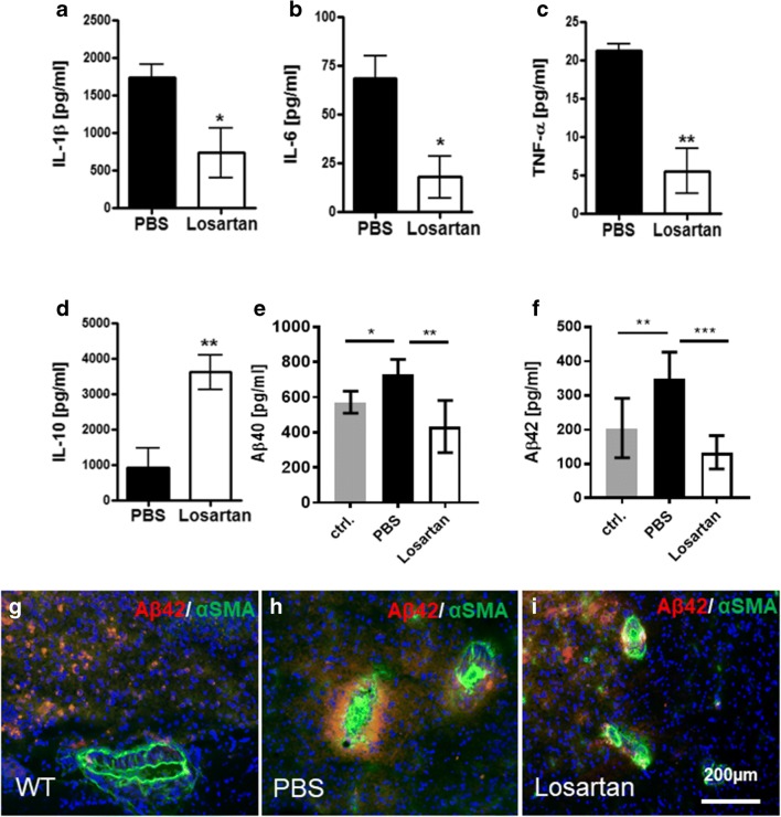
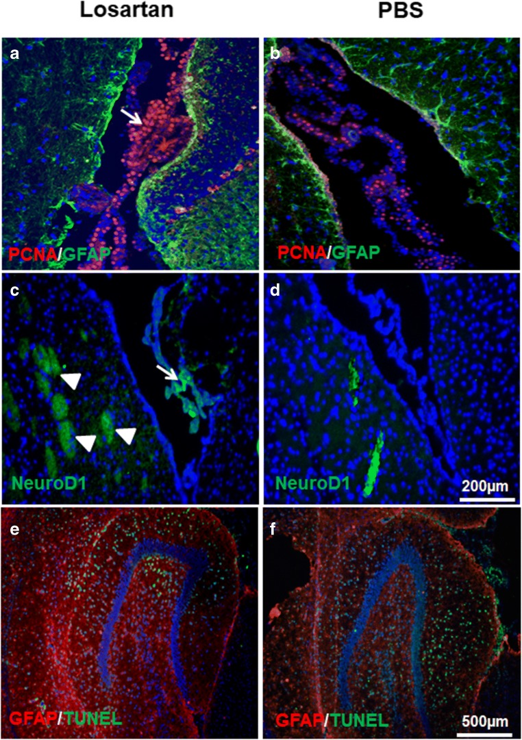
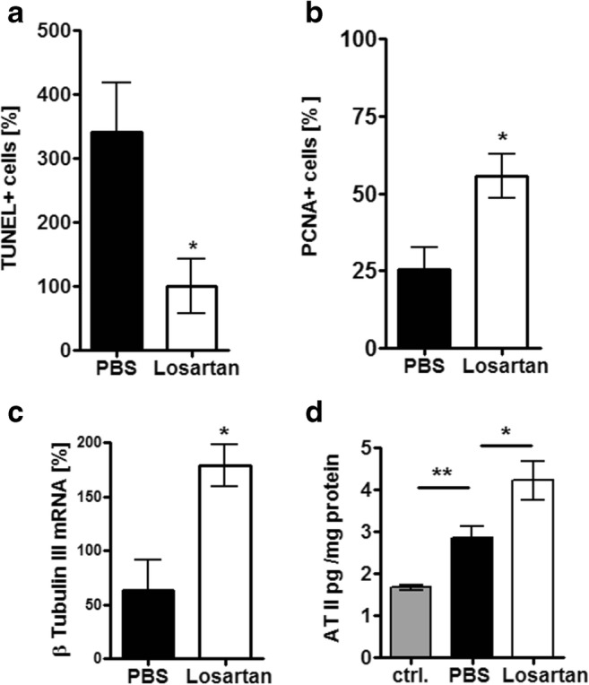
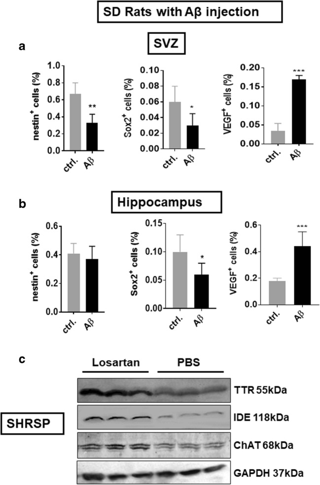
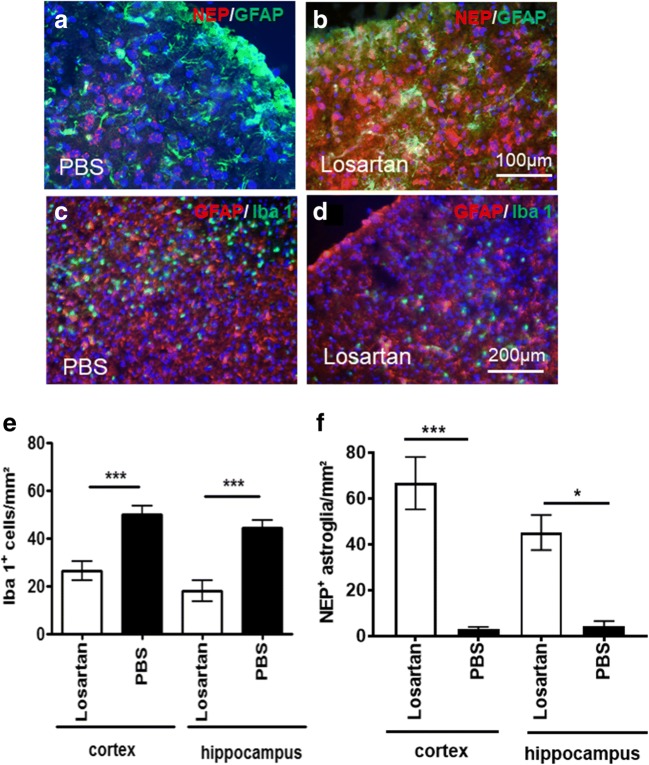
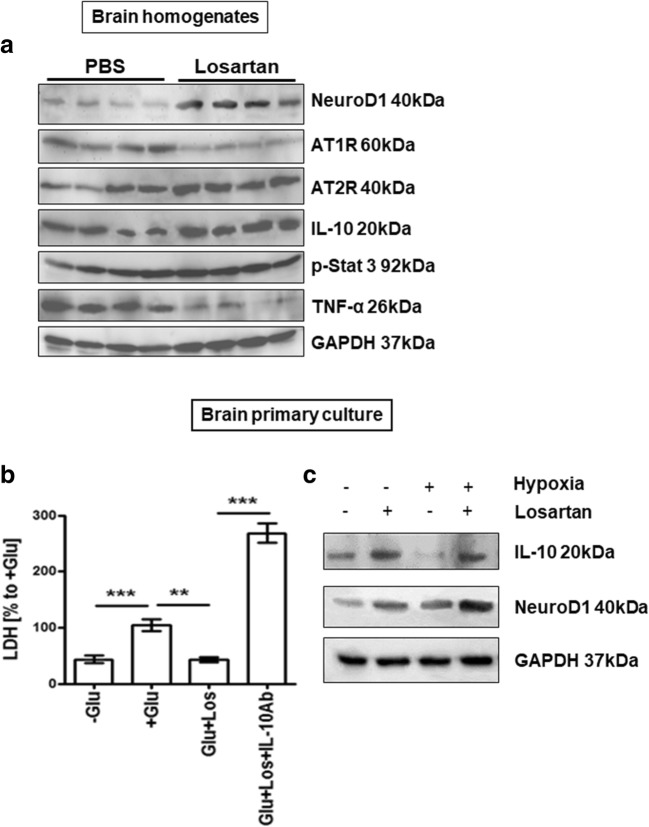
The contribution of the local angiotensin receptor system to neuroinflammation, impaired neurogenesis, and amyloid beta (Aβ) accumulation in Alzheimer's disease (AD) and in hypertension is consistent with the remarkable neuroprotection provided by angiotensin receptor blockers (ARBs) independent of their blood pressure-lowering effect. Considering the causal relationship between hypertension and AD and that targeting cerebrovascular pathology with ARBs does not necessarily require their systemic effects, we tested intranasal losartan in the rat model of chronic hypertension (spontaneously hypertensive stroke-prone rats, SHRSP). Intranasal losartan at a subdepressor dose decreased mortality, neuroinflammation, and perivascular content of Aβ by enhancing key players in its metabolism and clearance, including insulin-degrading enzyme, neprilysin, and transthyretin. Furthermore, this treatment improved neurologic deficits and increased brain IL-10 concentration, hippocampal cell survival, neurogenesis, and choroid plexus cell proliferation in SHRSP. Losartan (1 μM) also reduced LDH release from cultured astroglial cells in response to toxic glutamate concentrations. This effect was completely blunted by IL-10 antibodies. These findings suggest that intranasal ARB treatment is a neuroprotective, neurogenesis-inducing, and Aβ-decreasing strategy for the treatment of hypertensive stroke and cerebral amyloid angiopathy acting at least partly through the IL-10 pathway.

Keywords: Alzheimer’s disease; Angiotensin; Angiotensin receptor blocker; Cerebral amyloid angiopathy; Hemorrhagic stroke; Intranasal.


Conflict of interest statement

The authors declare that they have no conflict of interest.

Figures

Fig. 1
Fig. 1
Effects of intranasal losartan on survival, neurological deficits, and BP in SHRSP. (A) Survival of animals (n = 9 per group) from the beginning of drug treatment until the animals were sacrificed. Log–rank (**p = 0.0093) and Gehan–Breslow–Wilcoxon (**p = 0.0069) tests. The data were normalized to the total amount of animals assigned to the groups prior to losartan administration. (B) Motor and behavioral performance represented in the time course starting from randomization (day 47) until day 83 when the animals were sacrificed. (C) Mean systolic BP before and during losartan treatment. (D) Mean systolic BP within the losartan-treated group. Systolic BP was monitored 6 times prior to assignment of animals into the groups (once a week) and 5 times after assignment during the drug treatment period (once a week) 30 min and 24 h after losartan administration
Fig. 2
Fig. 2
Losartan effects on inflammatory cytokines and Aβ in SHRSP. Multiplex analysis of the inflammatory cytokines in brain homogenates of SHRSP demonstrates that IN-losartan decreased IL-1β (A), IL-6 (B), and TNF-α (C) while IL-10 was prominently upregulated (D) in comparison to PBS-treated SHRSP controls. (E) and (F) Aβ40 and Aβ40 multiplex analysis in the brain homogenates (n = 5) of SHRSP (black and white bars) and age-matched Wistar rats (ctrl., grey bar). (GI) Immunofluorescent analysis of Aβ42 (red) and vascular smooth muscle cell marker αSMA (green) in the brain sections of SHRSP (PBS and losartan) and age-matched Wistar rats (WT). Aβ42 is strongly upregulated in vehicle-treated SHRSP (H) in comparison with normotensive controls (G), while losartan diminished the Aβ load around the vessels in SHRSP (I). 4′,6 Diamidino-2-phenylindole (DAPI) counts in blue. The data shown are representative for brain sections obtained from n = 3 animals per treatment group
Fig. 3
Fig. 3
Intranasal losartan increases the population of NeuroD1+ cells and cell proliferation in the choriod plexus and decreases hippocampal cell death in SHRSP. PCNA-positive (red in A and B) cells in the plexus choroideus (arrow) of IN-losartan- (A) and vehicle-treated (B) SHRSP. The population of NeuroD1-positive cells (green in C and D) in the SVZ (arrowheads) and the plexus choroideus (arrow) of losartan-treated SHRSP (C) is greater than that of vehicle-treated SHRSP control. (E, F) TUNEL staining (green) in the hippocampus of losartan- (E) and vehicle-treated (F) SHRSP. GFAP (green in A and B) expression is demonstrated in green in (A and B) and in red in (E) and (F). Cell nuclei are shown with DAPI (blue in AF). The data shown are representative for brain sections obtained from n = 3 rats per treatment group. Scale bars in (D) (200 μm) and (F) (500 μm) apply to (AD) and (EF) respectively
Fig. 4
Fig. 4
Losartan effects on cell survival and proliferation and angiotensin II production in the brain of SHRSP. (A) Quantification of TUNEL+ cells in the hippocampus of SHRSP. The data are normalized to the mean of TUNEL+ cell number in the losartan-treated group. (B) Quantification of PCNA+ cells in the choroid plexus demonstrates that IN losartan doubles the number of proliferating cells in comparison to controls. The data are normalized to the total number of cell nuclei counted within the area of choroid plexus. (C) Losartan increases the expression of β-tubulin III mRNA in the brain homogenates of SHRSP (the mean of PBS-treated control group samples are considered as 100%). (D) Measurement of angiotensin II in brain homogenates shows a higher level of angiotensin II in vehicle-treated SHRSP in comparison to normotensive control rats. Losartan induced a further increase in angiotensin in SHRSP
Fig. 5
Fig. 5
Aβ42 influence on neurogenesis markers and VEGF in vivo and losartan’s effect on Aβ-degrading enzymes and ChAT in SHRSP. (A, B) Flow cytometry of hippocampal and SVZ cells after bilateral injection of fibrillar Aβ42 into the lateral ventricle of SD rats (n = 5). Aβ42 decreased the expression of nestin in the subventricular zone (SVZ) and Sox2 in the SVZ and hippocampus. A prominent upregulation of VEGF was seen in the SVZ and hippocampus of Aβ42-treated rats. (C) Western blot of TTR, IDE, and ChAT in the brain homogenates of SHRSP showing losartan-induced increase of all 3 marker proteins. GAPDH serves as a loading control
Fig. 6
Fig. 6
Losartan enhances NEP+ cells and decreases Iba+ microglia/macrophages in the cortex and hippocampus of SHRSP. (A) Weak expression of NEP (red) in GFAP (green)-negative neurons in the cortex of PBS-treated SHRSP. (B) Prominent increase in expression intensity and in number of NEP+ cells in losartan-treated SHRSP in both GFAP+ astroglia and GFAP-negative neurons. (C) High number of Iba1+ microglia/macrophages in the vehicle-treated SHRSP in the cortex. (D) Decrease in Iba1+ microglia/macrophages in the cortex of losartan-treated SHRSP, while GFAP+ astroglia (red in C and D) remained unaffected. DAPI+ cell nuclei are shown in blue. (E) Quantification of Iba-1+ cells in the cortex and hippocampus of vehicle- (black bars) and losartan-treated (white bars) SHRSP (n = 3 per group). (F) Number of NEP+ cells in the cortex and hippocampus of SHRSP. The data in (E) and (F) were obtained from 10 brain sections/animal (n = 3 animals per treatment group) and normalized to square millimeters. Scale bar in (A, B) 100 μm, in (C, D) 200 μm
Fig. 7
Fig. 7
Influence of IN-losartan on angiotensin receptors (ATR) 1 and 2, phosphorylated STAT3, IL-10, NeuroD1, TNF-α, and Abeta1–40 in SHRSP and on the survival of astroglia in vitro. (A) Western blot of AT1R and AT2R, phosphorylated STAT3, NeuroD1, IL-10, and TNF-α in the brain homogenates of losartan- and vehicle-treated SHRSP. GAPDH serves as a loading control. (B) Astroglial primary culture (day 14 in vitro) incubated with 1 mmol/L Glu (+Glu in B) showed a prominent increase in LDH release in comparison to the control without Glu (cf. −Glu with +Glu), whereas IN-losartan decreased LDH down to the level of the Glu-free control (cf. Glu+Los with +Glu and −Glu). Losartan-treated cells exposed simultaneously to IL-10 antibodies, losartan, and Glu (Glu+Los+IL-10Ab) showed a dramatic increase in LDH release over the control culture exposed to Glu only (cf. +Glu with Glu+Los+IL-10Ab). The data are normalized to the control exposed to Glu (+Glu). (C) Western blot of IL-10 and NeuroD1 from untreated and losartan-treated astroglial primary culture in 48-h normoxic and hypoxic conditions. Cell lysates from n = 5 different culture dishes were pooled prior to the Western blot analysis. GAPDH serves as a loading control
Fig. 8
Fig. 8
In vitro effects of losartan on rat astroglial primary culture. Losartan increased the expression of IL-10 (A), PSD95 (B), Neprilysin (NEP) (C), and ChAT mRNA (D) in APC upon hypoxic culture conditions. qPCR analysis of IL-10, PSD95, Neprilysin, and ChAT in astroglial primary culture (APC) under normoxic (white bars) and hypoxic culture conditions (grey bars)

Similar articles

Cited by

References

    1. Daulatzai MA. Cerebral hypoperfusion and glucose hypometabolism: key pathophysiological modulators promote neurodegeneration, cognitive impairment, and Alzheimer’s disease. J Neurosci Res. 2017;95:943–72. doi: 10.1002/jnr.23777. - DOI - PubMed
    1. Kalaria RN, Ballard C. Overlap between pathology of Alzheimer disease and vascular dementia. Alzheimer Dis Assoc Disord. 1999;13:S115–23. doi: 10.1097/00002093-199912003-00017. - DOI - PubMed
    1. Sironi L, Gelosa P, Guerrini U, et al. Anti-inflammatory effects of AT1 receptor blockade provide end-organ protection in stroke-prone rats independently from blood pressure fall. J Pharmacol Exp Ther. 2004;311:989–95. doi: 10.1124/jpet.104.072066. - DOI - PubMed
    1. Danielyan L, Klein R, Hanson LR, Buadze M, Schwab M, Gleiter CH, et al. Protective effects of intranasal losartan in the APP/PS1 transgenic mouse model of Alzheimer disease. Rejuvenation Res. 2010;13:195–201. doi: 10.1089/rej.2009.0944. - DOI - PubMed
    1. Joglar B, Rodriguez-Pallares J, Rodriguez-Perez AI, Rey P, Guerra MJ, Labandeira-Garcia JL. The inflammatory response in the MPTP model of Parkinson’s disease is mediated by brain angiotensin: relevance to progression of the disease. J Neurochem. 2009;109:656–69. doi: 10.1111/j.1471-4159.2009.05999.x. - DOI - PubMed

Publication types

Substances