. 2019 Dec 17;29(11):4831-4849.
doi: 10.1093/cercor/bhz018.

Dlx1/2 are Central and Essential Components in the Transcriptional Code for Generating Olfactory Bulb Interneurons

Affiliations

Dlx1/2 are Central and Essential Components in the Transcriptional Code for Generating Olfactory Bulb Interneurons

Teng Guo et al. Cereb Cortex. .

Abstract

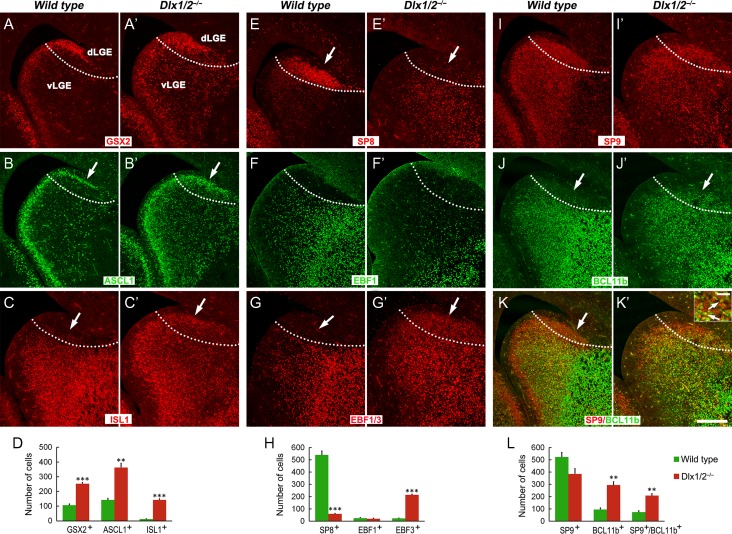
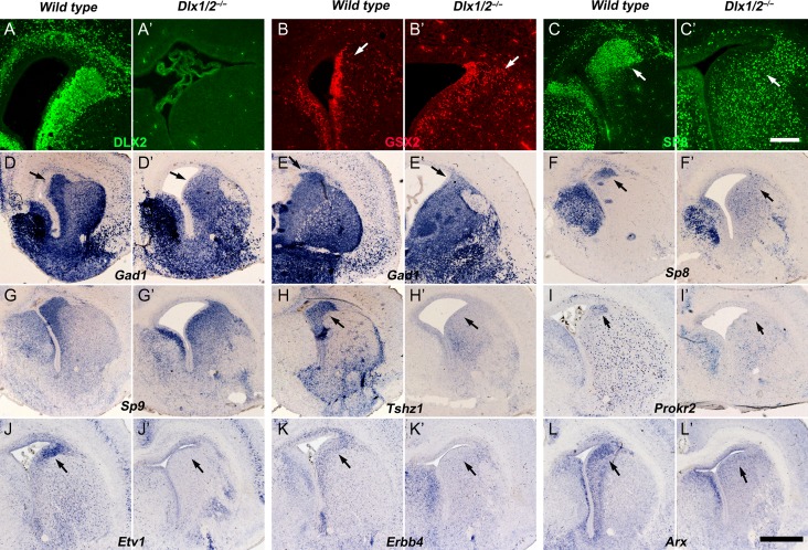
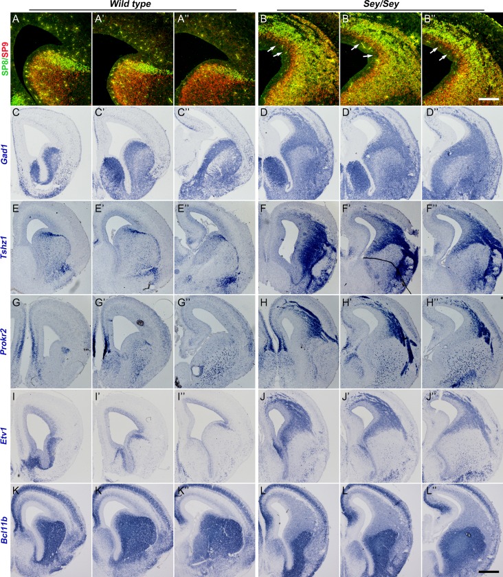
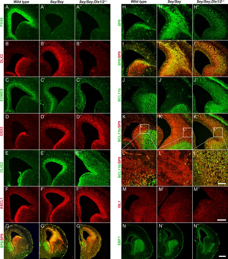
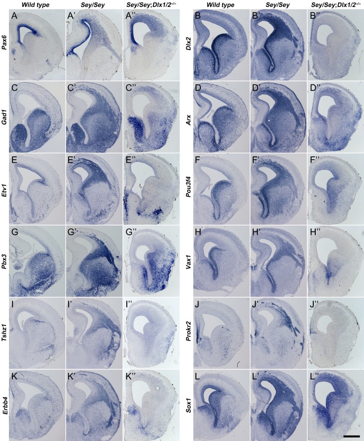
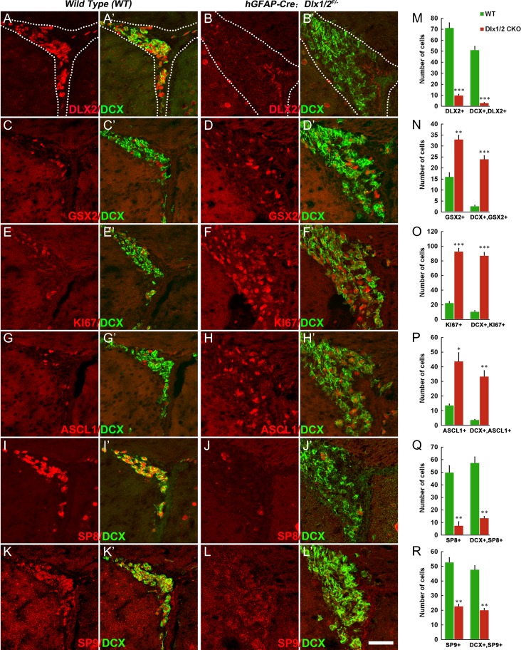
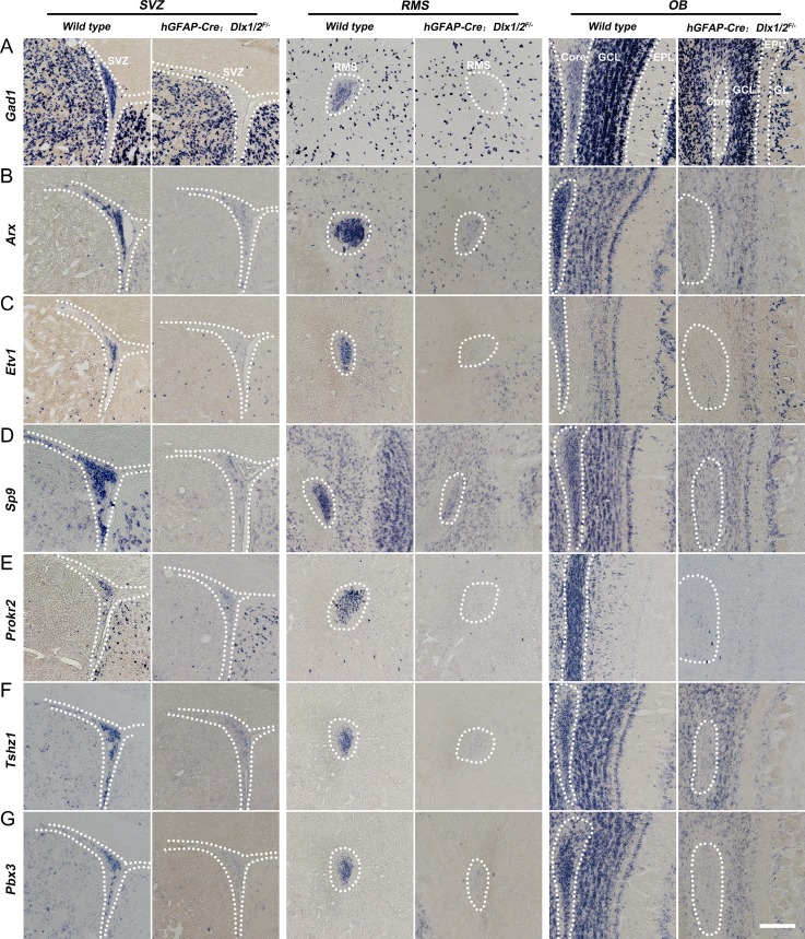
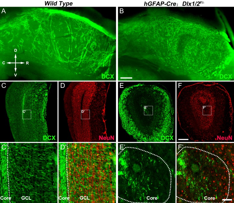
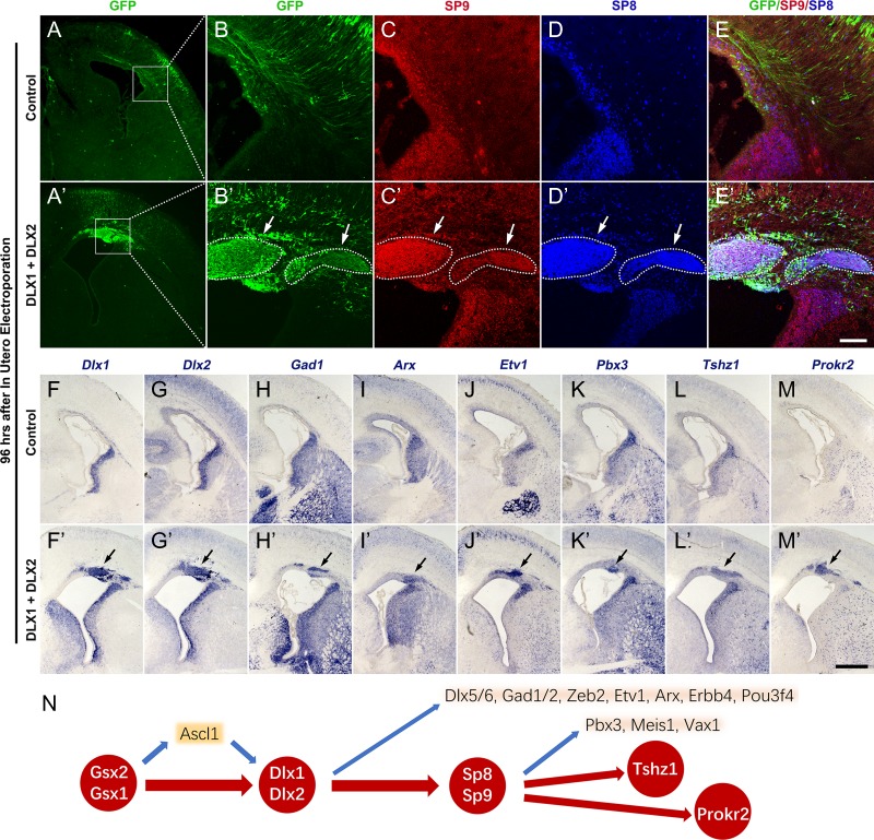
Generation of olfactory bulb (OB) interneurons requires neural stem/progenitor cell specification, proliferation, differentiation, and young interneuron migration and maturation. Here, we show that the homeobox transcription factors Dlx1/2 are central and essential components in the transcriptional code for generating OB interneurons. In Dlx1/2 constitutive null mutants, the differentiation of GSX2+ and ASCL1+ neural stem/progenitor cells in the dorsal lateral ganglionic eminence is blocked, resulting in a failure of OB interneuron generation. In Dlx1/2 conditional mutants (hGFAP-Cre; Dlx1/2F/- mice), GSX2+ and ASCL1+ neural stem/progenitor cells in the postnatal subventricular zone also fail to differentiate into OB interneurons. In contrast, overexpression of Dlx1&2 in embryonic mouse cortex led to ectopic production of OB-like interneurons that expressed Gad1, Sp8, Sp9, Arx, Pbx3, Etv1, Tshz1, and Prokr2. Pax6 mutants generate cortical ectopia with OB-like interneurons, but do not do so in compound Pax6; Dlx1/2 mutants. We propose that DLX1/2 promote OB interneuron development mainly through activating the expression of Sp8/9, which further promote Tshz1 and Prokr2 expression. Based on this study, in combination with earlier ones, we propose a transcriptional network for the process of OB interneuron development.

Keywords: Ascl1, Pax6; Dlx1; Dlx2; Gsx2; Sp8; Sp9; interneuron; olfactory bulb.


Figures

Figure 1.
Figure 1.
SP8, SP9, and DLX2 are coexpressed in the E16.5 dLGE SVZ. (A, B) GSX2 was strongly expressed in the dLGE VZ and SVZ1; only a few GSX2+ cells were in the SVZ2, whereas DLX2 was mainly expressed in the SVZ1&2. Note weak expression of GSX2 in the vLGE. (C) The section was stained with DAPI. (D, E) Most ASCL1+ cells expressed DLX2. (FI) DLX2 was strongly expressed in the SVZ1 and relatively weakly expressed in the SVZ2 and DLX2 expression began before SP8/9 expression. Most cells in the SVZ2 expressed DLX2/SP8/SP9. (J, K) Quantification of DLX2+ cells that expressed SP8, SP9, or SP8/9 in the dLGE SVZ1 and SVZ2 at E16.5. Scale bar: 100 μm in G for (AI).
Figure 2.
Figure 2.
Neural progenitors accumulate in the dLGE of Dlx1/2−/− mice at E16.5. (AB′) Compared with controls, there were more GSX2+ and ASCL1+ progenitors in the d/vLGE of Dlx1/2−/− mice (arrows). (C, C′) ISL1 expression was increased in the mutant dLGE (arrows). (E, E′) SP8 was lost in the mutant dLGE (arrows). (FG′) EBF1 was not expressed in the control and mutant dLGE, but the pan EBF antibody immunostaining showed that EBF3 was expressed in the mutant dLGE (arrows). (IK′) SP9 expression was reduced in the dLGE; most SP9+ cells coexpressed the immature MSN marker BCL11b (arrows in the inset of K′). (D, H, L) Quantification of cell numbers in the dLGE. Scale bars: 200 μm in K′ for AK′; 20 μm in the inset of K′.
Figure 3.
Figure 3.
Immature OB interneurons are not generated in the dLGE of Dlx1/2−/− Mice at E18.5. (AA′) No DLX2 protein was detected in the DLX1/2 mutant telencephalon. (BB′) More GSX2+ cells were in the mutant dLGE SVZ (arrows). (CC′, FF′) The expression of Sp8 mRNA and SP8 protein was greatly reduced in the mutant dLGE (arrows). (DE′) In situ hybridization showed Gad1 expression was reduced in the dLGE SVZ of mutants compared with controls (arrows). (GG′) Sp9 expression was maintained in the mutant dLGE. (HL′) Expression of OB interneuron lineage markers, Tshz1, Prokr2, Etv1, Erbb4, and Arx, were either lost or severely reduced in the mutant dLGE (arrows). Scale bars: 200 μm in C′ for AC′; 600 μm in L′ for DL′.
Figure 4.
Figure 4.
Pax6 Sey/Sey mice have a large ectopia containing OB interneurons in the cortex and dLGE at E16.5. (AB″) SP8 and SP9 were ectopically expressed in the cortex of Pax6Sey/Sey mice. Note SP9 expression appears in more immature cells (i.e., are closer to the ventricle) than SP8 in the cortical SVZ (arrows). (CJ″) Increased Gad1, Tshz1, Prokr2, and Etv1 expression was seen in the mutant dLGE and cortex. (KL″) Strong Bcl11b expression was not observed in the region of the ectopia mutant cortex. Scale bars: 200 μm in (B″) for (AB″); 400 μm in (L″) for (CL″).
Figure 5.
Figure 5.
More subpallial neural progenitors are in the cortex of Pax6Sey/Sey; Dlx1/2/ Mice at E16.5. (AB″) Immunostaining of PAX6 and DLX2 confirmed the genotypes of mice used. (CC″) EOMES expression was greatly reduced in both Pax6Sey/Seyand Pax6Sey/Sey; Dlx1/2/ mice. (DF″) GSX2+, OLIG2+, and ASCL1+ progenitors ectopically accumulated in the neocortex of Pax6Sey/Sey mice; their expression increased further in Pax6Sey/Sey; Dlx1/2/ mice. (GI″) The ectopic SP8/9 cortical expression in the Pax6Sey/Sey mice was reduced in the Pax6Sey/Sey; Dlx1/2/ cortex; furthermore, the SP8/9+ cells showed a scattered pattern. (JL″) Most SP9/8+ cells coexpressed BCL11b in the cortex of Pax6Sey/Sey; Dlx1/2/ mice. (MN″) ISL1 and EBF1 expression was not observed in the neocortex of these mutants. Scale bars: 70 μm in L″ for LL″; 250 μm in M″ for AF″, HK″, MM″; 500 μm in N″ for GG″, NN″.
Figure 6.
Figure 6.
Immature OB interneurons are not generated in the cortex of Pax6Sey/Sey; Dlx1/2/ mice at E16.5. (AL″) In situ RNA hybridization indicated that immature OB interneuron markers (Gad1, Etv1, Pbx3, Tshz1, Erbb4, Arx, pou3f4, Vax1, Prokr2) were not detected in Pax6Sey/Sey; Dlx1/2/ mice compared with Pax6Sey/Sey mice. Note that Pax6 transcripts and Sox1 expression were increased in the cortex of these mutants. Scale bar: 600 μm in L″ for AL″.
Figure 7.
Figure 7.
Neuronal differentiation of stem/progenitors in the adult SVZ (P50) of hGFAP-Cre; Dlx1/2F/ conditional knockout (Dlx1/2-CKO) mice was blocked. (AL′) DLX2+, SP8+, SP9+, DLX2+/DCX+, SP8+/DCX+, and SP9+/DCX+ cells were greatly reduced whereas GSX2+, Ki67+, ASCL1+, GSX2+/DCX+, Ki67+/DCX+, and ASCL1+/DCX+ cells were significantly increased in the SVZ of Dlx1/2-CKO mice compared with wild type control mice at P50. (MR) Quantification of these experiments. Scale bar: 50 μm in L′ for AL′.
Figure 8.
Figure 8.
Loss of immature OB interneurons in the adult (P50) Dlx1/2-CKO SVZ, RMS and the core of the OB. (AG) In situ RNA hybridization showed that expression of Gad1, Arx, Etv1, Sp9, Prokr2, Tshz1, and Pbx3 in the adult SVZ, RMS, and OB core of Dlx1/2-CKO mice were either severely reduced or undetectable. Scale bar: 100 μm in G for AG.
Figure 9.
Figure 9.
Tangential and radial migration defects of DCX+ cells in the adult SVZ and OB of Dlx1/2-CKO mice. (A, B) Wholemount DCX immunostaining of the lateral wall of the lateral ventricle showed DCX+ cells in the SVZ of Dlx1/2-CKO mice (P50) did not form migration chains. (CF) OB coronal sections stained for NeuN and DCX. Note that Dlx1/2-CKO mouse OB was smaller than wild type OB. (C′–F′) High magnification images of boxed areas in (CF) showing the defect of the radial migration of DCX+ cells. R, rostral; C, caudal; D, dorsal; V, ventral. Scale bars: 300 μm in B for AB′; 350 μm in F for CF; 50 μm in F′ for C′–F′.
Figure 10.
Figure 10.
Overexpression of Dlx1&2 in the dorsal pallium (cortex) induced the generation of immature OB interneurons. (AE′) Overexpression of Dlx1&2 in the neocortex by IUE of plasmids (pCAGIG-Dlx1-Ires-GFP + pCAGIG-Dlx2-Ires-GFP) at E14.5 induced the ectopic expression of SP8&9 (arrows) in the cortical SVZ at E18.5, whereas overexpression of GFP alone (pCAGIG-Ires-GFP) did not. (FM′) In situ RNA hybridization showed that ectopic expression of Dlx1&2 induced Gad1, Arx, Etv1, Pbx3, Tshz1, and Prokr2 (arrows). (N) Based on Dlx1/2 loss of function and gain of function analysis, we propose a transcriptional regulatory network that underlies OB interneuron development. Note that loss of Gsx2/1, Dlx1/2, Sp8/9, or Prokr2 function results in loss of all or most OB interneurons (see discussion). Scale bars: 500 μm in M′ for A, A′, FM′; 100 μm in E′ for BE′.

Similar articles

Cited by

References

    1. Allen ZJ 2nd, Waclaw RR, Colbert MC, Campbell K. 2007. Molecular identity of olfactory bulb interneurons: transcriptional codes of periglomerular neuron subtypes. J Mol Histol. 38:517–525. - PubMed
    1. Alvarez-Buylla A, Kohwi M, Nguyen TM, Merkle FT. 2008. The heterogeneity of adult neural stem cells and the emerging complexity of their niche. Cold Spring Harb Symp Quant Biol. 73:357–365. - PubMed
    1. Anderson SA, Qiu M, Bulfone A, Eisenstat DD, Meneses J, Pedersen R, Rubenstein JL. 1997. Mutations of the homeobox genes Dlx-1 and Dlx-2 disrupt the striatal subventricular zone and differentiation of late born striatal neurons. Neuron. 19:27–37. - PubMed
    1. Arlotta P, Molyneaux BJ, Jabaudon D, Yoshida Y, Macklis JD. 2008. Ctip2 controls the differentiation of medium spiny neurons and the establishment of the cellular architecture of the striatum. J Neurosci. 28:622–632. - PMC - PubMed
    1. Bartolini G, Ciceri G, Marin O. 2013. Integration of GABAergic interneurons into cortical cell assemblies: lessons from embryos and adults. Neuron. 79:849–864. - PubMed

Publication types

Substances