Review
. 2019 Jun 1;115(7):1167-1177.
doi: 10.1093/cvr/cvz053.

Cardiac innervation in acute myocardial ischaemia/reperfusion injury and cardioprotection

Affiliations
Review

Cardiac innervation in acute myocardial ischaemia/reperfusion injury and cardioprotection

Derek J Hausenloy et al. Cardiovasc Res. .

Abstract

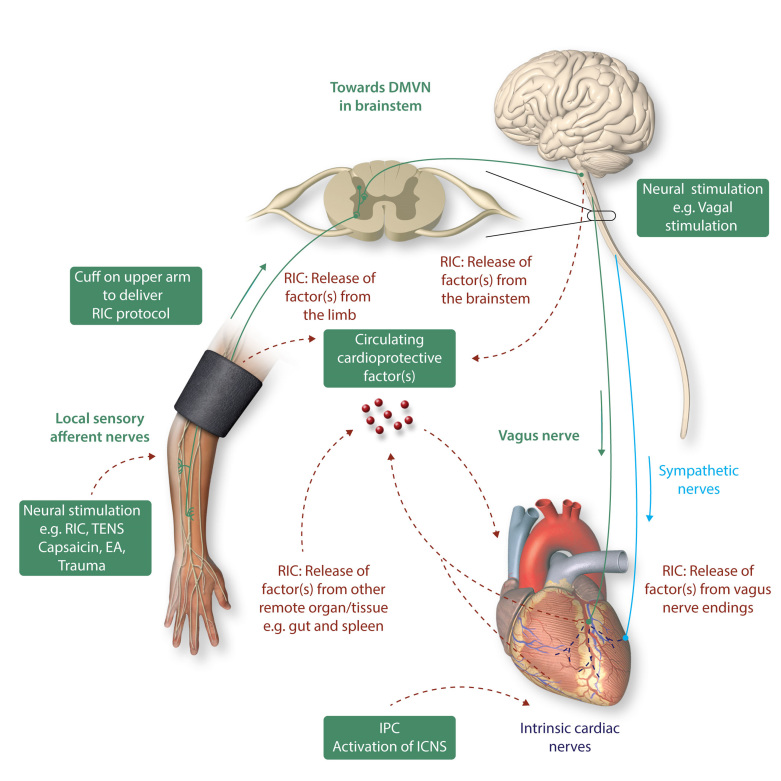
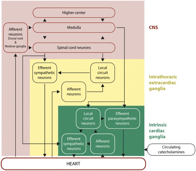
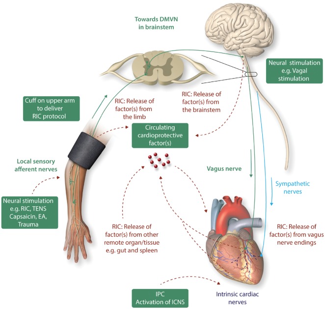
Acute myocardial infarction (AMI) and the heart failure (HF) that often complicates this condition, are among the leading causes of death and disability worldwide. To reduce myocardial infarct (MI) size and prevent heart failure, novel therapies are required to protect the heart against the detrimental effects of acute ischaemia/reperfusion injury (IRI). In this regard, targeting cardiac innervation may provide a novel therapeutic strategy for cardioprotection. A number of cardiac neural pathways mediate the beneficial effects of cardioprotective strategies such as ischaemic preconditioning and remote ischaemic conditioning, and nerve stimulation may therefore provide a novel therapeutic strategy for cardioprotection. In this article, we provide an overview of cardiac innervation and its impact on acute myocardial IRI, the role of extrinsic and intrinsic cardiac neural pathways in cardioprotection, and highlight peripheral and central nerve stimulation as a cardioprotective strategy with therapeutic potential for reducing MI size and preventing HF following AMI. This article is part of a Cardiovascular Research Spotlight Issue entitled 'Cardioprotection Beyond the Cardiomyocyte', and emerged as part of the discussions of the European Union (EU)-CARDIOPROTECTION Cooperation in Science and Technology (COST) Action, CA16225.

Keywords: Cardioprotection; Ischaemia/reperfusion injury; Myocardial infarction; Nervous system.


Figures

Graphical Abstract
Graphical Abstract
Figure 1
Figure 1
Hierarchy of cardiac innervation to the heart. This figure shows the complex and hierarchal interactions between the different components of the neural pathways of the CNS, intrathoracic extracardiac ganglia, and intrinsic cardiac ganglia of the intrinsic cardiac nervous system. This figure has been modified from Armour with permission.
Figure 2
Figure 2
Cardiac innervation and cardioprotection. The heart is innervated by the cardiac sympathetic and parasympathetic afferent and efferent neural pathways which interact with intrinsic cardiac nerves within the heart to modulate myocardial function, susceptibility to acute IRI, and cardiac arrhythmias. Cardioprotection induced by endogenous strategies such as IPC and RIC can modulate the intrinsic cardiac nerves and peripheral sensory afferent nerves in the limb and the vagus nerve, respectively. IPC cardioprotection in the isolated perfused has shown to be dependent on the function of intrinsic cardiac nerves within the heart. RIC which comprises brief non-lethal cycles of ischaemia and reperfusion to the limb, via cuff inflation/deflation, causes local autacoid release. This in turn activates sensory afferent neurons which relay, via the spinal cord, to the dorsal nucleus of vagal nerve (DMVN) in the CNS. Activation of nuclei within the DMVN results in increased vagal nerve firing to the heart which, via release of Ach and subsequent activation of muscarinic Ach receptors induces the cardioprotective phenotype. In addition, following activation of afferent sensory neurons in the conditioned limb, there is release of a dialysable cardioprotective factor into the systemic circulation. The source of this factor remains unknown, although possibilities include: (i) from the conditioned limb itself, (ii) from the central nervous system, (iii) from pre-/post-ganglionic parasympathetic nerve endings within the heart, and (iv) from a non-conditioned remote organ/tissue such as the gut or spleen. Neural stimulation of sensory afferent nerves [by RIC, transcutaneous nerve stimulation (TENS), trauma, EA, or topical capsaicin] or of the vagus nerve can induce cardioprotection. This figure has been modified from Sivaraman et al.

Similar articles

Cited by

References

    1. Armour JA. Potential clinical relevance of the ‘little brain’ on the mammalian heart. Exp Physiol 2008;93:165–176. - PubMed
    1. Kingma JG, Simard D, Rouleau JR.. Influence of cardiac nerve status on cardiovascular regulation and cardioprotection. World J Cardiol 2017;9:508–520. - PMC - PubMed
    1. Wake E, Brack K.. Characterization of the intrinsic cardiac nervous system. Auton Neurosci 2016;199:3–16. - PubMed
    1. Allen E, Coote JH, Grubb BD, Batten TF, Pauza DH, Ng GA, Brack KE.. The electrophysiological effects of nicotinic and electrical stimulation of intrinsic cardiac ganglia in the absence of extrinsic autonomic nerves in the rabbit heart. Heart Rhythm 2018;15:1698–1707. - PMC - PubMed
    1. Gatti PJ, Johnson TA, Phan P, Jordan IK III, Coleman W, Massari VJ.. The physiological and anatomical demonstration of functionally selective parasympathetic ganglia located in discrete fat pads on the feline myocardium. J Auton Nerv Syst 1995;51:255–259. - PubMed

Publication types

MeSH terms